抄録
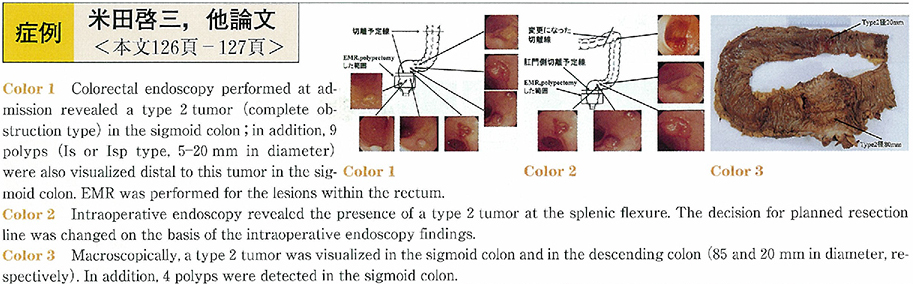
A 59-year-old man visited a nearby clinic with the chief complaint of abdominal pain, and was diagnosed to have cancer of the sigmoid colon Colorectal endoscopy revealed a complete obstruction type in the sigmoid colon ; in addition, 9 polyps were also visualized distal to this tumor in the sigmoid colon. Considering the planned resection line, EMR was performed for the polyps in the rectum Within the area of observation, however, the presence of lesions on the proximal side of the intestine could not be ruled out, considering the presence of multiple lesions on the distal side. Intraoperative endoscopy was therefore performed, which revealed the presence of a type 2 tumor in the descending colon. We performed extended left hemicolectomy low anterior resection, partial cystectomy, and regional D3 lymph node dissection. Histopathologically, the tumors resected from the sigmoid and descending colon were rated as type 2 tumors. The tumor in the sigmoid colon was diagnosed as moderately differentiated adenocarcinoma, with an invasion level of Si (urinary-bladder) . The tumor in the descending colon was also diagnosed histopathologically as a moderately differentiated adenocarcinoma, with an invasion level of SE (stage IIIa) . Thus, intraoperative endoscopy was useful for determining the extent of resection in this case.