抄録
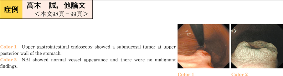
A 42-years-old woman was referred to our hospital for the treatment of submucosal tumor of the stomach which detected by medical examination. Upper gastrointestinal endoscopy showed a submucosal tumor measuring about 2cm at the upper posterior wall of the stomach. The ulcer was accompanied with the center of tumor. Computed tomography (CT) examination was performed and early phase showed that central portion of the mass was enhanced. Positron emission tomography (PET) showed no abnormal accumulation. On endoscopic ultrasonography, submucosal tumor was found in the forth layer and its size was 15×18mm. The color doppler showed blood flow in the tumor. Endoscopic ultrasonography-fine-needle aspiration biopsy was taken. And the histopathological examination showed proliferation of the cells which had oval nuclei and pale eosinophilic cytoplasm. Immunocytochemical staining was positive in the SMA, negative in the c-kit, CD34, S-100. Preoperative diagnosis was the gastric glomus tumor. We performed laparoscopy assisted partial resection of the stomach. The histopathologic findings of the resected specimen were characteristic of glomus tumor. Gastric glomus tumors are basically benign tumor. But preoperative diagnosis of gastric glomus tumor is difficult. In our case, we could diagnose the glomus tumor preoperatively, and less invasive therapy, such as laparoscopic surgery, could be selected.