抄録
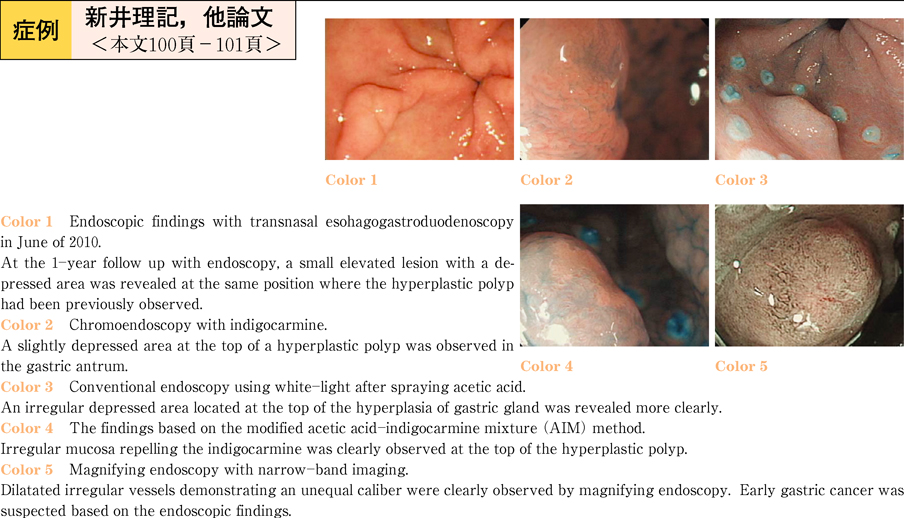
The present case was a 68-year-old female. She had been diagnosed to have a hyperplastic polyp in the posterior wall of the gastric antrum by a routine check-up with transnasal esohagogastroduodenoscopy (EGD) and biopsy. At the 1-year follow-up with EGD, a small elevated lesion with a depressed area supposed to early gastric adenocarcinoma was revealed at same position where the hyperplastic polyp had been detected. The diagnosis based on biopsy specimens was group IV, adenocarcinoma suspected. It was difficult to diagnose the existence and range of the lesion by conventional endoscopy with white-light and/or chromoendoscopy with indigocarmine. However, the modified acetic acid-indigocarmine mixture (AIM) method clearly revealed a small IIc cancerous lesion at the top of the elevated lesion. Magnifying endoscopy with narrow-band imaging showed an irregular mucosal structure, and dilatated irregular vessels demonstrating an unequal caliber. Because early gastric cancer was suspected based on the endoscopic findings, an endoscopic mucosal dissection (ESD) was selected for both diagnostic and treatment purposes. The lesion was completely resected, and there were no complications due to the ESD procedures. A histopathological examination revealed well differentiated intra-mucosal adenocarcinoma without vascular invasion. This case showed us the importance of performing image enhanced endoscopy and magnifying endoscopy to accurately rule out the presence of carcinoma even after biopsy specimens have previously indicated the presence of hyperplastic polyps.