抄録
Rectal mucosal prolapse syndrome is often difficult to differentiate from neoplastic lesions─such as advanced rectal carcinoma or rectal malignant lymphoma─using conventional colonoscopy. We report here two cases of rectal mucosal prolapse syndrome diagnosed using endoscopic ultrasonography.
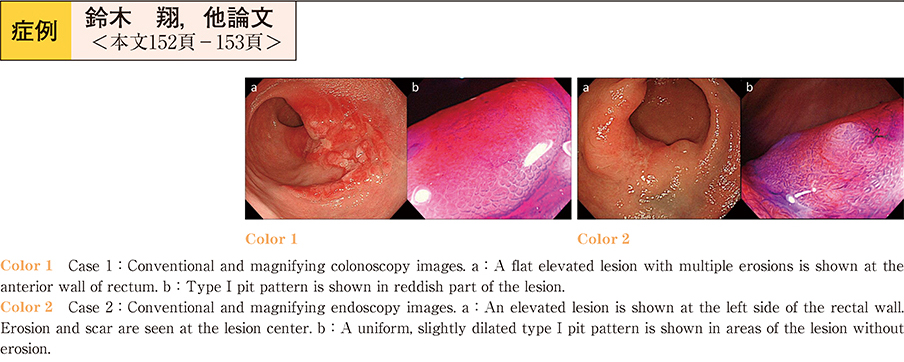
Case 1 : A 35-year-old man. Colonoscopy showed a flat elevated lesion about 5 cm diameter in the rectum. Multiple erosions were present on the surface of the lesion.
Case 2 : A 51-year-old woman. Colonoscopy showed an elevated lesion about 4 cm diameter in the rectum. Erosion and scarring were seen at the center of the lesion.
Endoscopic ultrasonography of both lesions showed smooth, diffuse thickening of the second and third layers of the rectal wall. Neither a solid hypoechoic mass nor a transmural infiltrating lesion was visible, and the five-layer structure of the rectal wall was preserved except for the scarred region. Histological examination of several biopsy specimens obtained from each lesion revealed fibromuscular obliteration. Based on these findings, the lesions were diagnosed as rectal mucosal prolapse syndrome. These findings suggest endoscopic ultrasonography is useful in diagnosis of rectal mucosal prolapse syndrome.