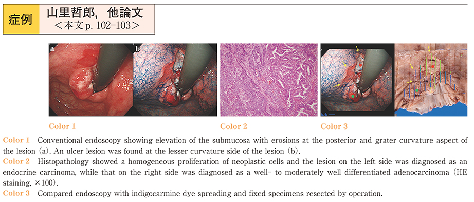
A-70-year-old man presented to our center with dysphagia. Upper gastrointestinal endoscopy revealed a type 3 lesion in the area between the lesser curvature and the posterior wall of the cardia. A shallow depressed area with a strong reddish color was found at the anal side of the ulceration and a marked elevation of the submucosal part with erosions was found at the posterior and greater curvature side of the lesion. Biopsy of the type 3 lesion revealed well-differentiated adenocarcinoma. The second lesion, a IIa lesion, was found in the lesser curvature of the lower gastric body. The two lesions were not contiguous with each other. A barium examination was performed, which revealed only the type 3 lesion mentioned above. Complete gastric resection was performed. Histopathological examination of the resected specimen revealed a well-differentiated adenocarcinoma involving the mucosa and submucosal layer. Endocrine cells of different sizes and shapes were found in the area of the ulceration. Immunohistochemical staining showed a strongly positive reaction for chromogranin A and synaptophysin. The final pathological diagnosis was endocrine carcinoma. The case is reported with a review of the literature.