抄録
This subject was a 50-year-old male who underwent routine health check-up at another clinic. Upper gastrointestinal endoscopy performed during the check-up revealed a flat, slightly reddish lesion measuring about 5 mm in diameter, that appeared malignant. Hiatal hernia, reflux esophagitis, and several fundic gland polyps measuring less than 5 mm in diameter each were also observed, however, there was no evidence of atrophic gastritis. Biopsy of the reddish lesion revealed a moderately differentiated adenocarcinoma. The cancerous lesion was resected by endoscopic submucosal resection (ESD) . Histopathologic examination revealed gastric adenocarcinoma of the fundic gland type.
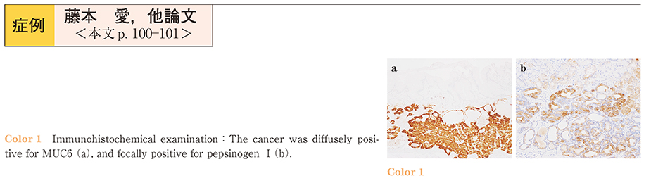
Gastric adenocarcinoma of the fundic gland type is a neoplastic lesion mainly composed of highly differentiated columnar cells mimicking the fundic gland cells, mainly chief cells, with nuclear atypia. The most important clinicopathologic feature is cancer invasion of the submucosal layer, however, most recurrent lesions tend to be less than 10 mm in diameter, as they were in our case. Immunohistochemical examination was useful for the diagnosis. The cancer cells were diffusely positive for MUC6 (a marker of fundic gland cells) and pepsinogen I (a marker of chief cells) .
We have reported the clinicopathologic findings of a case of gastric adenocarcinoma of the fundic gland type that was completely resected by ESD.