抄録
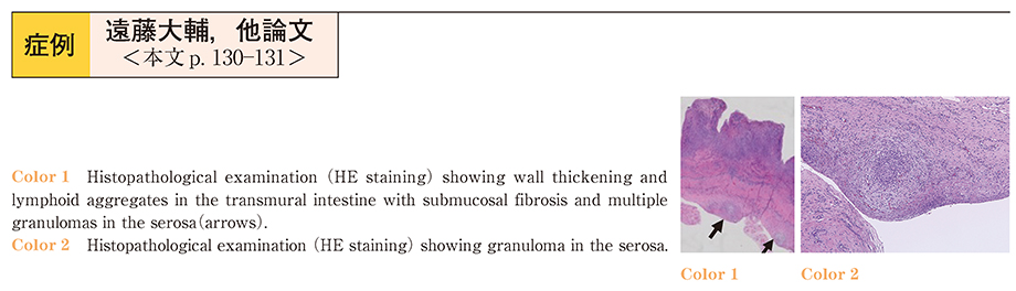
A 74-year-old man visited our hospital with complaints of abdominal discomfort and diarrhea. Upper gastrointestinal endoscopy and colonoscopy showed no evidence of disease that could explain his symptoms. He had a medical history of inguinal hernia repair by laparoscopic surgery five years ago and blood transfusion treatments for obscure gastrointestinal bleeding (OGIB) two years ago. Since some unknown small intestinal disease was suspected, capsule endoscopy was performed with a patency capsule used prior to the capsule endoscopy. At 54 hours after the capsule ingestion, he developed ileus. Computed tomography showed obstruction of the small intestine around the right inguinal region, and the patency capsule remained in the proximal extended small intestine. Ileo─jejunal resection with re-anastomosis was performed at 75 hours after the capsule ingestion. The stenosis was hard and measured 1 cm. The patency capsule was not incarcerated and had not dissolved. Histopathological (hematoxylin-eosin staining) examination showed infiltration of lymphocytes in the transmural layers within the limits of the stenosis, and to our surprise, multiple granulomas infiltrating the serosa, although the result of acid-fast bacterial staining was negative. There were no abnormal findings of the vessel walls. There are several case reports of paravesical granulomas whose formation is triggered by foreign bodies several years after inguinal hernia repair. In this case, the intestinal stenosis was adjacent to the narrow space between the adhesive cord and the abdominal wall. We thought that temporary and repeated incarcerations of the small intestine into that narrow space may have been responsible for the severe stenosis with granulomas in the serosa and the chronic ischemic inflammation of the intestine.