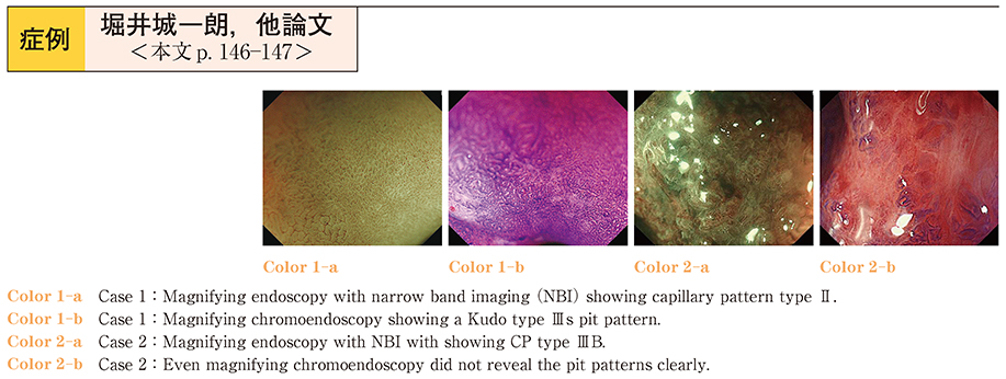
Case 1 : A 70-year-old man was referred for endoscopic resection, as a screening colonoscopy had revealed a flat neoplasm in the sigmoid colon measuring 40 mm in diameter without a granular pattern on the surface (laterally spreading tumor, non-granular type [LST-NG]) . Magnifying endoscopy with narrow-band imaging (NBI) revealed capillary pattern (CP) type II, whereas magnifying chromoendoscopy revealed a Kudo type IIIs pit pattern.
Case 2 : A 70-year-old woman was referred for endoscopic resection of a 40-mm LST-NG located in the rectum. Magnifying endoscopy with NBI revealed CP type IIIB, and even magnifying chromoendoscopy failed to reveal the pit pattern clearly, although the endoscopic examination revealed no hardness of the tumor.
Thus, the neoplasms were diagnosed as intramucosal carcinoma or cancer with superficial submucosal invasion, and we decided to attempt endoscopic submucosal dissection (ESD) to remove them. In Case 1, the histopathologic assessment showed a cancer with deep submucosal invasion, which was fully covered by the cancer with low-grade atypia in the mucosal layer. In Case 2, the histopathologic assessment showed tubular adenoma with a large quantity of mucus on the surface of the tumor. Thus, the histopathologic assessment also revealed the depth of the tumor invasion and the reason for the difficulty in the preoperative diagnosis. This case report highlights the importance of ESD, which enabled a precise histopathologic diagnosis of the LSTs in which estimation of the depth of submucosal invasion was difficult even with the use of magnifying NBI and chromoendoscopy.