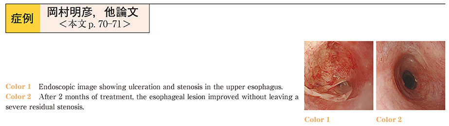
抄録
Pemphigoid is an autoimmune bullous disease characterized by subepithelial blistering of the skin and mucosa. The lesions are caused by autoantibodies against components of the hemidesmosomes, the junctional adhesion complex. Various subtypes of pemphigoid are recognized, such as bullous pemphigoid, mucous membrane pemphigoid and epidermolysis bullosa acquisita. Mucosal lesions are usually located in the oral and pharyngeal mucosa. Although the esophagus is also covered by squamous epithelium, there are only a few case reports of esophageal lesions of pemphigoid. From Jul/2011 to Jul/2013, we performed gastrointestinal endoscopy in 23 pemphigoid patients and found esophageal lesions in four (17%) ; in all four cases, blisters, erosions, ulceration and stenosis were found in the esophagus. While oropharyngeal lesions coexist in many cases, we should pay attention to cases without skin lesions, depending on the subtypes. Therefore, the gastrointestinal endoscopist has an important role in the diagnosis of pemphigoid.