抄録
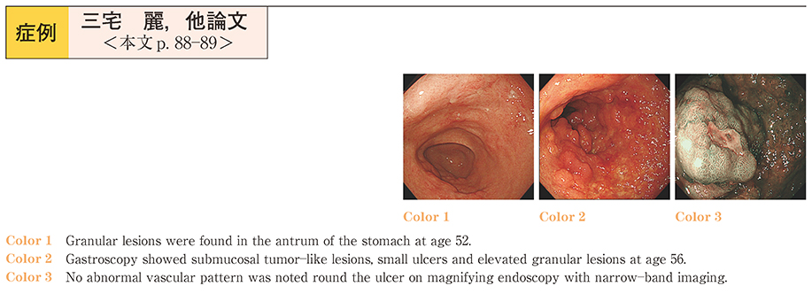
A 56-year-old-woman diagnosed as having primary gastric amyloidosis at age 52 was followed up by annual gastroscopy. Although small granular lesions in the antrum of the stomach were seen at age 52, a variety of lesions, such as small ulcers, submucosal tumor-like lesions and elevated granular lesions in the antrum and cardia of the stomach were found at age 56. With narrow-band imaging (NBI) enhancement, the mucosa surrounding the ulcers appeared to be intact. The histological examination findings were consistent with gastric amyloidosis. She was continued on treatment with a proton pomp inhibitor.
Primary gastric amyloidosis is rare and only 17 case reports with a description of the endoscopic findings have been published in Japan. Primary gastric amyloidosis manifests with a variety of endoscopic findings. We divided these findings into the following four types, submucosal tumor-like, early cancer-like (IIc-like) , advanced cancer-like (Borrmann typeII-like) and elevated granular lesions. In our case, we observed two types of endoscopic findings over the course of 5 years. Surgical interventions have been reported in some cases because of uncontrolled hemorrhage or stenosis. We would perform endoscopy annually in our case.