抄録
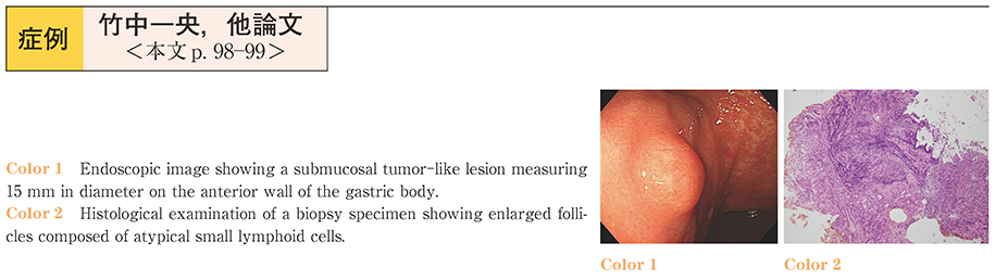
A 65-year-old woman with liver cirrhosis (type C) underwent esophagogastroduodenoscopy (EGD) for examination of esophago-gastric varices. EGD revealed a submucosal tumor (SMT) -like appearance in the anterior wall of the gastric body. Endoscopic ultrasonogaraphy indicated that the lesion was a low-echoic mass localized within the muscularis mucosa. Histopathology of a biopsy specimen from the lesion showed enlarged follicles composed of atypical small lymphoid cells, however, the atypical cells did not infiltrate or destroy the glandular epithelium, i.e., there were no lympho-epithelial lesions. On immunohistochemical analysis, the atypical cells was positive for CD10, CD20, CD79a and BCL-2, and negative for Cyclin D1. We diagnosed the SMT-like lesion as a follicular lymphoma. There was no obvious accumulation on FDG-PET. However, a bone marrow aspirate and biopsy revealed CD10-and BCL-2-positive cells. Based on these findings, the patient was diagnosed as having Stage IV primary gastric lymphoma, and was adiministered chemotherapy with rituximab. EGD after three courses showed that the SMT-like lesion had disappeared. We report a rare case of gastric follicular lymphoma presenting with a SMT-like appearance.