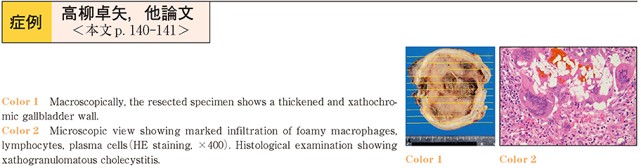
抄録
Xanthogranulomatous cholecystitis (XGC) is difficult to differentiate from malignant tumors, such as gallbladder and bile duct carcinoma. We report a case of XGC which was diagnosed preoperatively by recognizing the continuity of the mucosal layer in the gallbladder wall on multi-detector computed tomography (MDCT) . A 63-year-old man was admitted to the hospital because of jaundice. Abdominal ultrasound showed intrahepatic bile duct dilatation and stones in the gallbladder. MDCT showed diffuse thickening of the gallbladder wall and stenosis of the upper biliary tract. ERC showed bile duct stricture extending from the common bile duct to both hepatic ducts. Positron emission tomography (PET) showed abnormal accumulation in the bile duct stricture, with a standardized uptake value (SUV) of 5.0. The serum levels of the tumor markers CA19-9, DUPAN2 and SUPAN1 were also elevated. Based on these findings, the diagnosis of gallbladder carcinoma infiltrating the bile duct was suspected.
On the other hand, ERCP brush cytology and EUSFNA did not reveal any evidence of malignancy. Furthermore, we recognized continuity of the mucosal layer in the gallbladder wall on MDCT. Although XGC was strongly suspected based on the imaging findings, we performed resection of the gallbladder bed. Intraoperative pathological examination confirmed the diagnosis of XGC and ruled out malignancy. The findings on MDCT allowed us to avoid unnecessary extended operation.