Abstract
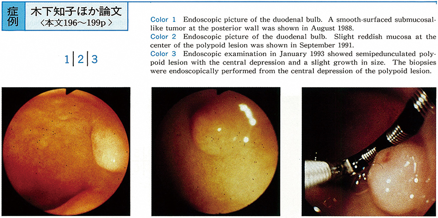
A 39-year-old male visited our hospital with a complaint of upper abdominal discomfort in 1988. The upper GI series showed the hemispherical polypoid lesion. Endoscopic examination showed a smooth-surfaced submucosal-like tumor (φ 6mm) at the posterior wall of the duodenal bulb. Afterward, endoscopic exmination was performed once a year.
The third endoscopic examination showed no remarkable change of the polypoid lesion, however, endoscopic examination performed 4 years and 5 months later showed semipedunculated polypoid lesion with the central depression and a slight growth in size (φ 8mm) . Carcinoid tumor was diagnosed by the endoscopic biopsy from the central depression.
The tumor was removed by surgical operation (hemiduodenectory) . Microscopic exanimation revealed a carcinoid composed of tumor cells with round shaped nuclei and trabecular structure. Carcinoid tumor invaded the duodenal submucosa massively. The tumor tissue was stained positively for Leu 7, Chg A, NSE, gastrin, insulin and somatostatin immunohistochemically.
We present our experience of a very rare case which showed morphological changes of carcinoid tumor after the observation period of 4 years and 5 months. We also reviewed 22 cases of carcinoid tumor from the literature which were diagnosed by endoscopic biopsy.