Progress of Digestive Endoscopy(1972)
Online ISSN : 2189-0021
Print ISSN : 0389-9403
Volume 43
Displaying 1-50 of 57 articles from this issue
Technology and instrument
Clinical study
  • Nobuhiko Nagamine, Norio Ueno, Yukiko Oota, Masami Fukuda, Takeshi Tom ...
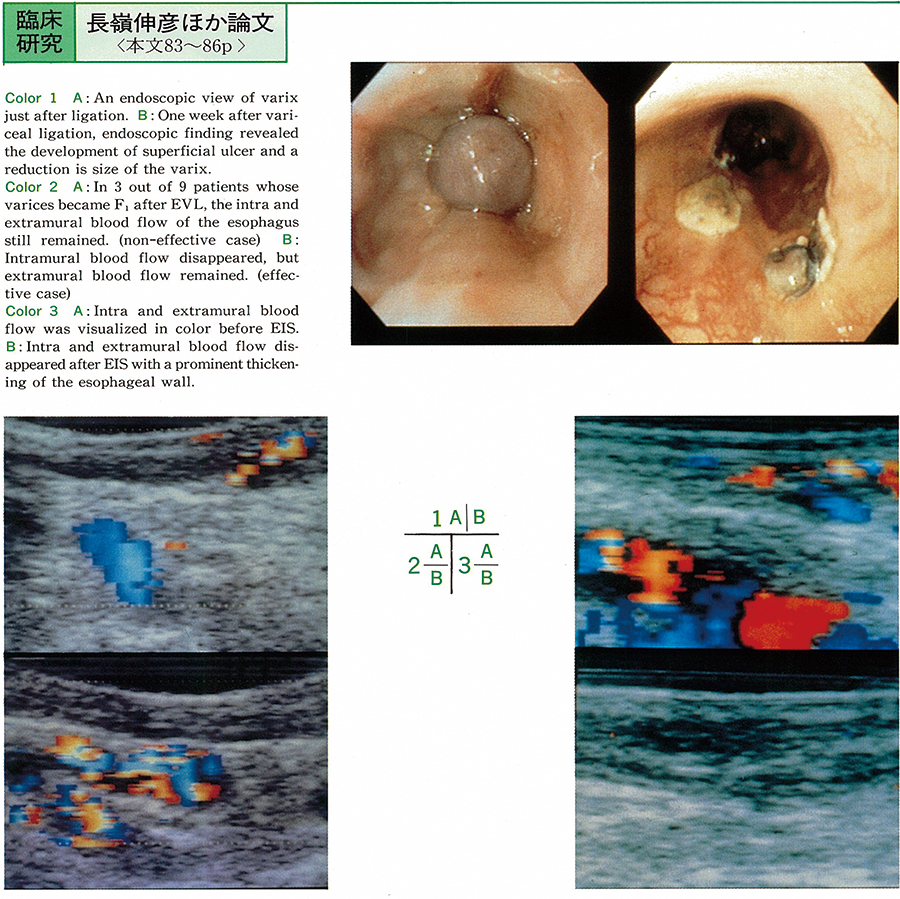
    1993 Volume 43 Pages 83-86
    Published: December 01, 1993
    Released on J-STAGE: July 15, 2015
    JOURNAL FREE ACCESS
    We evaluated the hemodynamics of the variceal venous flow in patients with esophageal varices before and after endoscopic variceal ligation (EVL) by using color Doppler endoscopic ultrasonography (CD-EUS) . We tried to detect intramural, extramural and supplying blood flow of the esophagus of 11 (varices form F3 in 2 patients, F2 in 9 patients) out of 24 patients who had been performed EVL using Stiegmann-Goff ligator kit (Bard, USA) by the method according to Stiegmann from November 1992 to May 1993.
    Color display indicating blood flow in varices could be observed in all cases before EVL by CD-EUS.
    Endoscopic examination revealed that the forms of varices were improved to F0 in 2 patients and F1 in 9 patients after EVL. CD-EUS examination showed that intramural blood flow disappeared after EVL in 8 out of 11 patients. On the other hand, the intramural blood flow had still remained in 3 out of 11 patients after EVL. There were no evidence of definite changes in the extramural or supplying venous blood flow in all patients. In 2 patients, they were treated with EIS in the past several years, prominent thickening of the esophageal wall was observed. In conclusion, CD-EUS examination was clinically useful in evaluation of blood flow after EVL.
    Download PDF (439K)
  • Norie Ishigaki, Shigehiro Kokubu, Masato Murakami, Masahiro Takada, Hi ...
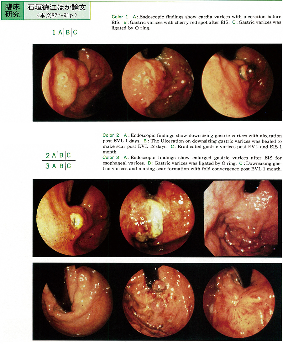
    1993 Volume 43 Pages 87-91
    Published: December 01, 1993
    Released on J-STAGE: July 15, 2015
    JOURNAL FREE ACCESS
    A newly developed“Endoscopic variceal ligation (EVL) ”is a method which has been evaluated for its usefulness, safety and easyhandling for the treatment of esophageal varices. However, no study on its use in cardia varices has been reported.
    We have recently evaluated the EVL (Stiegmann's method) and the results are reported before. A small elastic rubber band was used to occlude and eradicate the varices, with an adjunct therapy of endoscopic injection sclerotherapy (EIS) for cardia varices. Five patients with esophago-gastric varices (three male and two female) were treated with EVL for cardia varices. The timing of therapeutic indication for these cases was : urgent in two, elective in one and prophylactic in two cases.
    All the cases had good clinical course without post EVL rebleeding. We have determined the following indications of EVL in the present study : 1. Aged patient having multiple complications or other organic diseases ; 2. Patients having allergic reaction to sclerosant ; 3. Patients with poor condition who requires urgent therapy ; 4. Patients with rebleeding from the ulcer after EIS ; and 5. Patients who achieved no downsizing cardia varices though injection of sclerosant was conducted blood supply route. EVL alone could not have completely eradicated of cardia varices because a large amount of blood flows towards the cardia varices, as a results of endoscopic varicealography during injection sclerotherapy (EVIS) performed after EVL.
    We have concluded that EVL can achieve therapeutic efficacy by combination use of EIS as an adjunct therapy.
    Download PDF (1229K)
  • Tekeshi Matsuhisa, Masahide Itoh, Hiroshi Oshima
    1993 Volume 43 Pages 92-95
    Published: December 01, 1993
    Released on J-STAGE: July 15, 2015
    JOURNAL FREE ACCESS
    Relation between refluxed taurocholic acid into the stomach and atrophic gastritis, endoscopic forms of the pylorus were studied.
    1) A frequency of refluxed cases of taurocholic acid in the stomach occupied 41.0% out of 39 cases.
    2) We used Oshima's criteria in order to observe the relation between taurocholic acid and gastric mucosa. Gastric mucosa was classified into 3 groups, which were (+) and (±) group, (+) group and (⧺) group. Refluxed frequency and concentration in (+) and (±) group showed low level (each 18.8%, 1.82nmol/ml) . On the other hand, these levels were high in (+) group which has atrophic change and (⧺) group which has remarkable atrophic change ( (+) group : 57.1%, 17.43nmol/ml, (⧺) group : 55.6%, 21.45nmol/ml) . We considered the effect of taurocholic acid to atrophic change in gastric mucosa.
    3) We couldn't find difference between taurocholic acid and form of pyloric ring, peristalsis.
    Download PDF (420K)
  • Hisao Nakai, Masahito Ohida, Sayuri Yamagata, Tomoya Kan, Satoshi Tana ...
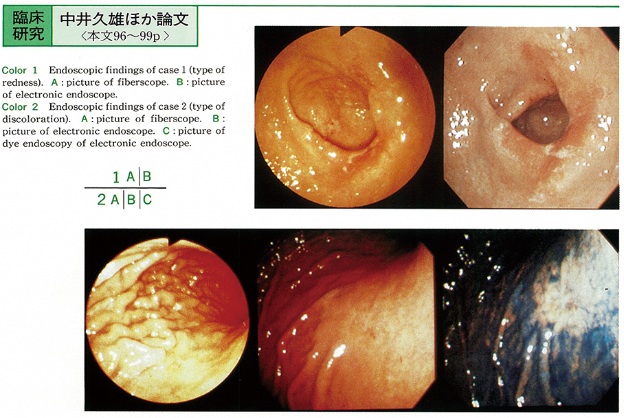
    1993 Volume 43 Pages 96-99
    Published: December 01, 1993
    Released on J-STAGE: July 15, 2015
    JOURNAL FREE ACCESS
    Forty-two depressed lesions of early gastric cancer which were resected at this hospital during the 1 year and 11 months from November 1990 to September 1992 and underwent examination by both fiberscope and electronic endoscope were studied to diagnose the extent of infiltration.
    The basic color tone of the lesions was classified into three types : discolored (22 lesions) , same color (5 lesions) and reddish (15 lesions) . The usefulness of electronic endoscopy was compared with that of fiberscopy in regard to the definition of color tone clarity, capillary transparence, gastric mucosal pattern, lesion margin and luster. In addition, resected specimens were studied histopathologically.
    In about half of the reddish lesions, electronic endoscopy was effective for defining lesion characteristics, and no major differences were noted between fiberscopy and electronic endoscopy. Discolored lesions were frequently unable to be effectively examined by electronic endoscopy and showed remarkable signs of superficial mucosal infiltration of cancer. However, the concurrent use of dye endoscopy resulted in improved clarity, thereby enhancing performance.
    Download PDF (530K)
  • Hideyuki Kishi, Shunya Ishii, Shunichi Nakajima, Shuji Matsumura, Hiro ...
    1993 Volume 43 Pages 100-103
    Published: December 01, 1993
    Released on J-STAGE: July 15, 2015
    JOURNAL FREE ACCESS
    Twenty four cases of pseudomembranous colitis diagnosed by endoscopy were divided into 2 : hemispherical type and membranous one. Most of hemispherical type were in poorly general condition while membranous one good. Oral administration of third generation of cephems was highly related with occurrence of membranous type in recent years.
    Histopathologically, hemispherical types showed Type 2 and 3, and membranous types showed Type 1 according to Price. Days from oncet to endoscopy and days until asymptomatic phase were almost same in both groups.
    It was suggested that endoscopy had been need to even in case with mild symptom when pseudomembranous colitis was suspected.
    Download PDF (491K)
  • Shichiroku Watanabe, Hirokazu Inoue, Hiroyuki Kobayashi, Hideyuki Kish ...
    1993 Volume 43 Pages 104-107
    Published: December 01, 1993
    Released on J-STAGE: July 15, 2015
    JOURNAL FREE ACCESS
    Four cases with ulcerative colitis were performed one shot intraarterial administration of corticosteroid whose intensive intravenous administration failed previously. For three cases with severe stage it was not effective, and one of them died in spite of emergent colectomy. But in one performed continuous arterial administration for three days, remission has been was maintained for two years. For one case with moderate stage it was quite effective.
    The prompt estimation of the intraarterial administration should be decided because the patient condition was critical. For shortning the judgement time of indication for surgical treatment, intraarterial administration should be selected earlier than intravenous one. Clinical symptoms, ie, abdominal pain, frequency of bowel movement, degree of melena, and body temperature were more helpful to decide the effectivity of intraarterial administration than laboratory data or endoscopy.
    Download PDF (520K)
  • Sumio Fujinuma, Yoshihiro Sakai, Shunichi Nakajima, Shuji Matsumura, S ...
    1993 Volume 43 Pages 108-111
    Published: December 01, 1993
    Released on J-STAGE: July 15, 2015
    JOURNAL FREE ACCESS
    From May 1989 to December 1992, 214 superficial lesions were obtained by endoscopic resection with prior injection into submucosa and 21 lesions (9.8%) of those were hyperplastic polyp.
    In this paper, these hyperplastic polyps were studied endoscopically and pathologically. They were divided histologically into 2 : the simple enlongated type (10 lesions) and the cystic dilated type (11 lesions) . The simple enlongated type showed mostly low in height, small in size, pale or similar to the surrounding mucosa and smooth in surface. On the other hand, the cystic dilated type showed high, large, similar or hyperemic in color, and nodular to depressed in surface.
    The few lesions could not be diagnosed hyperplastic lesion even by dissecting microscopic observation after recovery.
    Download PDF (786K)
  • Shichiroku Watanabe, Hirokazu Inoue, Hiroyuki Kobayashi, Tadayoshi Kak ...
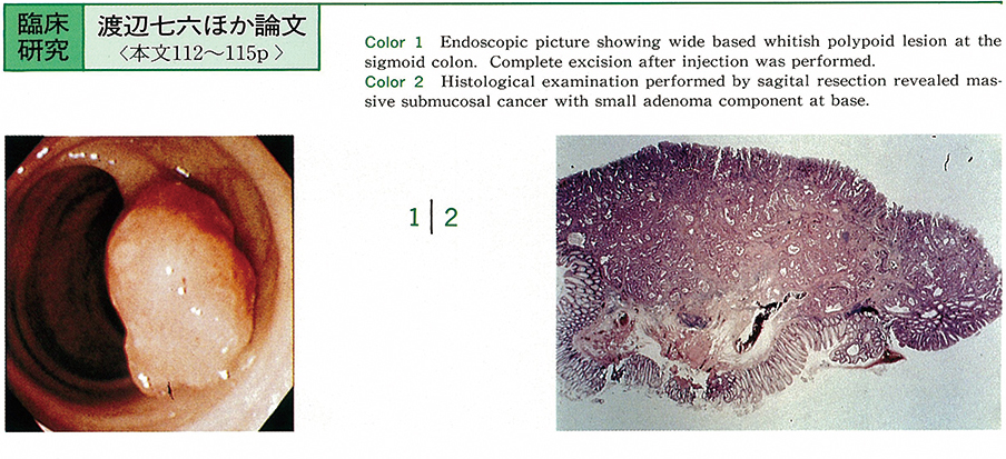
    1993 Volume 43 Pages 112-115
    Published: December 01, 1993
    Released on J-STAGE: July 15, 2015
    JOURNAL FREE ACCESS
    Endoscopically resected epithelial neoplastic lesions were studied to evaluate the carcinomatous change in them. Subjects were more than 10mm in size and more than 5mm in height. Histologically, they were 51 adenoma and 42 carcinoma with adenoma component. The latter were devided by the carcinoma area among the lesion in the maximal cut surface ; small (less than 10%) 17, medium (30-70%) 15, and large (more than 90%) 5 lesions. Remaining 5 lesions were already invaded into the submucosal layer (Table 1) .
    Macroscopically, sessile, and broad-based configuration suggested large amounts of carcinoma area of submucosal invasion (Table 2) . Concerning the head of the lesions, unusual shape revealed further involvement by carcinoma while spherical one showed adenoma or small amount of carcinoma (Table 3) . Asymmetric or manifested the high possibility of malignancy (Table 4) . The size and hyperemia however, did not correlate the amount of carcinoma (Table 5, 6) .
    Download PDF (490K)
  • Hideki Yoshioka, Shunya Ishii, Shunichi Nakajima, Shuji Matsumura, Hir ...
    1993 Volume 43 Pages 116-119
    Published: December 01, 1993
    Released on J-STAGE: July 15, 2015
    JOURNAL FREE ACCESS
    Eighteen lesions with minute colorectal cancer (5mm or less in diameter) out of 237 early colorectal cancers were identified and removed endoscopically from January 1982 to March 1993. They were devided into 3 groups according to the maximal diameter (Group 1 : smaller than 5mm, Group 2 : from 6mm to 10mm, Group 3 : larger than 11mm) . Group 1 and Group 2 were compared with studies of endoscopic findings (distribution, macroscopic forms, surface color, surface appearance) and histopathological findings (adenoma component, depth of cancer invasion) in this paper, and result were as follows ;
    1) Group 1 was 7.6% among all early cancer.
    2) Group 1 were distributed in the protruded type and the superficial type.
    3) Group 1 showed higher ratio of cancer accompanied without adenoma, distribution of transverse colon, and macroscopic forms of Is than those with Group 2.
    4) Five cases (36%) with superficial lesions of Group 1 could be diagnosed endoscopically by surface appearances with rough granules or central depression suggesting of malignacy.
    5) Histopathology revealed well differentiated adenocarcinoma and invasion limited into mucosa or submucosa in all lesions ; Even in cases of Group 1, both cancer adenoma sequence and“de novo”cancer theory were not denied.
    6) Two (29%) out of seven lesion cancer without adenoma component revealed submucosal invasion. In lesions of minute colorectal cancer, cancer without adenoma component was suggested more early invasion than cancer with adenoma component.
    Download PDF (509K)
  • Terumi Kamisawa, Tomoaki Isawa, Tu Yuyang, Nobuhiro Sakaki, Tsuyoshi T ...
    1993 Volume 43 Pages 120-122
    Published: December 01, 1993
    Released on J-STAGE: July 15, 2015
    JOURNAL FREE ACCESS
    We studied endoscopic findings of gastrointestine in 146 patients with pancreatic cancer.
    On gastrofiberscopy, abnormal findings were detected in 42 of 142 cases (33 findings of compression and 9 findings of invasion) . Pancreatic body and tail cancer showed high frequency (39% and 47%) of abnormality. On duodenofiberscopy, 43 of 122 cases showed abnormal findings in the duodenal second portion (15 findings of compression and 28 findings of invasion) . Pancreatic head and uncus cancer showed abnormal findings frequently (44% and 57%) . Four of 12 cases biopsied from stomach and nine of 23 cases from duodenum revealed cancer cells infiltrated from the pancreas.
    On colonofiberscopy, 6 of 28 cases showed stenosis or obstruction of transverse colon. It is necessary not to overlook advanced pancreatic cancer in routine panendoscopy.
    Download PDF (350K)
Case report
  • Izumi Hino, Fumihiko Tanaka, Hiroshi Asakawa, Mika Matsuoka, Takahiro ...
    1993 Volume 43 Pages 123-126
    Published: December 01, 1993
    Released on J-STAGE: July 15, 2015
    JOURNAL FREE ACCESS
    Since the history of continuous ambulatory peritoneal dialysis (CAPD) is only 13 years long, there are little known about the complication of gastrointestinal tract of CAPD patient.
    We experienced 2 patients whose chief complaints were dysphagia, and were diagnosed reflux esophagitis by endoscopic examination, in spite of having no esophageal hernia nor Barrett's esophagus. Manometric examination show that both of lower esophageal sphincter pressures were normal. By infusing CAPD solution into the peritoneal cavity, the intraabdominal pressure rises, and the gastric juice refluxes into the esophagus. This mechanism is thought to be one important cause of the reflux esophagitis.
    In our hospital, 2 patients had reflux esophagitis among the 32 CAPD patients (6.25%) . No reports were made about the patients of reflux esophagitis of CAPD patients till now. In recent 13 years, the number of CAPD patients is increasing up to 6,000. So reflux esophagitis is thought to be one of the important gastrointestinal complication of CAPD patients.
    Download PDF (973K)
  • Hiroshi Miyoshi, Tamami Hanashi, Yuji Hanatani, Tatsuo Asagoe, Tadahik ...
    1993 Volume 43 Pages 127-129
    Published: December 01, 1993
    Released on J-STAGE: July 15, 2015
    JOURNAL FREE ACCESS
    A 41-year-old male was admitted complaining of sudden cervico-thoracic pain and dysphagia. Esophagram and thoracic CT scan with barium meal showed barium retension round esophageal cavity, and the diagnosis of esophageal submucosal dissection was completed by emergent endoscopic examination. Conservative therapy including hyper-alimentation was indicated, but it took 2 months for complete remission.
    Esophageal submucosal dissection should be distinguished from exfoliative esophagitis, because the former usually need long-term treatment session.
    Download PDF (803K)
  • Hirofumi Miyamoto, Yoshitake Wakou, Minoru Hanashiro, Chiaki Abe, Tour ...
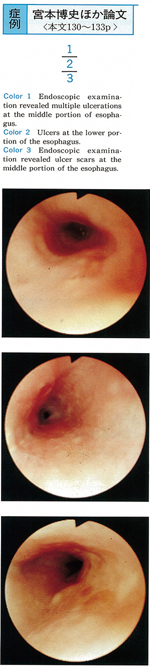
    1993 Volume 43 Pages 130-133
    Published: December 01, 1993
    Released on J-STAGE: July 15, 2015
    JOURNAL FREE ACCESS
    A 36-year-old woman was admitted to our hospital because of oral ulcers, genital ulcers, arthritis, and fever in Oct 1993. Being diagnosed with incomplete Behçet's disease, she was treated with on prednisolone, 30mg po. She had no particular subjective symptoms, but endoscopic examination of the upper gastrointestinal tract revealed multiple ulcers in the middle and lower esophagus. Biopsied specimens taken from lesions revealed granulation and infiltration by phlogocytes, yielding nonspecific inflammatory findings. On the 20th hospital day, the healing of ulcers was confirmed by endoscopy.
    It is said that Behçet's disease is rarely complicated by esophageal lesions, but several cases have recently been reported. In our case endoscopic findings improved with steroid treatment. This appears to be an interesting case of esophageal lesions supervening on Behçet's disease.
    Download PDF (829K)
  • Shigeo Yoshida, Manabu Asada, Takeshi Suzuki, Akira Nakamura, Ei Itoba ...
    1993 Volume 43 Pages 134-137
    Published: December 01, 1993
    Released on J-STAGE: July 15, 2015
    JOURNAL FREE ACCESS
    Dialatation of severe stricture at the site of esophago-gastric anastomosis was performed with coaxial dilator (Zeon Medical Inc) made of teflon, which was produced to dilate the bile duct stricture.
    The procedure was performed in two postoperative patients who had had esophageal cancer or idiopathic esophageal rupture. The size of the stricture of the former patient was 1 mm in diameter and 3 mm in length, and that of the later was 2 mm and 15 mm, respectively. Co-axial dilator of 2 mm in diameter was inserted under fluoroscope over a guide-wire which was previously passed through the stricture by endoscope. Then co-axial dilater of 3, 4 and 5 mm in diameter were inserted in succession. Then Rigiflex baloon dilator (Microvasive Inc) and SBM baloon dilator (Sumitomo Bakelite Inc) were used further to dilate the stricture.
    The anastomosis was dilated enough, and they had become able to ingest solid food. There was no symptom of restricture thereafter. Co-axial dilator was effective for the dilatation of severe esophageal stricture, especially in the cases in which dilatation technique using baloon cannot be indicated.
    Download PDF (1565K)
  • Yoshiyuki Suzuki, Hitomi Adachi, Yukihito Nemoto, Midori Ikeda, Kimie ...
    1993 Volume 43 Pages 138-142
    Published: December 01, 1993
    Released on J-STAGE: July 15, 2015
    JOURNAL FREE ACCESS
    A 49-year-old female with epigastralgia and abdominal tumor is described. The endoscopic examination of upper gastrointestinal tract revealed co-presence of elevated lesions and flat mucosa on the greater curvature of the lower to upper gastric body. Biopsy specimens of the elevated lesions demonstrated hyperplasia of fundic glands, and specimens of flat mucosa showed severe atrophic gastritis. Discolored annular zone was seen in the lower portion of the gastric body with congo red test.
    This case is apparently different from the known type A or B gastritis, and may represent an interesting case in considering gastritis progression.
    Download PDF (1751K)
  • Fumio Hirota, Takahiko Mori, Kazuhiro Abe, Hisashi Nakamura, Hiroshi K ...
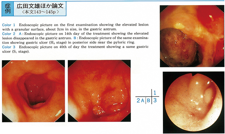
    1993 Volume 43 Pages 143-145
    Published: December 01, 1993
    Released on J-STAGE: July 15, 2015
    JOURNAL FREE ACCESS
    A 54-year-old male underwent upper gastrointestinal endoscopy due to chief complaint of epigastralgia. An elevated lesion with a granular surface, about 2 cm in size, was observed in the gastric antrum. A hepatic tissue was obtained by biopsy from the lesion and the patient was diagnosed as having gastric ulcer penetrated to the liver. Because of scarce inflammatory findings, conservative therapy was started with intravenous hyperalimentation and H2-receptor antagonist administration. On day 14 of the treatment, the elevated lesion disappeared and the ulcer showed a tendency to healing.
    A penetrating gastric ulcer in which the liver protruded is rare and interesting moreover with the successful conservative healing.
    Download PDF (1167K)
  • Kei Nakamura, Noboru Mizobuchi, Yasumitsu Katsuura, Takeo Maekawa, Nob ...
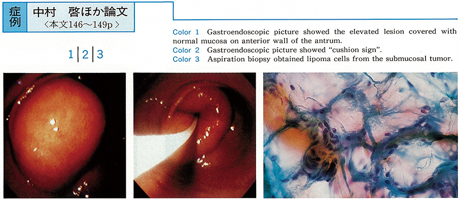
    1993 Volume 43 Pages 146-149
    Published: December 01, 1993
    Released on J-STAGE: July 15, 2015
    JOURNAL FREE ACCESS
    A 43-year-old man was referred to a near clinic with epigastralgia. He was admitted to our hospital with the diagnosis of gastric submucosal tumor.
    The upper GI series showed a protruded lesion of about 5cm in diameter on the anterior wall of the antrum. The gastroendoscopical finding showed the same lesion. It had the so-called“cushion sign”. EUS showed a mature tumor of a high echo pattern and heterogenous in the submucosal space. It was diagnosed as lipoma of the stomach. But the diagnosis of the liposarcoma was rejected because the EUS and CT scan showed liposarcoma's sign.
    On Jan 22, 1993, an antrectomy was underwent. The pathologic diagnosis was lipoma. It is a relatively rare case of lipoma of the stomach. There have been 167 reported cases of lipoma of the stomach in Japan. The mean age is 58.5 years old. The cases of women were more than cases of men. Most of the lipoma of the stomach was located within the A area, and most of the diameter was smaller than 5cm. Most of the lipoma of the stomach was underwent gastrectomy because lipoma of the stomach had no characteristic symptoms and preoperative diagnosis was difficult.
    Download PDF (1869K)
  • Shunya Ishii, Shunichi Nakajima, Shuji Matsumura, Hirokazu Inoue, Shun ...
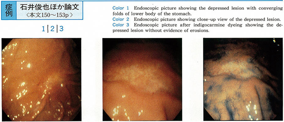
    1993 Volume 43 Pages 150-153
    Published: December 01, 1993
    Released on J-STAGE: July 15, 2015
    JOURNAL FREE ACCESS
    A patient was 58-year-old man, who was admitted to our hospital due to loss of appetite and upper abdominal pain. Gastroscopy revealed an irregularly depressed lesion with converging folds in anterior wall of lower body of the stomach. Biopsy specimen showed marked amyloid deposition in the mucosa and submucosa. All biopsied specimens from duodenal bulb, terminal ileum, sigmoid colon, rectum, and gastric mucosa except depressive lesion, were negative in amyloid deposition. Serum amyloid A protein was high, but localized amyloidosis of the stomach was diagnosed owing to blood examinations, urinalysis, physical finding, chest-abdominal X-ray films, abdominal ultrasonography, abdominal CT scan, ECG, and cardiac ultrasonography.
    Localized amyloidosis of the stomach with depression seemed to be very rare according to the previously published reports in the world.
    Download PDF (757K)
  • Takeo Arakawa, Masato Ohida, Satoshi Tanabe, Wasaburo Koizumi, Yasushi ...
    1993 Volume 43 Pages 154-157
    Published: December 01, 1993
    Released on J-STAGE: July 15, 2015
    JOURNAL FREE ACCESS
    A 40-year-old female visited our hospital complaining of epigastric discomfort. Physical examination and laboratory findings revealed iron deficiency anemia and hypoproteinemia due to protein losing gastropathy. Upper gastrointestinal series and endoscopic examination revealed numerous polyps from the gastric angle to the pylorus. Histological examination of the polyps showed characteristic features of juvenile polyposis, and adenomatous pattern was found.
    This patient is considered to be the rare case in which juvenile polyposis involved the stomach alone. Family history is positive. Her sister have multiple juvenile polyps, and several polyps have adenocarcinoma in situ. Follow-up study with endoscopic examination is necessary in this patient because of development and malignancy.
    Download PDF (1353K)
  • Hitoshi Yoshimoto, Makoto Futaki, Katsuhiko Iwakiri, Hideo Sawada, Nob ...
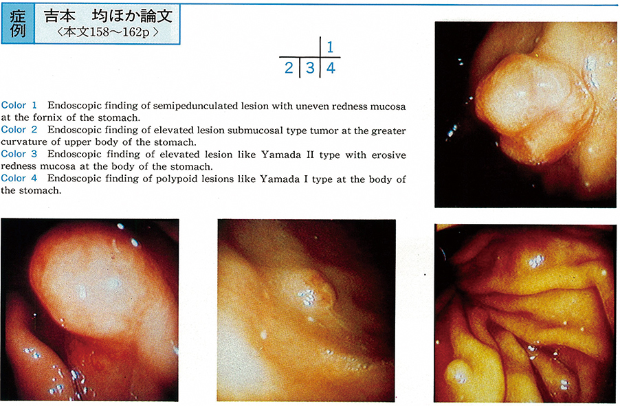
    1993 Volume 43 Pages 158-162
    Published: December 01, 1993
    Released on J-STAGE: July 15, 2015
    JOURNAL FREE ACCESS
    Endoscopic examination of the stomach was performed on a 33-year-old woman. Multiple elevated lesions with various appearance were observed from the cardia to the body. They included two semipedunculated tumors with uneven mucosa, erosion and redness, one submucosal tumor and one Yamada type II tumor with slightly redness and three Yamada type I tumors. Histological diagnosis of biopsy specimens of three of them was carcinoid tumor. Examination of totally resected stomach revealed 8 carcinod tumors.
    Micronests of endocrine cells were observed in the mucosal layer. There was no atrophy of the mucosa macroscopically, but the atrophy of fundic mucosa was confirmed on histological examination compatible with type A gastritis. She was hyper-gastrinemic preoperatively. She had no carcinoid symptom.
    Download PDF (2412K)
  • Mari Saito, Mikio Matsuoka, Kohki Mitsuya, Masahiko Takao, Kotaroh Mat ...
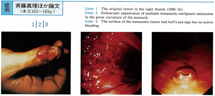
    1993 Volume 43 Pages 163-165
    Published: December 01, 1993
    Released on J-STAGE: July 15, 2015
    JOURNAL FREE ACCESS
    The patient is 63-year-old man, who had had resection of the right thumb for malignant melanoma in 1989. Lung and brain metastasis had been detected with X-ray film and MRI in 1992.
    On December 1992, he complained of massive tarry stool and severe anemia. Gastrointestinal endoscopy revealed multiple elevated lesions showing so-called bull's eye sign. Biopsy under endoscopy revealed metastasis of malignant melanoma in the submucosal layer of the stomach. It was thought that the massive bleeding did occur from the surface of the submucosal tumor.
    Metastatic gastric tumor is thought to grow in the submucosal layer and have little bleeding from its surface. In that mean, it can be said that this is a rare case of metastatic gastric tumors in malignant melanoma.
    Download PDF (1471K)
  • Osamu Motohashi, Atsushi Kiyohashi, Hideya Sano, Seiichi Takagi, Hisah ...
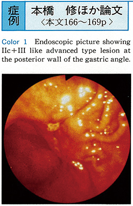
    1993 Volume 43 Pages 166-169
    Published: December 01, 1993
    Released on J-STAGE: July 15, 2015
    JOURNAL FREE ACCESS
    The case was a 60-year-old female, complaining of epigastralgia. Endoscopic and barium studies revealed IIc+III-like advanced type lesion at the posterior wall of the gastric angle. Subtotal gastrectomy was done.
    Histopathologically, the diagnosis of adenosquamous cell carcinoma of the stomach was established in the resected specimen. Well differentiated adenocarcinama with neoplastic esophageal duct's like polylayer epithelium and neoplastic cells containing many acidphil granules, invaded mainly in the submucosal layer. Moderately differentiated squamous cell carcinoma occupied at the center of the adenocarcinoma. And the intermingling areas, which lied between neoplastic adenomatous and squamous components, was existed with signet ring cells and mucin-containing cells.
    Download PDF (1988K)
  • Takahiko Mori, Kozo Nagai, Hisashi Nakamura, Kazuhiro Abe, Hiroshi Kim ...
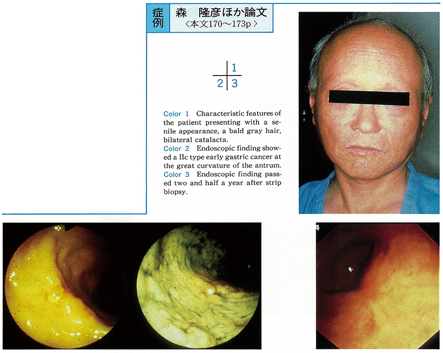
    1993 Volume 43 Pages 170-173
    Published: December 01, 1993
    Released on J-STAGE: July 15, 2015
    JOURNAL FREE ACCESS
    A 53-year-old male was admitted with the left lower limb lateral condylar osteomyelitis. He was diagnosed as having diabetes mellitus with alopecia, cataract, and scleroderma at 39 years of age. Endocrinological examinations revealed insulin resistant diabetes and primary hypogonadism, and the Werner's syndrome was diagnosed.
    During the course, he complained of epigastric discomfort and the endoscopic examination of the upper gastrointestinal tract revealed a IIc type early gastric cancer about 8 mm in size at the greater curvature side of the antrum, while a biopsy revealed a moderately-differentiated adenocarcinoma. The case was treated with endoscopic mucosal resection (EMR) because the healing of injury tends to prolong in the Werner's syndrome and the focus was considered to be a mucosal carcinoma smaller than 1 cm. As of now, after 2 years and 6 months, no recurrence has been observed.
    The Werner's syndrome is highly complicated with malignant tumors, but most of them are melanoma and sarcoma and the complication of gastric cancer is rare. In fact, as far as we know, this is the first case in Japan in which such complications were seen with early cancer. Moreover, the indication for this case was considered to be EMR.
    Download PDF (909K)
  • Chikako Yasuda, Nobuhiko Jyoki, Shunichi Nakajima, Shuji Matsumura, Hi ...
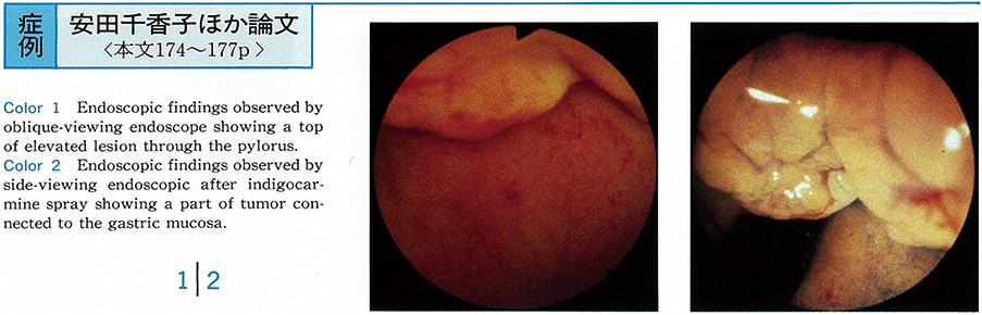
    1993 Volume 43 Pages 174-177
    Published: December 01, 1993
    Released on J-STAGE: July 15, 2015
    JOURNAL FREE ACCESS
    A 64-year-old man was referred to our hospital, because an elevated lesion at the duodenal bulb was pointed out in upper GI X-ray series by his doctor. In the initial endoscopy in our hospital, only small part of elevated lesion was just noted through the pylorus and biopsy from the lesion revealed well-defferentiated tubular adenocarcinoma.
    Subtotal gastrectomy was performed. In the removed specimen, early carcinoma (typeI+IIa) , 22×8 mm in size, was located on the pyloric ring, but not in the bulb. Although according to the general rules for the gastric cancer study in Japan, a border between stomach and duodenum is defined to be a pyloric ring (edge of the thickest pyloric sphincter) , it was also hard to decide from where the carcinoma was derived, as the carcinoma was located on a pyloric ring. Taking the possibility of deviation by the fixation into consideration, it should be appropriate to make a border between them by the marginal line which Brunnner's gland was appeared. Finally, we diagnosed the carcinoma located mainly in the stomach.
    We reported a case which suggested that a border between stomach and duodenum should be decided microscopically.
    Download PDF (1456K)
  • Masayoshi Nishina, Chiho Fujii, Akitsugu Kohama, Yoshinori Fujimura, T ...
    1993 Volume 43 Pages 178-180
    Published: December 01, 1993
    Released on J-STAGE: July 15, 2015
    JOURNAL FREE ACCESS
    We experienced a case of a duodenal foreign body (toothbrush) swallowed 43 years ago. A 57-year-old man was admitted to the hospital with hematemesis. Endoscopic examination showed acute gastritis and a stick like foreign body in the duodenum. On questioning, he was reminded of swallowing a toothbrush 43 years ago in the confusional mental state after the second world war. The toothbrush was removed endoscopically and he was discharged uneventful.
    Download PDF (1339K)
  • Yasuo Tanaka, Jun Tomita, Noriko Sudoh, Makiko Imamura, Yukifusa Igeta ...
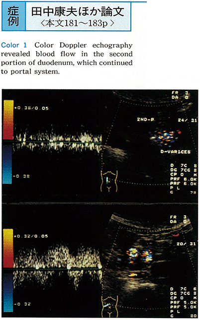
    1993 Volume 43 Pages 181-183
    Published: December 01, 1993
    Released on J-STAGE: July 15, 2015
    JOURNAL FREE ACCESS
    We experienced a case of duodenal varix. A 76-year-old female, followed up for 5 years as liver cirrhosis, was endoscopically pointed out of submucosal mass at the second portion of duodenum.
    As color Doppler echography revealed blood flow in the mass which continued to portal vein, the mass was diagnosed as duodenal varix. Duodenal varix is relatively rare disease. We reported a case of duodenal varix and showed the usefulness of color Doppler echography for examination of collateral flow around the portal system.
    Download PDF (960K)
  • Nobuhiko Jyoki, Shunichi Nakajima, Shuuji Matsumura, Chikako Yasuda, H ...
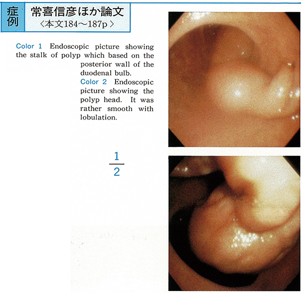
    1993 Volume 43 Pages 184-187
    Published: December 01, 1993
    Released on J-STAGE: July 15, 2015
    JOURNAL FREE ACCESS
    A 40-years-old female was found to have a polyp with long stalk based on the duodenal bulb at a screening X-ray examination of the upper gastrointestinal tract.
    Duodenoscopy on admission showed a pedunculated polyp with the stalk of which based on the posterior wall of the duodenal bulb. A head of the polyp located in the second part of duodenum with smooth surface. Duodenography showed that diameter of the head was about 30mm and stalk was about 70mm in length. Only head of the polyp was resected endoscopically.
    As the resected polyp could not pass through the pyloric ring. We administered polyethyleneglycol electrolyte lavage solution through the biopsy channel of endoscope and after 2 hours we recovered the resected polyp per anus. It was 34×24×15mm in sized and revealed thc Brunner's hyperplasia histologically.
    This was a largest size in the previously published reports in Japan.
    Download PDF (1119K)
  • Shuji Matsumura, Syunya Ishii, Syunichi Nakajima, Hirokazu Inoue, Syun ...
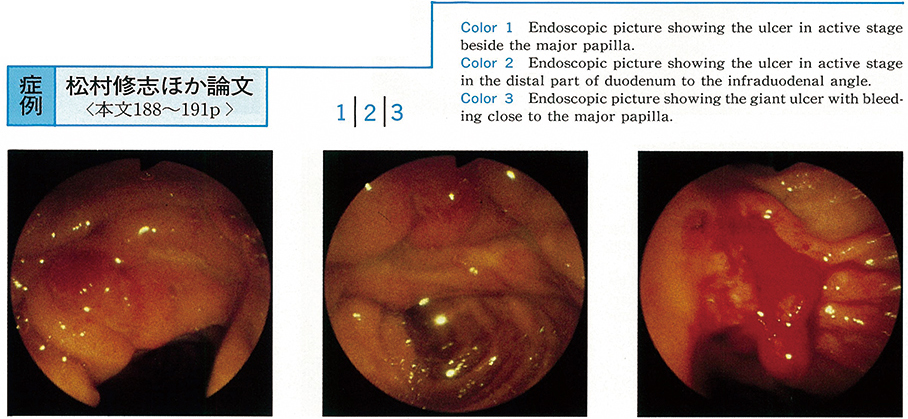
    1993 Volume 43 Pages 188-191
    Published: December 01, 1993
    Released on J-STAGE: July 15, 2015
    JOURNAL FREE ACCESS
    A 65-year-old man was admitted to our hospital for vitrectomy due to the diabetic retinopathy although he has stated to be hemodialyzed under diagnosis of diabetic renal failure for 1 year. After the surgery, vancomycin had administrated because fever and diarrhea were appeared and was diagnosed pseudomembranous colitis by colonoscopy. After while myocardial infarction and cardiac failure were detected, and intravenous heparin administration was added.
    A few days later massive tarry stool was noted and hemoglobin falled to 4.8 g/dl. The 4 ulcers were revealed in the descending part of duodenum by endoscopy, the two on the anterior and posterior wall close the bulb and the one beside the major papilla and remaining large one below the major papilla over the inferior duodenal angulus. But the active bleeding source was unknown throughout GI tract. The anemia was improved only after large amount of blood transfusion. Finally, repeated endoscopy found the enlarged ulcer with continous bleeding area close to the major papilla to the inferior duodenal angulus and various hemostatic procedures were undergone duodenoscopically.
    In cases with serious background, ulcer located in the second part of duodenum induced severe hemorrhage, probably due to unusual micro circulation and poor protecting disorder. Side viewing instrument was quite effective to stop the bleeding using various procedures.
    Download PDF (1089K)
  • Minami Yamada, Noritsugu Umeda, Shigeru Yamato, Mikio Yanase, Yuhko Sh ...
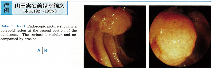
    1993 Volume 43 Pages 192-195
    Published: December 01, 1993
    Released on J-STAGE: July 15, 2015
    JOURNAL FREE ACCESS
    Duodenal cancers are relatively rare, especially early duodenal cancers, but we recently experienced the following case of early duodenal cancer.
    A 69-year-old man was admitted to our institution for further evaluation of the duodenal lesion. Panendoscopy revealed a polypoid lesion at the second portion of the duodenum. Its pathological evaluation disclosed adenoma with severe atypical epithelium. Hypotonic duodenography showed the polypoid lesion with irregular surface, 27×20 mm in size, at middle of the second portion of the duodenum. Angiography did not reveal tumor staining or hypovascular lesion. Distant metastasis was not detected by abdominal ultrasonography and CT scan.
    Cancer of the papilla Vater was highly suspected, therefore the patient underwent surgical operation. It, however, disclosed the polypoid lesion with a wide stalk at 2 or 3 cm above the papilla Vater, so polypectomy was performed instead of duodenectomy. The resected specimen was 20×18×15 mm and early duodenal cancer in adenoma was detected by its histological examination (m, ly0, v0) .
    Download PDF (1277K)
  • Tomoko Kinoshita, Akihiko Kagami, Toshirou Usui, Yoshihiro Tanaka, Sei ...
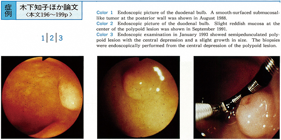
    1993 Volume 43 Pages 196-199
    Published: December 01, 1993
    Released on J-STAGE: July 15, 2015
    JOURNAL FREE ACCESS
    A 39-year-old male visited our hospital with a complaint of upper abdominal discomfort in 1988. The upper GI series showed the hemispherical polypoid lesion. Endoscopic examination showed a smooth-surfaced submucosal-like tumor (φ 6mm) at the posterior wall of the duodenal bulb. Afterward, endoscopic exmination was performed once a year.
    The third endoscopic examination showed no remarkable change of the polypoid lesion, however, endoscopic examination performed 4 years and 5 months later showed semipedunculated polypoid lesion with the central depression and a slight growth in size (φ 8mm) . Carcinoid tumor was diagnosed by the endoscopic biopsy from the central depression.
    The tumor was removed by surgical operation (hemiduodenectory) . Microscopic exanimation revealed a carcinoid composed of tumor cells with round shaped nuclei and trabecular structure. Carcinoid tumor invaded the duodenal submucosa massively. The tumor tissue was stained positively for Leu 7, Chg A, NSE, gastrin, insulin and somatostatin immunohistochemically.
    We present our experience of a very rare case which showed morphological changes of carcinoid tumor after the observation period of 4 years and 5 months. We also reviewed 22 cases of carcinoid tumor from the literature which were diagnosed by endoscopic biopsy.
    Download PDF (1359K)
  • Makoto Nakano, Atsushi Murata, Shuntaro Yoshida, Hiroaki Nakaya, Shige ...
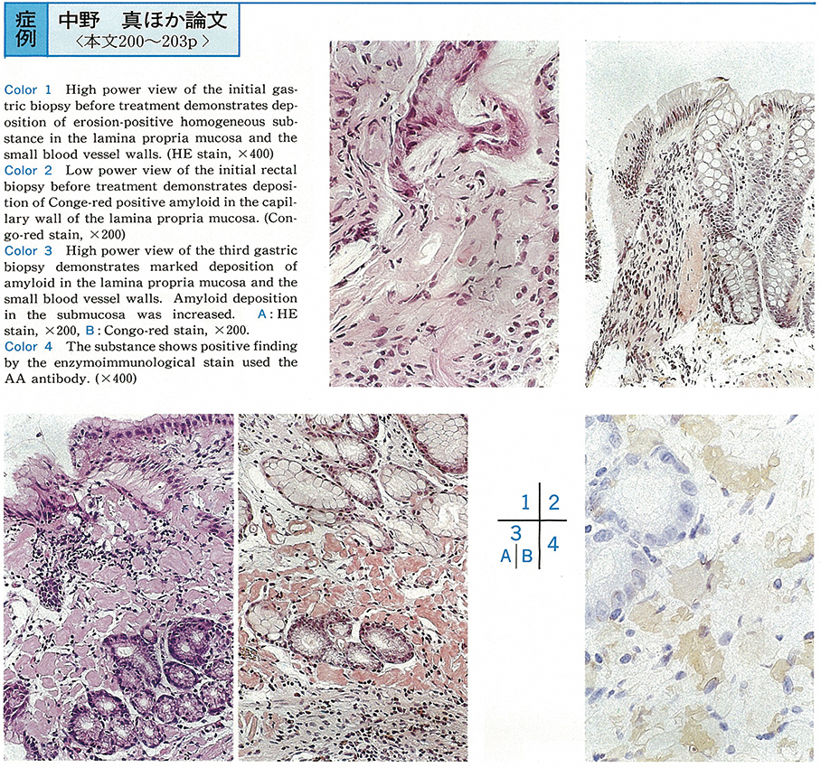
    1993 Volume 43 Pages 200-203
    Published: December 01, 1993
    Released on J-STAGE: July 15, 2015
    JOURNAL FREE ACCESS
    The patient was a 56-year-old female with the chief complaint of watery diarrhea and abdominal pain. She had undergone right thoracoplasty due to pulmonary tuberculosis at the age of 19 and had been treated for rheumatoid arthritis from the time she was 48 years old.●改行● She showed apparent hypoproteinemia with total protein of 4.4g/dl and albumin of 2.2g/dl on admission. The condition was improved by either antidiarrheal drugs or intravenous hyperalimentation (IVH) and her general status worsened. Biopsy of both the rectal and gastric mucosa due to suspected gastrointestinal amyloidosis revealed amyloid deposition.●改行● Treatment with prednisolone at a daily dosage of 60mg was initiated, and as this was being tapered, oral dimethyl sulfoxide (DMSO) at a daily dosage of 4ml was instituted. The hypoproteinemia improved gradually and the diarrhea disappeared. The patient has been followed-up at our clinic without recurrence of symptoms.
    Download PDF (1155K)
  • Motoko Oka, Hiroshi Hashimoto, Ikuo Ikeda, Shinichi Nakamura, Motoko C ...
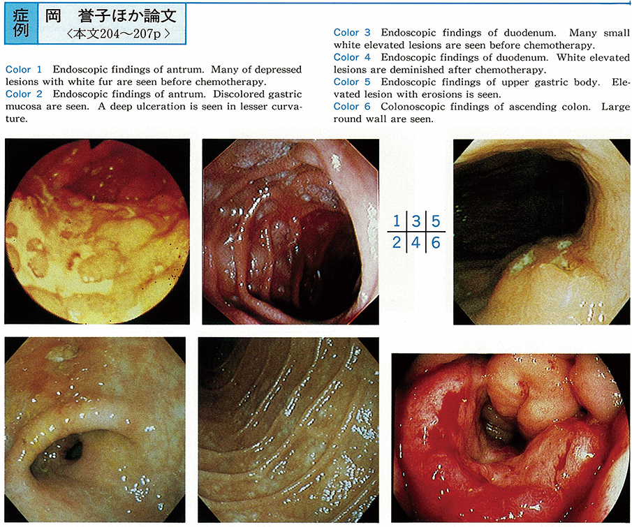
    1993 Volume 43 Pages 204-207
    Published: December 01, 1993
    Released on J-STAGE: July 15, 2015
    JOURNAL FREE ACCESS
    We reported five years follow up rare case of systemic malignant lymphoma on endoscope and colonoscope.
    A 58-year-old male was admitted with appetite loss. We diagnosed systemic malignant lymphoma by biopsy under endoscope and immunological monoclonalities, and performed chemotherapies for five years.
    On early stage, alimentary tract lesions were located in only superficial layers, and these lesions were deminished by chemothrapies. While five years, elevated lesions with thickened alimentary tract wall were appeared, accompanied with swelling of submandibular and cervical lymph nodes and swelling of tonzil.
    This case suggested the progress of alimentary tract lesions in systemic malignant lymphoma showed same tendency to that of other lymphatic lesions.
    Download PDF (923K)
  • Hideki Sakurai, Hirofumi Gonnda, Yuji Fujii, Seiroh Shiomi, Akinori Hi ...
    1993 Volume 43 Pages 208-210
    Published: December 01, 1993
    Released on J-STAGE: July 15, 2015
    JOURNAL FREE ACCESS
    A 71-year-old woman was admitted to our hospital with the tumor at the right lower abdominal area.
    On barium enema study, a deformity from the cecum to the ascending colon was demonstrated and the appendix itself was not visualized. The colonoscopy disclosed that the submucosal tumor like lesion was visible from the cecum to the ascending colon, the orifice of the appendix was not detected. The tumor infiltrated into the ascending colon, and the biopsies histologically showed well differentiated adenocarcinoma with product of mucin. The CT and the ultrasound study revealed the cystic tumor at the right lower abdomen. These findings suggested that the tumor was suspected the cystic type appendeceal cancer.
    The curative right hemicolectomy with lymphnode dissection was performed. The preoperative diagnosis of the appendiceal cancer is extremely difficult because of its growing pattern, but in our case histopathological diagnosis was obtained by colonoscopic study with biopsy.
    Download PDF (898K)
  • Yoshiaki Kohata, Takeo Miyahara, Naoki Shimizu, Kouichi Watanabe, Kazu ...
    1993 Volume 43 Pages 211-214
    Published: December 01, 1993
    Released on J-STAGE: July 15, 2015
    JOURNAL FREE ACCESS
    The endoscopic removal of colonic foreign body was underwent in three cases. In two cases the foreign bodies were a needle and an artificial tooth, which passed down from the upper intestinal tract. In the another case, it was a vibrator, which was introduced through the anus. They were each removed endoscopically from the transvers colon, ascending colon and rectum. Polypectomy snare or biopsy forceps were used removals. No complications were observed.
    A foreign body sometimes brings on the perforation, bleeding, and etc. Surgical procedures are necessary for these cases. Endoscopic removal of foreign body is popular method for upper gastrointestinal tract. But it is rare for lower intestinal tract. We concluded that the endoscopic removal of colonic foreign body is a safe and effective method.
    Download PDF (1753K)
  • Kazunari Yahagi, Kaori Kaneko, Tatsuhiko Matsumoto, Jun Masuda, Yoshia ...
    1993 Volume 43 Pages 215-217
    Published: December 01, 1993
    Released on J-STAGE: July 15, 2015
    JOURNAL FREE ACCESS
    A 40-year-old woman was admitted to our hospital because of hematochezia on the next day of the onset. Colonoscopy revealed hematoma covered with the stripped mucosa in the sigmoid colon. Four days after the admission, the hematoma was discharged. Immediately after that, barium enema and colonoscopy revealed sharply domarcated girdle ulcer, about 10cm in long, in the sigmoid colon. The conservative treatment including elemental diets was performed. After 10 months the ulcer healed over with little stenosis.
    The mucosal abrasion of the large intestine was rarely reported. In our case, the mucosal abrasion might be related to bleeding tendency owing to ticlopidine hydrochloride which she had taken until the onset.
    Download PDF (1161K)
  • Masaaki Fujita, Michimasa Ohi, Kazuo Kotaka, Kiyoshi Mizushima, Yasuyu ...
    1993 Volume 43 Pages 218-220
    Published: December 01, 1993
    Released on J-STAGE: July 15, 2015
    JOURNAL FREE ACCESS
    Ofloxacin (300 mg/day) was administered to a male case, aged 68, with urinary tract infection. On about the 12th day of drug administration, however, he started to show symptoms such as abdominal pain, diarrhea together with pyrexia, and was sent to hospital finally on the 12th day.
    On the very day of admission, this case was diagnosed by endoscopy as pseudomembranous enterocolitis. Accordingly, administration of ofloxacin was discontinued, and instead oral administration of vancomycin was started at a dose of 1,000 mg/day. As the result, the symptom was alleviated rapidly, and the disappearance of pseudomembrane was confirmed by endoscopy on the 5th day of administration of vancomycin.
    Six cases, including one observed by the author, have so far reported to have developed pseudomembranous enterocolitis associated with ofloxacin. The number of reports is now on the increase with respect to pseudomembranous enterocolitis caused by new antibacterial quinolones such as ofloxacin. It was thus considered that the greatest care must be taken in treating patients with ofloxacin.
    Download PDF (321K)
  • Shunichi Nakajima, Shunya Ishii, Shuji Matsumura, Hirokazu Inoue, Shun ...
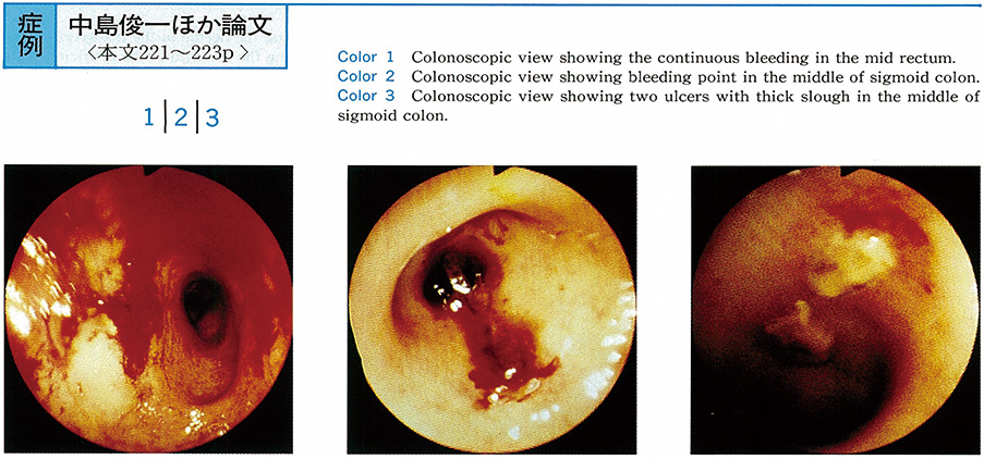
    1993 Volume 43 Pages 221-223
    Published: December 01, 1993
    Released on J-STAGE: July 15, 2015
    JOURNAL FREE ACCESS
    A patient was 79-year-old female. She recieved radiation therapy for cause of uterine cervix in July 1990. Bloody stool was noticed occasionally since Nov 1992. She admitted to our hospital due to of heartfailure in Feb 1993. Colonoscopy revealed diffuse coarse mucosa upto mid sigmoid colon, induced by irradiation and bleeding points was noted in the mid rectum and mid sigmoid. Local injection of pure ethanol was undergone and complete hemostasis was obtained.
    Download PDF (828K)
  • Toshiyuki Baba, Yasushi Akita, Tetsuya Inoue, Hirotaka Yamada, Hitoshi ...
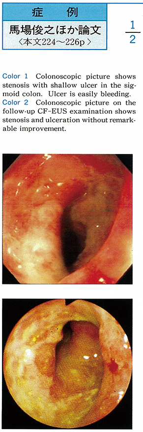
    1993 Volume 43 Pages 224-226
    Published: December 01, 1993
    Released on J-STAGE: July 15, 2015
    JOURNAL FREE ACCESS
    Ischemic colitis is similar to type 4 of colonic cancer on the macroscopical findings and the differential diagnosis is very difficult.
    A 66-year-old woman visited our hospital with complaints of lower abdominal pain and bloody stool. Barium enema and colonoscopy revealed a stenotic lesion with shallow ulcer in the sigmoid colon.
    CF-EUS findings showed thickness of the colonic wall and hypoechoic changes with unclearness of boundary form from the first to fourth layer. But boundary form was not destructed. Histological findings of colonic mucosa showed non-specific chronic inflammatory changes and malignant cell was not found. We finally diagnosed as ischemic colitis and denied colonic cancer.
    CF-EUS was useful for differential diagnosis of ischemic colitis from other malignant disease showing stenosis, such as type 4 of colonic cancer.
    Download PDF (1223K)
  • Tadao Taguchi, Toshiaki Miyamoto, Takeshi Ishihara, Toshiki Ehata, Sho ...
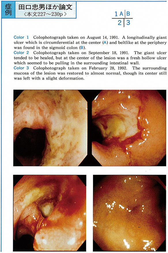
    1993 Volume 43 Pages 227-230
    Published: December 01, 1993
    Released on J-STAGE: July 15, 2015
    JOURNAL FREE ACCESS
    Colonic ulcer that is complicated by systemic lupus erythematosus (SLE) is known to be liable to penetrate the intestinal wall, thus leading to a fatal outcome. This paper describes an ulcer that was found penetrating the sigmoid colon in an SLE patient, but with increased dosage of steroid, was cicatrized.
    The patient was 41-year-old woman. She had been diagnosed as having SLE 19 years before. During prolonged maintenance steroid therapy diarrhea and melena appeared. Colonoscopic studies revealed a longitudinally giant ulcer which was localized in the sigmoid colon and was circumferential at the center and beltlike at the periphery. After one month of hospitalization and steroid therapy with increased dosage the giant ulcer further exacerbated, following out at its center. After five months of steroid therapy, however, the ulcer was cicatrized, though left with slightly converging folds. The ulcer has not recurred to date.
    No definitive histopathological diagnosis of the ulcer lesion was made in this case, but from an immunological viewpoint it appears that vasculitis was concerned with the development of this ulcer, considering it developed during the aggravation of SLE and that increased dosage of steroid elicited a favorable response.
    Download PDF (1499K)
  • Hironori Tachikawa, Makoto Kotoyori, Atsushi Tatsuguchi, Motoko Nachi, ...
    1993 Volume 43 Pages 231-233
    Published: December 01, 1993
    Released on J-STAGE: July 15, 2015
    JOURNAL FREE ACCESS
    We reported a case of intestinal tuberculosis with a submucosal tumor like lesion.
    A 66-year-old female, complaining of right lower abdominal pain and continuous low grade fever, visited our hospital. Barium enema examination revealed intestinal tuberculous lesion and a small elevated lesion in the cecum and she was admitted for further examination and treatment.
    Colonoscopic examination revealed the ulcer and stenosis of terminal ileum, the deformity of the ileocecal valve and multiple small ulcers in the ascending colon and cecum. Additionally, a submucosal tumor liked lesion about 1cm in size was found in the cecum and endoscopical polypectomized for the histological diagnosis. At the polypectomized specimen, a Langhans giant cell and a caseous necrosis existed in the submucosal layer.
    Consequently she was confirmedly diagnosed as intestinal tuberculosis by these findings.
    Download PDF (1027K)
  • Seiichi Yamawaki, Ken Adachi, Toshikazu Nomura, Yoko Nakamura, Tetsu H ...
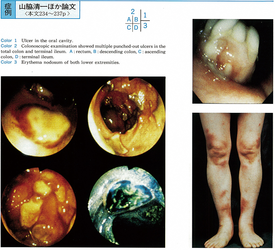
    1993 Volume 43 Pages 234-237
    Published: December 01, 1993
    Released on J-STAGE: July 15, 2015
    JOURNAL FREE ACCESS
    The patient was a 50-year-old female who was admitted to our department with the chief complaints of diarrhea and high fever. She had recurrent oral ulcer, and also had acne-like eruptions. She had past histories of genital ulcer and erythema nodosum of both lower extremities.
    Multiple punched-out ulcers were revealed in the total colon and terminal ileum by the colonoscopic examination. Those ulcerative lesions of the colon and ileum were diagnosed as the intestinal lesions of Behçet's disease. Her symptoms, inflammatory reactions including CRP and intestinal ulcers were improved by total parenteral nutritional therapy.
    It is very uncommon to find multiple ulcers in the total colon and terminal ileum in Behçet's disease.
    Download PDF (692K)
  • Masato Nakajima, Asako Katayama, Morihito Igawa, Yumiko Taguchi, Jun H ...
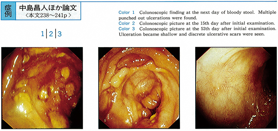
    1993 Volume 43 Pages 238-241
    Published: December 01, 1993
    Released on J-STAGE: July 15, 2015
    JOURNAL FREE ACCESS
    A 31-year-old male admitted in the Department of Hematology of this hospital for the treatment of osteomyelodysplasia since September 1988 had fever and elevated erythema with pain in the back. The diagnosis of Sweet disease was made when skin biopsy showed diffuse neutrophil infiltration from the superficial to deep dermal layers. He was re-admitted from October 1989 due to exacerbation of Sweet disease but was referred to us when massive bloody stools occurred on January 17, 1990.
    Multiple punched-out ulcers were discovered in the cecum upon large bowel endoscopy. Biopsy showed non-specific inflamatory findings. Improvement was seen when complete iv alimentation and oral administration of azelastine hydrochloride were conducted. Colonic ulcers similar to Behçet disease associated with Sweet disease are rare. Our case appears to provide valuable information in determining the casual relationship between these two disease.
    Download PDF (1142K)
  • Tazuko Ohishi, Hiroshi Serizawa, Yoshiki Hamada, Naoto Iwabuchi, Naoki ...
    1993 Volume 43 Pages 242-246
    Published: December 01, 1993
    Released on J-STAGE: July 15, 2015
    JOURNAL FREE ACCESS
    A 68-year-old female patient with primary Sjögren's syndrome who developed multiple rectal erosions is described.
    She admitted to our hospital because of bloody stool and“dry eye”. The diagnosis of Sjögren's syndrome was confirmed by Schirmer's test, Rosebengal test, sialography and lip biopsy. Barium enema X-ray and colonoscopic studies revealed multiple tiny erosions in the rectum. The intervening mucosa was slightly hypervascular and biopsy specimens showed the erosive changes with marked lymphocyte infiltration, mucosal edema and hemorrhage. After 6 months' symptomatic treatment, bloody stool disappeared and erosions were not revealed by the 2nd colonoscopic examination. However, mucosa was still hypervascular and biopsy specimens showed lymphocyte infiltration with lymphoid follicle formation.
    Immunohistochemical study of rectal mucosa revealed the increase in the number of IgA containing cells and the decrease in the ratio of CD4+cells to CD8+cells (CD4/CD8) in the lamina propria. The decrease of CD4/CD8 was also demonstrated in peripheral blood. These results suggested that the immunological abnormalities lying in the basis of Sjögren's syndrome may contribute to the induction of rectal regions in this patient.
    Download PDF (2515K)
  • Masato Nakajima, Mitsuhide Gotou, Asako Katayama, Keisuke Sasaki, Kazu ...
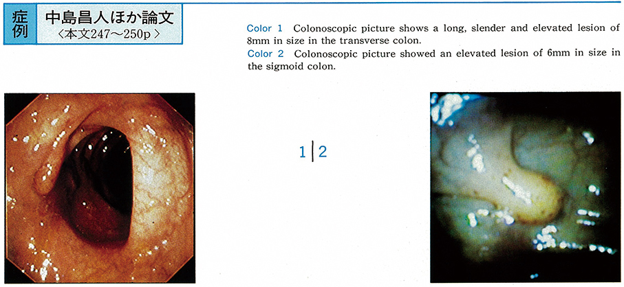
    1993 Volume 43 Pages 247-250
    Published: December 01, 1993
    Released on J-STAGE: July 15, 2015
    JOURNAL FREE ACCESS
    Case 1 : A 41-year-old male. A polyp was discovered in large bowel X-rays conducted for the chief complaint of hemorrhage after defecation. A long, slender and elevated lesion (8mm) was discovered on the left of the transverse colon. Endoscopic polypectomy was conducted. In histological examination, spindle-shaped cells with clear cell body showed linear or curved trabecular alignment and were stained with S-100 protein and NSE but not with vimentin. The diagnosis of neurinoma was made.
    Case 2 : A 56-year-old female. Large bowel endoscopy was carried out upon onset of melena during ambulatory treatment for idiopathic thrombocytopenic purpura and hypertension. The diagnosis of ischemic colitis was made. Although ischemic change were relieved in the re-examination conducted 40 days later, an elevated lesion of 6mm was discovered in the sigmoid colon. Endoscopic polypectomy was carried out. In histological examination, fascicular hypertrophy and ganglion cells were observed, with cells being stained with S-100 protein and NSE but not with vimentin. The diagnosis of neuroganglioma was made based on these findings.
    These cases are reported with some reference to the literature because of endoscopic excision of neurogenic tumors with primary lesions in the large bowel is rare.
    Download PDF (1324K)
  • Yoshitaka Hirahara, Hidemichi Takahashi, Kouji Tsurui, Fuminori Horimu ...
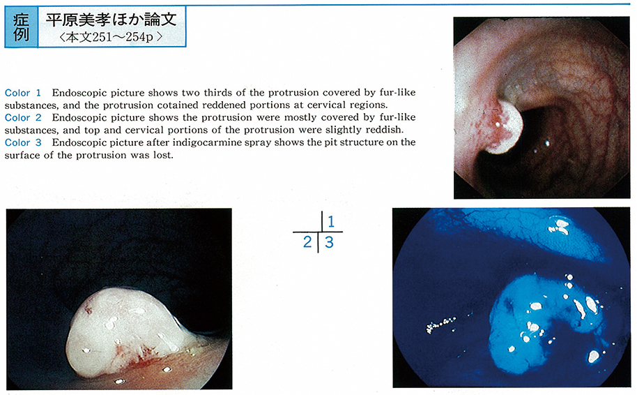
    1993 Volume 43 Pages 251-254
    Published: December 01, 1993
    Released on J-STAGE: July 15, 2015
    JOURNAL FREE ACCESS
    A 51-year-old female was stool occult blood test revealed positive reactions during mass health screening. The patient visited our cancer center for further examinations.
    Enema examinations revealed a protrusion of approximately 7 mm in size in taransverse colon. Endoscopic examinations of colon were performed and revealed that the protrusion were mostly covered by fur-like substances and top and cervical portions of the protrusion were slightly reddish. Dye tests revealed that pit structure on the surface of the protrusion was lost. Small, dense luminal structures and dense prolification of capillary arteries were observed inside of the tumor by histological examinations. The surface layers were covered by necrotic substances. Based on these finding, a diagnosis of capillary hemangioma was made.
    Retrospectively speaking, we should have considered the possibillity of hemangioma from cervical findings of the protrusion, but it was difficult to diagnose it preoperatively because endoscopic image were obscured due to fur covered the protrusion. Thus, we reported a case with capillary hemangioma of colon.
    Download PDF (1393K)
  • Hiroyuki Ishito, Fuyuhiko Inoue, Hiroaki Sugisaka, Takaaki Matsui, Tam ...
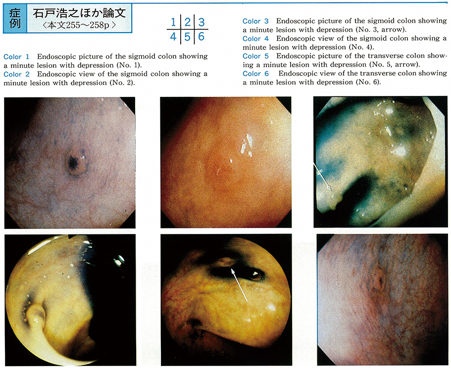
    1993 Volume 43 Pages 255-258
    Published: December 01, 1993
    Released on J-STAGE: July 15, 2015
    JOURNAL FREE ACCESS
    A 41-year-old male, presented here, had six minute colonic neoplasms of superficial type with depression. He was admitted because of abdominal pain. Colonoscopy revealed four minute lesions with obvious depression in the sigmoid colon. Another minute lesion with depression was detected in the transverse colon by the second examination. Twelve months later, the other lesion was found in the transverse colon.
    All six lesions were removed by means of endoscopic mucosal resection, and these samples were examined by stereomicroscope, and then histopathologically. The lesions were 2 to 5 mm in size, which were consisted of two IIc, one IIc+IIa, two IIa+IIc, and one (IIa+IIc) ′. We define (IIa+IIc) ′as a relatively depressed lesion whose elevated margin is not neoplastic. One lesion was adenocarcinoma, and the others were adenomas with moderate to severe atypia.
    During the past three years, we experienced 42 cases (60 lesions) who had minute colorectal neoplasms of superficial type with depression. Among them, each of 11 cases had two or more neoplasms. Twenty one of the 60 neoplasms were diagnosed as adenocarcinomas. Careful examination and vigorous follow-up study are recommended, since minute colorectal neoplasms of superficial type with depression can be multiple and malignant.
    Download PDF (2050K)
  • Sachio Hirama, Kiyonori Kobayashi, Arata Honda, Yoshirou Watanabe, Aki ...
    1993 Volume 43 Pages 259-262
    Published: December 01, 1993
    Released on J-STAGE: July 15, 2015
    JOURNAL FREE ACCESS
    A 63-year-old female visited our hospital because of positive occult blood in stool. Endoscopic and barium enema studies demonstrated multiple flat elevated lesions with small conglomerated nodular surface (nodule-aggregating lesion) in the ascending, transvers and descending colon. Total six nodule-aggregating lesions were detected in the colon, and the avarage size of the lesions was 31±14 mm in diameter (ranged 17-55 mm in diameter) .
    Strip biopsy was performed to one of the lesions in the transverse colon, and histological examination revealed tubular adenoma with moderate atypia. And other lesions were diagnosed tubular adenoma with mild and moderate atypia by biopsied specimen histologically.
    In our knowledge, there have been few case reports of multiple nodular-aggrregating lesions of the colon such as in this case.
    Download PDF (1554K)
  • Yoshiro Tamegai, Nobutaka Sato, Eita Morikoshi
    1993 Volume 43 Pages 263-266
    Published: December 01, 1993
    Released on J-STAGE: July 15, 2015
    JOURNAL FREE ACCESS
    The Patient, a 65-year-old man, was found by a barium enema X-ray study to have type 2 advanced carcinoma of the cecum and an elevated lesion in the splenic flexure.
    He was admitted to our hospital on July 1, 1992. Colonoscopic examination revealed that the cecum had a type 2 advanced lesion and the splenic flexure a nodular aggregated type lesion, both of which were classified as group V when biopsies were taken.
    On July 13, colonoscopic examination was made again and the nodular aggregated type lesion was resected en bloc by the method of strip biopsy, removing a block of tissue 31mm wide including normal mucosa. The lesion measured 25mm×19mm and was classified as highly differentiated sm1 adenocarcinoma. Typical surgery could be performed with success on a patient with a type 2 advanced carcinoma of the cecum without resort to colectomy which would have involved extensive resection.
    Download PDF (2061K)
feedback
Top