Abstract
Forty-two depressed lesions of early gastric cancer which were resected at this hospital during the 1 year and 11 months from November 1990 to September 1992 and underwent examination by both fiberscope and electronic endoscope were studied to diagnose the extent of infiltration.
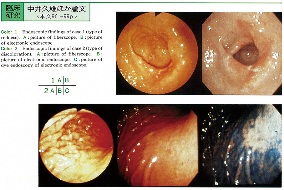
The basic color tone of the lesions was classified into three types : discolored (22 lesions) , same color (5 lesions) and reddish (15 lesions) . The usefulness of electronic endoscopy was compared with that of fiberscopy in regard to the definition of color tone clarity, capillary transparence, gastric mucosal pattern, lesion margin and luster. In addition, resected specimens were studied histopathologically.
In about half of the reddish lesions, electronic endoscopy was effective for defining lesion characteristics, and no major differences were noted between fiberscopy and electronic endoscopy. Discolored lesions were frequently unable to be effectively examined by electronic endoscopy and showed remarkable signs of superficial mucosal infiltration of cancer. However, the concurrent use of dye endoscopy resulted in improved clarity, thereby enhancing performance.