抄録
This paper describes a case of granular cell tumor (GCT) of the esophagus, of which was endoscopically diagnosed and resected.
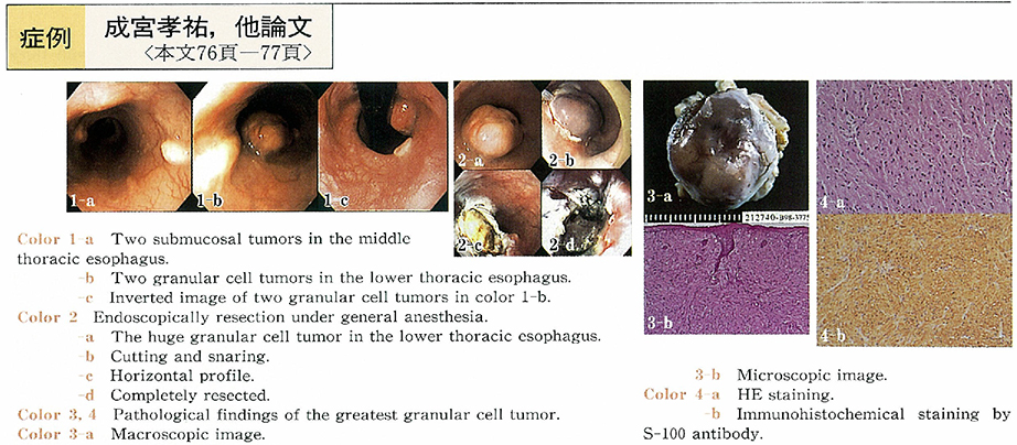
A 43-year-old with multiple GCTs In lower esophagus had been followed up for 7 years and was admitted to our hospital with earnest patient wish for tumor removal. Endoscopy revealed five GCTs with diameter of 7, 10, 20, 42, and 12mm respectively. Endoscopic biopsy showed S-100 positive tumor cell with characteristic eosinophilic granules, of which nuclei were small and moderately hyperchromatic. EUS represented that the tumors had no invasion to proper muscle layer, thus we adopted endoscopic treatment for GCT removal.
In literature views esophageal GCT is a relatively rare disease and so far a total of 170 patient were reported in Japan. The size of the tumor is mostly less than 30mm and only 2% of the tumors behave as malignant tumor. Our case had seven years follow-up period and seemed to have had the biggest tumor among the cases treated endoscopically.
Further observation would be required to avoid unfavorable regrowth and/or tumor relapse.
