Abstract
Cryo-focused ion beam scanning electron microscopy (cryo-FIB-SEM) combined with cryo-electron tomography (cryo-ET) enables visualization of chloroplast architecture close to its native state, but vitrifying isolated organelles remains challenging. We outline practical considerations for applying cryo-FIB-SEM and cryo-ET to plant chloroplasts and compare osmolyte conditions that support vitrification without distorting membranes. Using isolated chloroplasts from an AtVIPP1-GFP transplastomic Nicotiana tabacum line to aid fluorescence-guided targeting, we found that trehalose (0.33 M) and betaine (0.66 M) maintained lamella quality and preserved grana organization, whereas mannitol and sucrose frequently produced ice artifacts. Tomograms from trehalose-treated samples showed well-resolved grana stacks and intact thylakoid networks consistent with native architecture. These observations offer practical guidance for laboratories adopting chloroplast cryo-ET and highlight sample preparation choices that improve reproducible 3D imaging at nanometer resolution.
INTRODUCTION
Chloroplasts are the vital organelles within plant cells responsible for photosynthesis, the process that converts light energy into chemical energy. Their internal structure is highly organized, consisting of a complex network of membranes called thylakoids (reviewed in Sakamoto, 2025 and references therein). These thylakoids are arranged into dense stacks known as grana and interconnected by stroma lamellae. The precise three-dimensional arrangement of these membranes is fundamental to the efficiency of photosynthesis. Therefore, visualizing this architecture in its near-native state is crucial for understanding the structure-function relationships that govern the principles of life at a molecular level. Thylakoid membranes are dynamic structures whose architecture is maintained by coordinated biogenesis, remodeling, and repair pathways, often described as thylakostasis. In vascular plants, grana, stroma lamellae, grana margins, and curvature domains partition photosystems and enable photoprotection and membrane turnover, linking structural organization to photosynthetic performance (Staehelin, 2003; Jarvis and Lopez-Juez, 2013; Kirchhoff, 2019; Bussi et al., 2019; Floris and Kuhlbrandt, 2021; Kreis et al., 2023; Li et al., 2023; Ostermeier et al., 2024; Perez-Boerema et al., 2024).
For decades, our view of chloroplast ultrastructure has been shaped by traditional electron microscopy techniques. While these methods provided foundational knowledge, they rely on chemical fixation, dehydration, and plastic embedding, which are known to introduce significant artifacts. These processes can alter membrane morphology, create artificial spacing, and cause the extraction or aggregation of proteins, compromising the structural integrity of the thylakoid network (McDonald, 2014). Similar structural distortions have been described in detailed chloroplast ultrastructure surveys (Staehelin, 2003). While high-pressure freezing followed by freeze-substitution has improved preservation, it still involves chemical steps that can obscure the native molecular landscape (Studer et al., 2008) and benefits from rapid protocols to limit processing artifacts (McDonald and Webb, 2011). The advent of cryo-electron tomography (cryo-ET) offers a solution by imaging biological samples in a vitrified, near-native state, free from chemical artifacts (Dubochet et al., 1988). When combined with cryo-focused ion beam scanning electron microscopy (cryo-FIB-SEM), it is possible to mill electron-transparent windows (lamellae) into vitrified cells and organelles, enabling 3D visualization of their internal architecture at nanometer resolution (Rigort et al., 2012). This workflow has revolutionized structural cell biology, with optimized protocols for biological lamellae (Schaffer et al., 2017) and correlative workflows (Villa et al., 2013), but its application to plant organelles like chloroplasts has been hindered by specific sample preparation challenges.
Three-dimensional analysis of thylakoid membranes has progressed from conventional electron tomography (Shimoni et al., 2005; Daum and Kuhlbrandt, 2011) to cryo-preserved approaches. In situ cryo-ET of Chlamydomonas chloroplasts provided the first native 3D view of thylakoids (Engel et al., 2015), and recent cryofixed tomography in Arabidopsis captures prolamellar body-to-grana transitions during greening (Liang et al., 2022). Despite these advances, cryo-ET of vascular-plant thylakoids remains limited, motivating optimized sample preparation and targeting workflows. Recent syntheses from Engel’s group highlight emerging cryo-ET insights into thylakoid structural diversity (Perez-Boerema et al., 2024; Engel, 2024). Complementary single-molecule mapping in Chlamydomonas further refines thylakoid organization at native resolution (Wietrzynski et al., 2020). The primary obstacle is achieving successful vitrification. Isolated chloroplasts are highly sensitive to osmotic stress, and the aqueous buffer they are suspended in must be vitrified without forming damaging ice crystals. Conventional osmolytes used in chloroplast biochemistry, such as mannitol and sucrose, are poor cryoprotectants and tend to crystallize during rapid freezing, which often disrupts membrane structures and makes high-resolution imaging difficult (Wolkers and Oldenhof, 2015). Therefore, the selection of a suitable osmolyte that both maintains osmotic stability and promotes vitrification is a major step for successful chloroplast cryo-ET.
In this study, we address this key bottleneck by providing a systematic evaluation of different osmolytes for the cryo-preservation of isolated chloroplasts. We compare the performance of conventional osmolytes (mannitol) with superior glass-forming agents (trehalose and betaine). Using chloroplasts isolated from AtVIPP1-GFP transplastomic tobacco recently reported by us (Gachie et al., 2025), we observed that osmolyte choice is a major determinant of success. We then present a complete, optimized workflow, from chloroplast isolation and quality control to cryo-FIB milling and tomogram reconstruction, that reproducibly yields high-quality structural data. This work provides plant morphology laboratories with the practical guidance needed to overcome the initial hurdles of cryo-ET, facilitating the broader adoption of these techniques. The overall workflow is summarized in Figure 1.
PRINCIPLES OF CRYO-FIB-SEM AND CRYO-ET FOR PLANT ORGANELLES
Fundamental requirements for structural preservation
Cryo-electron tomography achieves near-native structural preservation through vitrification, the rapid cooling of biological samples to cryogenic temperatures at rates sufficient to prevent ice crystal formation, typically >105 K/s (Kasas et al., 2024). In vitreous ice, water molecules are trapped in a non-crystalline, glass-like state that preserves the native spatial relationships between cellular components without the distortions introduced by crystalline ice formation or chemical fixation (Dubochet et al., 1988).
For plant organelles and suborganelles, achieving successful vitrification presents unique challenges compared to mammalian cell samples. Chloroplasts possess highly ordered internal membrane systems, the thylakoid network, which are particularly sensitive to osmotic stress and mechanical disruption. The stromal matrix contains high concentrations of soluble proteins and metabolites that must remain in solution during vitrification. Additionally, isolated chloroplasts lack the protective cellular environment present in intact tissues, making them more vulnerable to handling stresses during sample preparation (Engel et al., 2015).
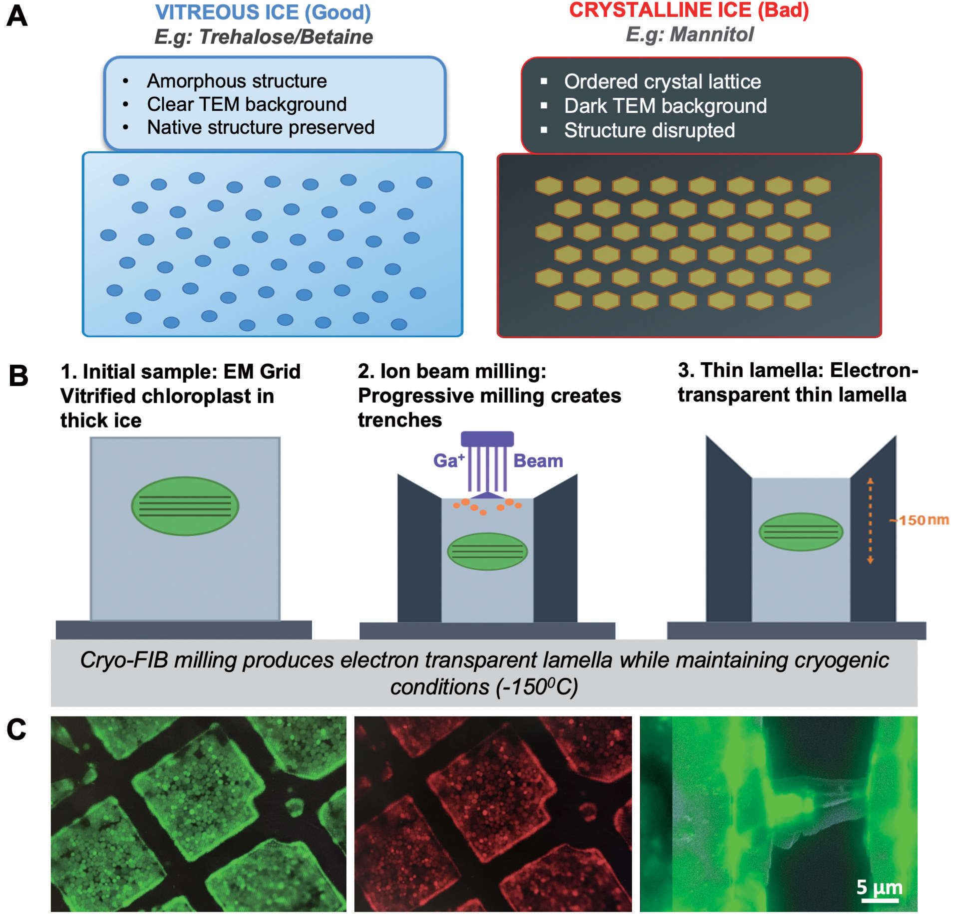
The thickness limitation of cryo-ET (typically <300 nm for 300 kV microscopes) necessitates the preparation of thin lamellae from larger biological samples. Cryo-FIB-SEM addresses this requirement by using a focused gallium ion beam to mill vitrified samples under cryogenic conditions, creating electron-transparent windows while maintaining the frozen-hydrated state (Rigort et al., 2012; Schaffer et al., 2017). The scanning electron beam allows real-time monitoring of the milling process, enabling precise control of lamella thickness and position (Schaffer et al., 2017).
Challenges specific to plant organelles
Plant organelles present unique challenges that make osmolyte choice particularly critical. Chloroplasts are osmotically sensitive, with grana membrane spacing of only 3-4 nm that is easily disrupted by crystallization artifacts (Austin and Staehelin, 2011). Unlike mammalian cells, isolated chloroplasts lack cellular buffering capacity, making them dependent on external osmotic support throughout sample preparation. The fundamental challenge is that conventional osmolytes (mannitol, sucrose) used successfully for chloroplast biochemistry often fail under vitrification conditions. These compounds have low glass transition temperatures and high nucleation rates, resulting in crystalline ice that scatters electrons and obscures structure. This necessitates systematic evaluation of alternative osmolytes with superior glass-forming properties.
Role of osmolytes in cryo-preservation
Osmolytes serve multiple critical functions in chloroplast cryo-ET workflows beyond simple osmotic balancing. During vitrification, the choice of osmolyte fundamentally influences whether the sample achieves true vitreous ice formation or undergoes crystallization. Effective cryoprotective osmolytes must possess a high glass transition temperature (Tg) to enable formation of a stable vitreous state during rapid cooling, extensive hydrogen bonding capacity to maintain membrane organization during dehydration stress, low membrane permeability to prevent osmotic disruption during buffer exchanges, protein-stabilizing effects that preserve native conformations of photosynthetic complexes, and minimal ice nucleation to prevent crystallization seeds that propagate through the sample. The mechanisms by which different osmolyte classes achieve these properties vary substantially, with important implications for their performance in cryo-FIB-SEM applications.
One significant advantage of modern cryo-FIB-SEM systems is the integration of integrated fluorescence light microscopy (iFLM), which enables targeted milling of specific organelles or structures within vitrified samples. For chloroplasts expressing fluorescent markers such as AtVIPP1-GFP (Zhang et al., 2012; Zhang et al., 2016; Gachie et al., 2025), iFLM allows precise localization by tracking both GFP signals and chlorophyll autofluorescence. In this study, the AtVIPP1-GFP transplastomic tobacco line served only as a locator for intact chloroplasts, with GFP signal distinguished from chlorophyll autofluorescence during targeting. For chloroplasts, fluorescence targeting provides rapid quality assessment via chlorophyll autofluorescence, enables functional correlation using genetic markers such as AtVIPP1-GFP, and reveals chloroplast orientation on the grid to guide milling toward relevant structural features.
VIPP1 is a conserved membrane remodeling factor in photosynthetic organisms, in which it assembles into large oligomeric rods and rings, and binds thylakoid membranes and adopts ESCRT-III-like assemblies that remodel membranes (Gupta et al., 2021; Junglas et al., 2021; Pan et al., 2024; Junglas et al., 2024; Naskar et al., 2024). In vascular plants, genetic and biochemical studies support roles in membrane integrity and thylakoid development (Kroll et al., 2001; Zhang et al., 2012; Zhang et al., 2016; Micol-Ponce et al., 2020; Gachie et al., 2025; Li et al. 2026). The GFP-tagged line preserves this localization and provides a reliable correlative marker for intact chloroplasts (Gachie et al., 2025). Key principles of cryo-FIB-SEM and correlative targeting are summarized in Figure 2.
OSMOLYTE SELECTION: MECHANISMS AND SYSTEMATIC EVALUATION
Trehalose: multi-mechanism cryoprotection
Trehalose, a non-reducing disaccharide (α-D-glucopyranosyl-α-D-glucopyranoside), has emerged as a superior cryoprotectant due to multiple complementary mechanisms. Its high glass transition temperature (Tg ≈ 115°C) enables formation of stable vitreous ice during rapid cooling, preventing crystallization that would disrupt membrane organization (Crowe et al., 1992). This glass-forming property is particularly important for cryo-FIB-SEM applications, where samples must remain vitreous not only during initial plunge-freezing but throughout subsequent handling, transfer, and milling operations at cryogenic temperatures.
At the molecular level, trehalose forms extensive hydrogen bonding networks with membrane phospholipids, effectively replacing water molecules at the membrane surface and maintaining membrane spacing during dehydration (water replacement hypothesis; Crowe et al., 1992). This direct interaction with membrane lipids provides a protective shell that preserves the bilayer structure even under the mechanical stress of ion beam milling. For chloroplast thylakoid membranes, where precise spacing between appressed grana membranes is critical for photosynthetic function, this membrane-stabilizing property is particularly valuable. Additionally, trehalose’s non-reducing nature prevents Maillard reactions with proteins during processing, preserving the native conformation of photosynthetic complexes. This chemical stability ensures that any observed structural features in cryo-ET tomograms represent authentic biology rather than artifacts from chemical modification. Trehalose’s low membrane permeability minimizes osmotic disruption during buffer exchanges (both when switching between different buffer compositions and during dilution steps), maintaining chloroplast structural integrity throughout sample preparation, a critical advantage when working with isolated organelles that lack the buffering capacity of intact cells.
Betaine: compatible osmolyte protection
Betaine (trimethylglycine), a quaternary ammonium compound, functions primarily as a compatible osmolyte through preferential exclusion mechanisms (Bohnert and Jensen, 1996). While we did not pursue extensive tomographic data acquisition with betaine-treated samples, initial results demonstrated superior vitrification quality compared to mannitol, positioning betaine as a viable alternative to trehalose for chloroplast cryo-ET applications. Its zwitterionic structure allows favorable interactions with both charged and hydrophobic regions of membrane proteins, stabilizing native conformations under osmotic stress without directly binding to protein surfaces. This preferential exclusion forces proteins into compact, native states by making the hydrated protein surface thermodynamically unfavorable (Yancey, 2005).
The mechanism of preferential exclusion has important implications for cryo-preservation: by stabilizing the folded state of proteins, betaine helps maintain the structural integrity of large protein complexes such as photosystems and ATP synthase during the rapid dehydration that occurs during vitrification. This protection extends to the protein-lipid interfaces critical for photosynthetic membrane function, where protein complexes must maintain specific orientations and spacing within the bilayer. Betaine also provides osmotic balance without permeating organellar membranes, making it particularly suitable for isolated chloroplast preparations where osmotic stability must be maintained during extended handling. Unlike small polyols that can cross membranes and cause osmotic imbalances, betaine remains in the external medium, providing consistent osmotic support throughout sample preparation. However, the different protective mechanism of betaine compared to trehalose suggests that optimal concentrations may differ, reflecting its lower intrinsic cryoprotective capacity per molecule compared to the direct membrane-stabilizing effects of trehalose.
Limitations of conventional osmolytes
In contrast, polyol osmolytes such as mannitol and disaccharides like sucrose, while widely used in chloroplast isolation protocols (Jarvis and Lopez-Juez, 2013), have properties that limit their effectiveness under cryo-FIB conditions. Both compounds have relatively low glass transition temperatures and high propensity for crystallization under rapid cooling (Wolkers and Oldenhof, 2015). Mannitol, being a small polyol (182 Da), can partially permeate organellar membranes, potentially causing osmotic imbalance during sample preparation. Its linear structure and relatively simple hydrogen bonding pattern provide less extensive membrane stabilization compared to the more complex trehalose molecule. Most critically for cryo-applications, mannitol solutions show high nucleation rates during cooling, making it difficult to achieve true vitrification even at rapid cooling rates. Sucrose, while forming hydrogen bonds with membranes, lacks the extensive stabilization networks characteristic of trehalose due to subtle differences in stereochemistry and hydroxyl group positioning. The alpha,alpha-1,1 glycosidic linkage in trehalose creates a more constrained, symmetric molecule compared to sucrose’s alpha,beta-1,2 linkage, resulting in different hydrogen bonding patterns with membrane lipids (Albertorio et al., 2007). Sucrose also shows greater tendency toward crystallization at the cooling rates achieved during plunge-freezing, potentially explaining the dark backgrounds observed in TEM imaging of sucrose-treated samples-a hallmark of ice crystal formation that scatters electrons and obscures structural detail.
EXPERIMENTAL EVALUATION APPROACH
Chloroplast isolation
To systematically compare osmolyte performance under conditions relevant to cryo-FIB-SEM workflows, we evaluated trehalose, betaine, and mannitol using isolated chloroplasts from AtVIPP1-GFP transplastomic Nicotiana tabacum. Chloroplasts were isolated using standard Percoll gradient centrifugation methods with mannitol at the established laboratory protocol concentration of 0.33 M. Following isolation, samples were divided into parallel treatments with different osmolytes. A systematic concentration series was tested for trehalose and betaine: 0.2, 0.3, 0.33, 0.4, 0.5, 0.55, 0.6, and 0.66 M. This comprehensive screening was necessary because optimal concentrations depend on the specific protective mechanisms of each osmolyte class, trehalose’s direct membrane stabilization may require different concentrations than betaine’s preferential exclusion effects. Each osmolyte condition was evaluated through a multi-stage quality control pipeline that included immediate post-isolation confocal fluorescence microscopy to assess chloroplast morphology and chlorophyll autofluorescence, post-vitrification low-magnification TEM screening to assess ice quality, and post-FIB SEM evaluation of lamella surface quality and thickness. This multi-stage evaluation ensures that osmolyte performance is assessed not only for initial chloroplast integrity but throughout the workflow, a critical distinction, as some osmolytes may preserve chloroplasts during isolation but fail during vitrification or FIB milling.

Pre-vitrification quality assessment revealed distinct differences in chloroplast structural preservation across osmolyte conditions. Trehalose treatment at 0.33 M maintained characteristic ellipsoidal chloroplast morphology with uniform chlorophyll autofluorescence distribution and strong GFP signal, indicating intact envelope membranes and functional thylakoid organization. Chloroplasts retained their normal size distribution (Kirschner et al., 2024) of 5-8 µm diameter without evidence of swelling or shrinkage. Pre-vitrification quality assessment and representative morphology are shown in Figure 3. Betaine showed concentration-dependent effects, with optimal preservation observed at 0.66 M, notably higher than the optimal trehalose concentration. At lower betaine concentrations (0.2-0.4 M), chloroplasts showed signs of osmotic stress including size heterogeneity and reduced chlorophyll fluorescence intensity. This concentration dependence reflects betaine’s osmotic stabilization mechanism: sufficient concentration is required to provide adequate preferential exclusion effects and maintain osmotic balance. Mannitol at 0.33 M (the conventional concentration used in chloroplast isolation buffers) showed acceptable initial chloroplast morphology, indicating that the failure of this osmolyte occurs specifically during the vitrification process rather than during initial isolation, an important distinction that explains why mannitol remains widely used for routine chloroplast biochemistry despite its unsuitability for cryo-applications.
Vitrification quality and crystallization artifacts
Post-vitrification TEM screening highlighted the importance of osmolyte selection (Figure 4). Mannitol-treated samples showed pronounced crystallization-dark backgrounds characteristic of hexagonal ice that scatter electrons and obscure structural detail. In contrast, trehalose and betaine samples consistently showed clear, uniform backgrounds characteristic of vitreous ice (n=8 grids for trehalose, n=5 for betaine). This difference shows that choosing glass-forming osmolytes greatly improves the likelihood of successful chloroplast cryo-ET. Trehalose and betaine-treated samples showed clear backgrounds characteristic of vitreous ice with only minor contamination. Grid squares containing thylakoids membranes were easily identifiable, and the envelopes maintained their morphology without distortion from ice crystal growth. This reproducible vitrification success across multiple independent experiments (n=8 grids for trehalose, n=5 for betaine) supports the glass forming properties of trehalose under cryo-ET conditions. Betaine at 0.66 M showed intermediate performance: backgrounds were clearer than mannitol but occasionally showed subtle texture compared to trehalose samples. This suggests that betaine achieves partial but not complete suppression of ice crystallization, possibly due to its lower glass-forming capacity compared to trehalose. Nevertheless, betaine-treated samples were suitable for FIB milling and cryo-ET data collection, making betaine a viable alternative when trehalose is not available or not compatible with specific experimental requirements.
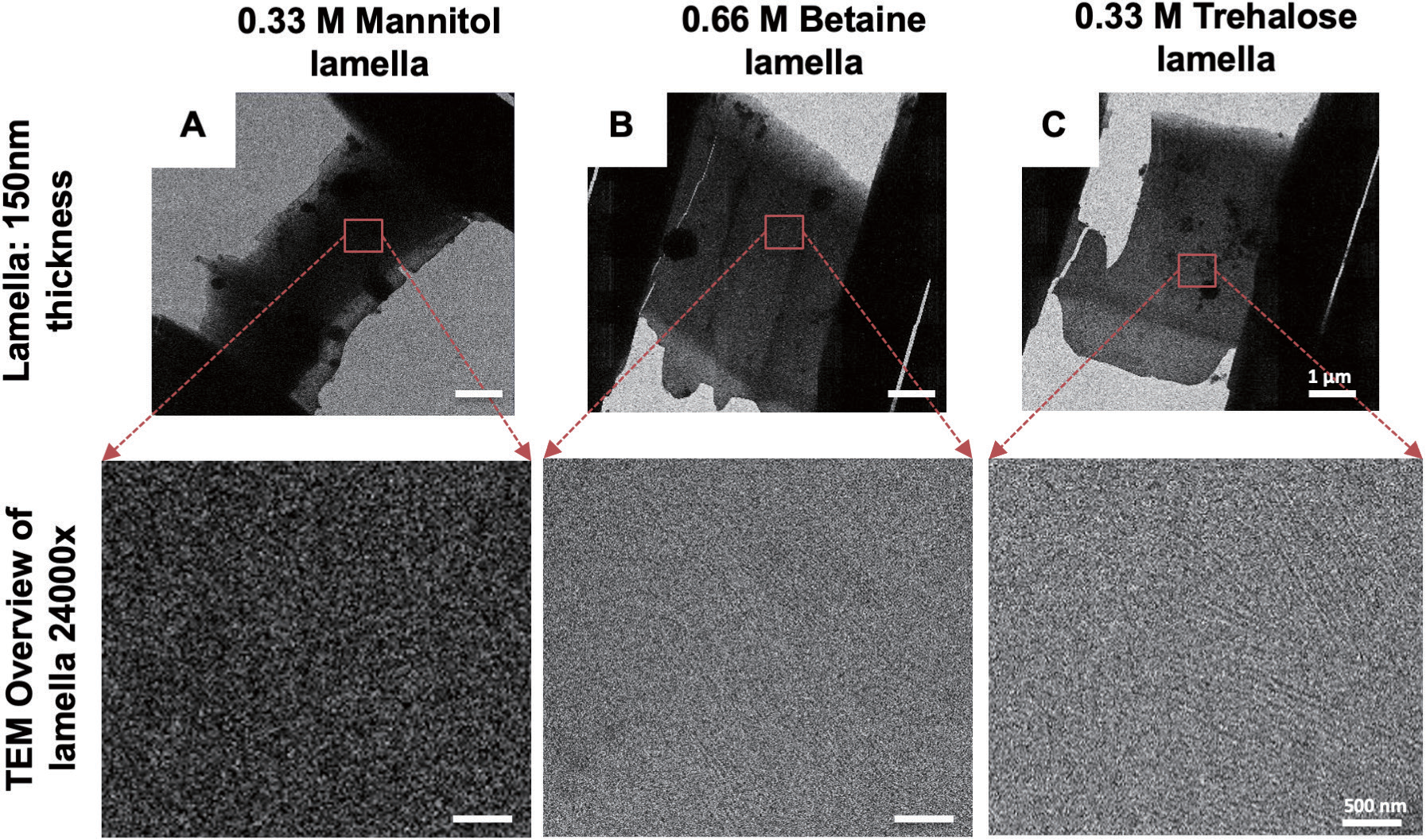
Cryo-FIB milling success rates directly correlated with vitrification quality. All samples were prepared at a chlorophyll concentration of 0.25 mg chl/mL, providing optimal grid loading density for target selection. Trehalose-treated samples yielded high-quality lamellae in the majority of milling attempts, with consistent thickness (~150 nm), smooth surfaces, and minimal curtaining artifacts. The integrated fluorescence light microscopy (iFLM) module of the Cryo-FIB-SEM system (Aquilos, Thermo Fisher Scientific) enabled precise targeting of GFP-positive, structurally intact chloroplasts, further improving success rates by avoiding damaged or partially lysed organelles. Betaine-treated samples showed good milling success, with occasional increased beam sensitivity during milling-possibly reflecting subtle differences in ice quality or organelle osmotic state. Final lamellae were suitable for cryo-ET but occasionally showed slightly reduced thickness uniformity compared to trehalose samples. Mannitol-treated samples proved unsuitable for cryo-ET data acquisition: the severe ice crystallization created dark backgrounds that made it impossible to identify intact chloroplasts for targeting under TEM conditions. The few attempted millings resulted in excessive beam damage and poor structural preservation. The prohibitively low success rate makes mannitol unsuitable for cryo-FIB-SEM workflows despite its continued utility for routine chloroplast biochemistry.
Tomogram quality and structural preservation
The ultimate test of osmolyte performance is the quality of final tomographic reconstructions and the structural information they reveal. Tomograms from trehalose-treated samples showed exceptional preservation of chloroplast ultrastructure. Grana stacks displayed the characteristic appressed thylakoid configuration with clear visualization of individual membrane bilayers. Quantitative analysis revealed thylakoid membrane thickness of 4.5 ± 1.2 nm and lumenal spacing (lumen width) of 13.8 ± 2.2 nm between adjacent grana membranes, consistent with native chloroplast architecture reported in previous cryo-EM studies and significantly improved over chemical fixation methods which typically show artifacts in membrane spacing (Austin and Staehelin, 2011). Stroma lamellae connecting different grana stacks were clearly resolved, demonstrating their crucial role in maintaining thylakoid network connectivity. The well-preserved membrane organization indicated successful osmotic balance throughout sample preparation. Three-dimensional reconstructions from trehalose-treated samples revealed the characteristic architecture of chloroplast grana stacks at nanometer resolution. The correlation between osmolyte choice, vitrification quality, and final tomogram resolution demonstrates that proper osmolyte selection is not merely a technical detail but a fundamental requirement for successful chloroplast cryo-ET. The mechanistic understanding of how different osmolytes protect (or fail to protect) membrane structure during vitrification provides rational guidance for researchers implementing these techniques.
OPTIMIZED WORKFLOW: PRACTICAL IMPLEMENTATION
Overview and decision points
The complete workflow from plant material to interpretable tomograms involves multiple steps, each with critical quality control checkpoints. Success depends not only on technical execution but on systematic quality assessment that identifies problems early, before investing expensive microscope time in samples that will ultimately yield poor results. The workflow and quality check (QC) decision points are summarized in Figure 5.
Sample preparation and quality assessment
AtVIPP1-GFP transplastomic Nicotiana tabacum plants were grown under standardized conditions (16 h light/8 h dark, 22-26°C, 100-150 µmol photons m−2 s−1) for approximately one month before harvesting mature leaves. Intact chloroplasts were isolated using two-step Percoll gradient centrifugation, a method that separates intact organelles from broken chloroplasts and cellular debris based on density. The isolation buffer composition is critical: 50 mM HEPES-KOH (pH 7.5), 0.33 M trehalose, 2 mM EDTA, 1 mM MgCl2, 0.1% BSA. The trehalose is included from the beginning of isolation to prevent osmotic shock during buffer exchanges. Achieving optimal loading density on EM grids proved critical for efficient FIB milling. Stock chloroplast suspensions (1 mg chl/mL) were diluted to working concentrations of 0.125-0.5 mg chl/mL. The 0.25 mg chl/mL concentration provided ideal distribution: sufficient chloroplasts per grid square for target selection without overcrowding that would complicate milling geometry or create overlapping structures in tomograms. Confocal fluorescence microscopy assessment (5-10 minutes per sample) provides immediate feedback on chloroplast integrity before committing to expensive cryo-processing. Samples showing <90% intact chloroplasts, evidence of broken chloroplast envelopes indicated by rhodamine B staining (An et al., 2021), free GFP signal outside intact organelles, or weak chlorophyll fluorescence were discarded.
Vitrification protocol
Quantifoil R2/2 holey carbon grids (200 mesh) were glow-discharged for 20 seconds immediately before use to improve hydrophilicity and ensure even sample spreading. While 200 mesh grids were used in this work, 300 mesh grids may be preferable when sample volume is limiting, as the smaller grid squares reduce material requirements. Vitrification was performed using a Leica EM GP2 automated plunge-freezing system under controlled environmental conditions (18°C, 95% relative humidity to prevent evaporation). A 3 µL aliquot of chloroplast suspension was applied to the grid, blotted for 3 seconds with Whatman No. 1 filter paper, and immediately plunged into liquid ethane cooled to approximately −183°C. The entire process from application to plunging takes <5 seconds, minimizing settling artifacts and ensuring uniform chloroplast distribution. Vitrified grids were briefly examined under low-dose TEM conditions (or using the Aquilos SEM mode) to assess ice quality before committing to FIB milling. This rapid screening (5-10 minutes per grid) identifies crystallization problems early, allowing optimization of vitrification parameters or osmolyte adjustments before proceeding with expensive FIB operations.
Cryo-FIB targeting and milling
To precisely locate chloroplasts for milling, an integrated fluorescence light microscopy (iFLM) step was employed. Fluorescence-to-SEM correlation typically achieved <2 µm accuracy, enabling confident targeting even of specific chloroplasts within clustered regions. The ability to pre-select high-quality organelles based on fluorescence improves overall workflow efficiency by focusing expensive FIB milling time on samples most likely to yield excellent tomograms. Lamella preparation followed a standardized multi-step protocol designed to minimize beam damage while achieving target thickness (150 nm). Initial rough milling created protective trenches on either side of the target area, followed by progressive thinning at decreasing beam currents. Throughout milling, sample temperature was maintained below −150°C, and beam-induced heating was minimized through intermittent milling cycles that allowed thermal dissipation.
Data Acquisition and tomogram reconstruction
Tilt series were acquired on a 300 kV cryo-TEM (Titan Krios, Thermo Fisher Scientific) equipped with a K3 direct electron detector and energy filter. The dose-symmetric acquisition scheme (Hagen et al., 2017) starts from 0° and tilts bidirectionally to minimize accumulated radiation damage while maximizing angular coverage. Tilt range of −51° to +69° provided sufficient angular sampling for high-quality reconstructions while remaining within the geometric constraints of the sample holder. Low-dose conditions were critical: total dose per tilt series was maintained below 120 e-/Å2 (Ni et al., 2022), distributed across all tilt images to minimize radiation damage. Tilt series motion correction, alignment using AreTomo2 (Zheng et al., 2022), tomogram reconstruction, and denoising were performed using standard protocols. Multiple software packages (e.g., RELION, IMOD, Dynamo) can be used depending on computational resources and specific requirements. The reconstructed tomograms were subjected to denoising and quality assessment before proceeding to detailed structural analysis. Only reconstructions showing clear membrane detail, minimal artifacts, and sufficient resolution were used for quantitative measurements; resolution was estimated at 6-8 nm for membrane features using Fourier shell correlation (Verbeke et al., 2024).
Validation of structural preservation quality
Tomograms from trehalose-treated chloroplasts demonstrated successful preservation of native ultrastructure, validating the optimized osmolyte selection. Grana stacks displayed the characteristic appressed thylakoid configuration with clear visualization of individual membrane bilayers. Quantitative analysis of multiple tomograms revealed a thylakoid membrane thickness of 4.5 ± 1.2 nm and a lumenal spacing (lumen width) of 13.8 ± 2.2 nm, consistent with native chloroplast architecture reported in previous cryo-EM studies (Austin and Staehelin, 2011). These measurements confirm that the trehalose-based protocol preserves authentic membrane spacing without the artifacts typically introduced by chemical fixation methods. Stroma lamellae connecting different grana stacks were clearly resolved, demonstrating that the optimized workflow maintains the structural continuity of the thylakoid network. The quality of preservation achieved with trehalose-treated samples provides a solid foundation for future detailed structural studies of chloroplast organization and photosynthetic membrane architecture. Representative tomograms and preservation metrics are shown in Figure 6.

FUTURE PERSPECTIVES
Core equipment required for cryo-ET includes a cryo-FIB-SEM system (e.g., Aquilos), a 300 kV cryo-TEM (e.g., Titan Krios), a direct electron detector, a plunge-freezing system, and cryogenic transfer and storage infrastructure. Because these instruments are highly specialized and expensive to maintain, cryo-ET workflows are often supported through shared university platforms, core facilities, or regional structural biology centers rather than within a single laboratory. Access to such facilities, together with technical support from experienced staff, enables broader adoption of cryo-ET in plant research.
The osmolyte optimization framework, systematic testing across concentration series, and multi-stage quality assessment can guide protocol development for other plastid types (chromoplasts, amyloplasts, etioplasts), mitochondria with buffer modifications, other plant species (including crop plants and non-vascular plants), and intact plant tissues using high-pressure freezing. The key insight, that osmolyte selection strongly influences vitrification quality, applies broadly. The optimized protocol presented here improves cost-effectiveness by increasing success rates and reducing wasted instrument time. Researchers implementing cryo-ET for new plant systems should prioritize osmolyte optimization early, rather than defaulting to conventional biochemical buffers that are unsuitable for cryo-applications.
The methodological framework presented here addresses a key bottleneck in plant structural biology. The principle that osmolyte choice is a primary determinant of vitrification quality is broadly applicable to other sensitive plant organelles and can guide protocol development for intact tissues using high-pressure freezing (Pöge et al. 2025). Future integration of this workflow with subtomogram averaging will allow for high-resolution analysis of photosynthetic complexes in their native membrane environment. As cryo-ET becomes more accessible, these foundational methods will facilitate its broader adoption across the plant sciences.
In conclusion, this work establishes that careful osmolyte selection is the most critical factor for successful cryo-ET of isolated chloroplasts. We demonstrate that trehalose (0.33 M) effectively promotes vitrification and preserves native thylakoid architecture, whereas conventional osmolytes like mannitol lead to crystallization artifacts that prevent high-resolution imaging. The optimized workflow and integrated quality control checkpoints presented here provide a reproducible pathway for obtaining high-quality tomograms. By validating our method with quantitative structural measurements (4.5±1.2 nm thylakoid membranes, 13.8±2.2 nm lumen width), we offer a practical guide for plant biology labs. This contributes to the “Seeing the Principles of Life” symposium’s goal by enabling the direct visualization of photosynthetic membrane organization at the molecular level.
ACKNOWLEDGEMENTS
We gratefully acknowledge the collaborative efforts of multiple research institutions and facilities that made this work possible. The cryo-FIB-SEM and cryo-ET data collection was performed at the Institute of Protein Research (IPR), Osaka University, with particular thanks to the technical support staff. We also thank the organizers of the “Seeing the Principles of Life” symposium for the opportunity to present this work and for the valuable discussions with participants. We thank Masanobu Iwasaki, Hiroko Takazaki, Akihiro Kawamoto, and Genji Kurisu (IPR) for collaboration and technical support.
This work was supported by the KAKENHI Grant from the Ministry of Education, Culture, Sports, Science and Technology (23H04959 to W.S.) and from the Japan Society for the Promotion of Science (21H02508, 24K02044 to W.S.), and by the research grant from Ohara Foundation. ET analysis performed in this study was in part supported under the Collaborative Research Program of IPR, Osaka University (CR-24-02).
COMPETING INTEREST
The authors declare that there are no competing interests.
AUTHOR CONTRIBUTIONS
W.S. conceived the research and designed the experiments. A.M. and S.W.G. performed experiments. A.M. performed data acquisition and analysis. A.M. and W.S. wrote the manuscript.
Acknowledgments
We gratefully acknowledge the collaborative efforts of multiple research institutions and facilities that made this work possible. The cryo-FIB-SEM and cryo-ET data collection was performed at the Institute of Protein Research (IPR), Osaka University, with particular thanks to the technical support staff. We also thank the organizers of the “Seeing the Principles of Life” symposium for the opportunity to present this work and for the valuable discussions with participants. We thank Masanobu Iwasaki, Hiroko Takazaki, Akihiro Kawamoto, and Genji Kurisu (IPR) for collaboration and technical support. This work was supported by the KAKENHI Grant from the Ministry of Education, Culture, Sports, Science and Technology (23H04959 to W.S.) and from the Japan Society for the Promotion of Science (21H02508, 24K02044 to W.S.), and by the research grant from Ohara Foundation. ET analysis performed in this study was in part supported under the Collaborative Research Program of IPR, Osaka University (CR-24-02).
References
- Albertorio, F., Chapa, V.A., Chen, X., Diaz, A.J., and Cremer, P.S. (2007) The α,α-(1→1) Linkage of Trehalose Is Key to Anhydrobiotic Preservation. J Am Chem Soc 129: 10567–10574. doi: 10.1021/ja0731266
- An, Y., Liu, L., Chen, L., and Wang, L. (2021) Visualizing the integrity of chloroplast envelope by rhodamine and Nile red staining. Front Plant Sci 12: 668414. doi: 10.3389/ fpls.2021.668414
- Austin, J. R., and Staehelin, L. A. (2011) Three-dimensional architecture of grana and stroma thylakoids of higher plants as determined by electron tomography. Plant Physiol 155: 1601-1611. doi: 10.1104/pp.110.170647
- Bohnert, H. J., and Jensen, R. G. (1996) Strategies for engineering water-stress tolerance in plants. Trends Biotechnol 14: 89-97. doi: 10.1016/0167-7799(96)80929-2
- Bussi, Y., Shimoni, E., Weiner, A., Kapon, R., Charuvi, D., Nevo, R., Efrati, E., and Reich, Z. (2019) Fundamental helical geometry consolidates the plant photosynthetic membrane. Proc Natl Acad Sci USA 116: 22366-22375. doi: 10.1073/ pnas.1905994116
- Crowe, J. H., Hoekstra, F. A., and Crowe, L. M. (1992) Anhydrobiosis. Annu Rev Physiol 54: 579-599. doi: 10.1146/ annurev.physiol.54.1.579
- Daum, B., and Kuhlbrandt, W. (2011) Electron tomography of plant thylakoid membranes. J Exp Bot 62: 2393-2402. doi: 10.1093/jxb/err034
- Dubochet, J., Adrian, M., Chang, J. J., Homo, J. C., Lepault, J., McDowall, A. W., and Schultz, P. (1988) Cryo-electron microscopy of vitrified specimens. Q Rev Biophys 21: 129-228. doi: 10.1017/S0033583500004169
- Engel, B. D., Schaffer, M., Kuhn Cuellar, L., Villa, E., Plitzko, J. M., and Baumeister, W. (2015) Native architecture of the Chlamydomonas chloroplast revealed by in situ cryo-electron tomography. eLife 4: e04889. doi: 10.7554/elife.04889
- Engel, B. D. (2024) Integrating cellular electron microscopy with multimodal data to explore biology across space and time. Cell 187: 563-584. doi: 10.1016/j.cell.2024.01.005
- Floris, D., and Kuhlbrandt, W. (2021) Molecular landscape of etioplast inner membranes in higher plants. Nat Plants 7: 514-523. doi: 10.1038/s41477-021-00896-z
- Gachie, S. W., Muhire, A., Li, D., Kawamoto, A., Takeda-Kamiya, N., Goto, Y., Sato, M., Toyooka, K., Yoshimura, R., Takami, T., et al. (2025) The thylakoid membrane remodeling protein VIPP1 forms bundled oligomers in tobacco chloroplasts. Plant Physiol 198: kiaf137. doi: 10.1093/ plphys/kiaf137
- Gupta, T. K., Klumpe, S., Gries, K., Heinz, S., Wietrzynski, W., Ohnishi, N., Niemeyer, J., Spaniol, B., Schaffer, M., Rast, A., et al. (2021) Structural basis for VIPP1 oligomerization and maintenance of thylakoid membrane integrity. Cell 184: 3643-3659.e23. doi: 10.1016/j.cell.2021. 05.011
- Hagen, W. J., Wan, W., and Briggs, J. A. (2017) Implementation of a cryo-electron tomography tilt-scheme optimized for high resolution subtomogram averaging. J Struct Biol 197: 191-198. doi: 10.1016/j.jsb.2016.06.007
- Jarvis, P., and Lopez-Juez, E. (2013) Biogenesis and homeostasis of chloroplasts and other plastids. Nat Rev Mol Cell Biol 14: 787-802. doi: 10.1038/nrm3702
- Junglas, B., Huber, S. T., Heidler, T., Schlösser, L., Mann, D., Hennig, R., Clarke, M., Hellmann, N., Schneider, D., and Sachse, C. (2021) PspA adopts an ESCRT-III-like fold and remodels bacterial membranes. Cell 184: 3674-3688.e18. doi: 10.1016/j.cell.2021.05.042
- Junglas, B., Kartte, D., Kutzner, M., Hellmann, N., Ritter, I., Schneider, D., and Sachse, C. (2024) Structural basis for Vipp1 membrane binding: from loose coats and carpets to ring and rod assemblies. Nat Struct Mol Biol 32: 555-570. doi: 10.1038/s41594-024-01399-z
- Kasas, S., Frenkel, D., Curnow, P., Phillips, M., Barnes, C. H. W., Isa, L., Topolancik, J., and Janka, Z. (2024) Direct measurement of the critical cooling rate for the vitrification of water. Phys Rev Res 7: 013095. doi: 10.1103/ physrevresearch.7.013095
- Kirchhoff, H. (2019) Chloroplast ultrastructure in plants. New Phytologist 223: 565-574. doi: 10.1111/nph.15730
- Kirschner, G. K., Xiao, T. T., and Blilou, I. (2024) Because size matters: measuring chloroplast volumes by confocal and scanning electron microscopy. Plant J 117: 899-913. doi: 10.1111/tpj.16603
- Kreis, E., Konig, K., Misir, M., Niemeyer, J., Sommer, F., and Schroda, M. (2023) TurboID reveals the proxiomes of Chlamydomonas proteins involved in thylakoid biogenesis and stress response. Plant Physiol 193: 1772-1796. doi: 10.1093/plphys/kiad335
- Kroll, D., Meierhoff, K., Bechtold, N., Kinoshita, M., Westphal, S., Vothknecht, U. C., ... and Westhoff, P. (2001) VIPP1, a nuclear gene of Arabidopsis thaliana essential for thylakoid membrane formation. Proc Natl Acad Sci USA 98: 4238-4243. doi: 10.1073/pnas.061500998
- Li, D., Gachie, S. W., Ozawa, S., Scholz, M., Hippler, M., and Sakamoto, W. (2026) Chloroplast heat shock protein cpHsc70-1 interacts with thylakoid membrane remodeling protein VIPP1 C-terminal tail and controls VIPP1 oligomer disassembly. PNAS Nexus 5: pgaf393. doi: 10.1093/pnasnexus/ pgaf393.
- Li, N., Wong, W. S., Feng, L., Wang, C., Wong, K. S., Zhang, N., Yang, W., Jiang, Y., Jiang, L., and He, J. X. (2023) The thylakoid membrane protein NTA1 is an assembly factor of the cytochrome b6f complex essential for chloroplast development in Arabidopsis. Plant Commun 4: 100509. doi: 10.1016/j.xplc.2022.100509
- Liang, Z., Yeung, W. T., Ma, J., Mai, K. K. K., Liu, Z., Chong, Y. F., Cai, X., and Kang, B. H. (2022) Electron tomography of prolamellar bodies and their transformation into grana thylakoids in cryofixed Arabidopsis cotyledons. Plant Cell 34: 3830-3843. doi: 10.1093/plcell/koac205
- McDonald, K. L., and Webb, R. I. (2011) Freeze substitution in 3 hours or less. J Microsc 243: 227-233. doi: 10.1111/j.1365-2818.2011.03526.x
- McDonald, K. L. (2014) Out with the old and in with the new: rapid specimen preparation procedures for electron microscopy of sectioned biological material. Protoplasma 251: 429-448. doi: 10.1007/s00709-013-0575-y
- Micol-Ponce, R., García-Alcázar, M., Capel, C., Yuste-Lisbona, F. J., Pineda, B., Atarés, A., García-Sogo, B., Capel, J., Moreno, V., and Lozano, R. (2020) The Tomato SlVIPP1 Gene Is Required for Plant Survival Through the Proper Development of Chloroplast Thylakoid Membrane. Front Plant Sci 11: 1305. doi: 10.3389/fpls.2020.01305
- Naskar, S., Merino, A., Espadas, J., Singh, J., Roux, A., Colom, A., and Low, H. H. (2024) Mechanism for Vipp1 spiral formation, ring biogenesis, and membrane repair. Nat Struct Mol Biol 32: 571-584. doi: 10.1038/s41594-024-01401-8
- Ni, T., Frosio, T., Mendonça, L. et al. High-resolution in situ structure determination by cryo-electron tomography and subtomogram averaging using emClarity. Nat Protoc 17, 421–444 (2022) https://doi.org/10.1038/s41596-021-00648-5
- Ostermeier, M., Garibay-Hernández, A., Holzer, V.J.C., Schroda, M., and Nickelsen, J. (2024) Structure, biogenesis, and evolution of thylakoid membranes. Plant Cell 36: 4014–4035. doi: 10.1093/plcell/koae102
- Pan, S., Gries, K., Engel, B. D., Schroda, M., Haselwandter, C. A., and Scheuring, S. (2024) The cyanobacterial protein VIPP1 forms ESCRT-III-like structures on lipid bilayers. Nat Struct Mol Biol 32: 543-554. doi: 10.1038/s41594-024-01367-7
- Perez-Boerema, A., Engel, B. D., and Wietrzynski, W. (2024) Evolution of thylakoid structural diversity. Annu Rev Cell Dev Biol 40: 169-193. doi: 10.1146/annurev-cellbio-120823-022747
- Pöge, M. et al. (2025) Making plant tissue accessible for cryo-electron tomography. eLife 14: e106455. doi: 10.7554/eLife. 106455
- Rigort, A., Bauerlein, F. J., Villa, E., Eibauer, M., Laugks, T., Baumeister, W., and Plitzko, J. M. (2012) Focused ion beam micromachining of eukaryotic cells for cryoelectron tomography. Proc Natl Acad Sci USA 109: 4449-4454. doi: 10.1073/pnas.1201333109
- Sakamoto, W. (2025) Thylakostasis: key factors in thylakoid membrane organization with emphasis on biogenesis and remodeling proteins in vascular plants. Plant Cell Physiol 66: 1602-1618. doi: 10.1093/pcp/pcaf098
- Schaffer, M., Mahamid, J., Engel, B. D., Laugks, T., Baumeister, W., and Plitzko, J. M. (2017) Optimized cryo-focused ion beam sample preparation aimed at in situ structural studies of membrane proteins. J Struct Biol 197: 73-82. doi: 10.1016/j.jsb.2016.07.010
- Shimoni, E., Rav-Hon, O., Ohad, I., Brumfeld, V., and Reich, Z. (2005) Three-dimensional organization of higher-plant chloroplast thylakoid membranes revealed by electron tomography. Plant Cell 17: 2580-2586. doi: 10.1105/tpc. 105.035030
- Staehelin, L. A. (2003) Chloroplast structure: from chlorophyll granules to supra-molecular architecture of thylakoid membranes. Photosynth Res, 76: 185-196. doi: 10.1023/ A:1024994525586
- Studer, D., Humbel, B. M., and Chiquet, M. (2008) Electron microscopy of high pressure frozen samples: bridging the gap between cellular ultrastructure and atomic resolution. Histochem Cell Biol 130: 877-889. doi: 10.1007/s00418-008-0500-1
- Verbeke, E. J., Zhou, Y., Horton, A. P., Mallam, A. L., Taylor, D. W., and Marcotte, E. M. (2024) Self Fourier shell correlation: properties and application to cryo-ET. Commun Biol 7: 144. doi: 10.1101/2023.11.07.565363
- Villa, E., Schaffer, M., Plitzko, J. M., and Baumeister, W. (2013) Opening windows into the cell: focused-ion-beam milling for cryo-electron tomography. Curr Opin Struct Biol 23: 771-777. doi: 10.1016/j.sbi.2013.08.014
- Wietrzynski, W., Schaffer, M., Tegunov, D., Albert, S., Kanazawa, A., Plitzko, J. M., et al. (2020) Charting the native architecture of Chlamydomonas thylakoid membranes with single-molecule precision. eLife 9: e53740. doi: 10.7554/elife.53740
- Wolkers, W. F., and Oldenhof, H. (2015) Cryopreservation and freeze-drying protocols. Methods Mol Biol 1257: 1-8. doi: 10.1385/0-89603-296-5:49
- Yancey, P. H. (2005) Organic osmolytes as compatible, metabolic and counteracting cytoprotectants in high osmolarity and other stresses. J Exp Biol 208: 2819-2830. doi: 10.1242/ jeb.01730
- Zhang L, Kato Y, Otters S, Vothknecht UC, Sakamoto W. (2012) Essential role of VIPP1 in chloroplast envelope maintenance in Arabidopsis. Plant Cell. 24: 3695-3707. doi.org/10.1105/ tpc.112.103606
- Zhang L, Kondo H, Kamikubo H, Kataoka M, Sakamoto W. (2016)VIPP1 has a disordered C-terminal tail necessary for protecting photosynthetic membranes against stress. Plant Physiol. 171: 1983-1995. doi.org/10.1104/pp.16.00532
- Zheng, S., Wolff, G., Greenan, G., Chen, Z., Faas, F. G. A., Bárcena, M., Koster, A. J., Cheng, Y., and Agard, D. A. (2022) AreTomo: An integrated software package for automated marker-free, motion-corrected cryo-electron tomographic alignment and reconstruction. J Struct Biol X 6: 100068. doi: 10.1016/j.yjsbx.2022.100068