Abstract
Introduction: Adolescent idiopathic scoliosis (AIS) with a major curve at the main thoracic (MT) area is classified as Lenke type 1, 2, or 3 depending on the flexibility of the proximal thoracic (PT) curve and lumbar curve. No definite classification has been established for a major curve at the PT spine. The purpose of this study is to investigate the radiographic characteristics before and after correction surgery for AIS with a major curve at the PT area.
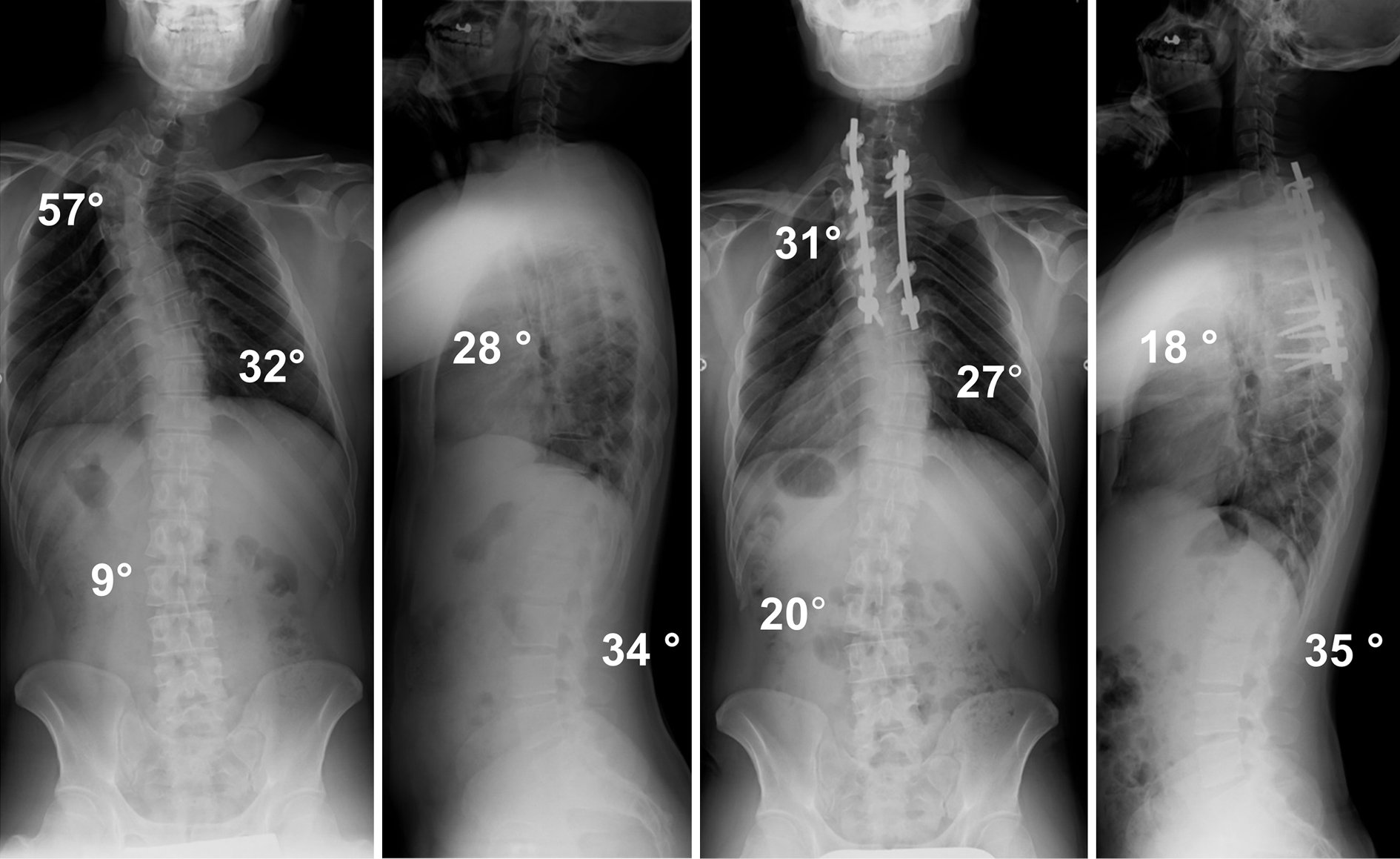
Methods: This is a retrospective cohort study at a single academic institution. Twelve patients with a major curve at the PT spine participated in our study and followed for at least two years after surgery. We evaluated the pre- and postoperative Cobb angles of the curve, curve range, location of the apex, sagittal parameters, and shoulder balance-related parameters. All patients were treated by posterior correction and fusion surgery using pedicle screw constructs.
Results: The patients were classified as having a double-curve (DC) type, in which the MT curve was structural, or a single-curve (SC) type, in which the MT curve was corrected to less than 25° on supine side-bending films. The mean correction rates for the PT curve were favorable in both groups (DC, 65.7%±9.6%; SC, 39.2%±4.9%). The mean Cobb angle of the lumbar curve improved in the DC group (preoperative, 17.1°±4.0°; postoperative, 5.0°±4.2°) but deteriorated in the SC group (preoperative, 7.1°±1.2°; postoperative, 12.4°±4.4°) after surgery.
Conclusions: We illustrated the postoperative radiographical changes of 12 consecutive patients with the major curve at the PT curve. Although posterior correction and fusion surgery corrected the PT curve satisfactorily in both DC and SC patients, the Cobb angle of the lumbar curve deteriorated after surgery in all SC patients. Surgeons need to pay attention to the fusion area, especially LIV, when operating the SC curve type.