Spine Surgery and Related Research
Online ISSN : 2432-261X
ISSN-L : 2432-261X
Volume 7, Issue 4
Displaying 1-21 of 21 articles from this issue
EDITORIAL
SPECIAL ARTICLE: GUIDELINES
REVIEW ARTICLE
  • Ryosuke Hirota, Masanori Sasaki, Osamu Honmou, Toshihiko Yamashita
    2023 Volume 7 Issue 4 Pages 319-326
    Published: July 27, 2023
    Released on J-STAGE: July 27, 2023
    Advance online publication: February 13, 2023
    JOURNAL OPEN ACCESS

    Since the 1990s, our group has been conducting basic research on regenerative medicine using various cell types to treat several central nervous system diseases, including spinal cord injury (SCI). We have reported many positive effects of the intravenous administration of mesenchymal stem cells (MSCs) derived from the bone marrow. In the current study, MSCs were administered intravenously to a rat model of severe SCI (crush injury) during the acute to subacute stages-considerable motor function recovery was observed. Furthermore, MSC transplantation in a chronic-phase SCI model improved motor function. In this review, we discuss recent updates in basic research on the intravenous infusion of MSCs and prospects for SCI research.

    Download PDF (902K)
ORIGINAL ARTICLE
  • Yuki Matsumoto, Tetsuo Hayashi, Yuichi Fujiwara, Kensuke Kubota, Munea ...
    2023 Volume 7 Issue 4 Pages 327-332
    Published: July 27, 2023
    Released on J-STAGE: July 27, 2023
    Advance online publication: March 13, 2023
    JOURNAL OPEN ACCESS

    Introduction: Aspiration pneumonia is one of the most frequent and fatal life-threatening complications among individuals with acute traumatic cervical spinal cord injury (CSCI). However, the mechanism of dysphagia among individuals with CSCI is not well understood. Morbidity and mortality associated with CSCI may result from the interplay between respiratory dysfunction and dysphagia. This study aimed to elucidate the effect of respiratory dysfunction on the swallowing function of individuals with acute traumatic CSCI.

    Methods: A prospective cohort study was conducted involving 54 individuals with acute traumatic CSCI who were admitted within 2 weeks following injury. Dysphagia was evaluated using the Dysphagia Severity Scale (DSS) and the Functional Oral Intake Scale (FOIS). Respiratory function was evaluated by measuring the cough peak flow (CPF), forced expiratory volume in 1 s (FEV1.0), FEV1.0/forced vital capacity (FEV1.0%), and percent vital capacity (%VC). We recorded these parameters at weeks 2, 4, 8, and 12 following injury and analyzed pertinent changes over time and significant correlations.

    Results: Among 54 individuals (46 men and 8 women) recruited in this study, 48 (88.9%) had restrictive ventilatory impairment and 17 (31.5%) had severe dysphagia (DSS level 1−4) 2 weeks following injury. However, respiratory function and swallowing function significantly improved thereafter. CPF, FEV1.0, and %VC were significantly correlated with the severity of dysphagia during each period.

    Conclusions: Restrictive ventilatory impairment, poor cough force, and dysphagia are closely related, and the evaluation of respiratory function plays an important role in evaluating dysphagia.

    Download PDF (110K)
  • Chikara Ushiku, Kota Suda, Takehiro Michikawa, Satoko Matsumoto Harmon ...
    2023 Volume 7 Issue 4 Pages 333-340
    Published: July 27, 2023
    Released on J-STAGE: July 27, 2023
    Advance online publication: March 13, 2023
    JOURNAL OPEN ACCESS

    Introduction: Pneumonia is one of the leading causes of acute- and chronic-phase mortality in patients with cervical spinal cord injury (CSCI) with quadriplegia. The risk factors for chronic-phase pneumonia recurrence in CSCI are still unknown. This study aimed to investigate the incidence of pneumonia in the chronic phase after injury and to identify its risk factors.

    Methods: This retrospective clinical observational study included patients with CSCI with American Spinal Injury Association Impairment Scale grades of A or B admitted to our center within 72 h of CSCI injury who started treatment and were available for follow-up for at least 90 days. The patients were assessed for incidences of pneumonia and its associations with clinical characteristics, including risk factors at the time of injury. Patients in whom pneumonia developed within 30 days postadmission and those after 30 days of hospitalization were comparatively examined using univariate and multivariate analyses.

    Results: Pneumonia occurred in 36% of the 69 enrolled patients throughout the study period and in 20% of all patients after 30 days of hospitalization. Multivariate analysis of risk factors for pneumonia showed that atelectasis (adjusted OR [aOR], 95% confidence interval [CI]: 4.9, 1.2-20.0), enteral feeding (aOR [95% CI]: 13.3 [3.0-58.9]), mechanical ventilation (aOR [95% CI]: 4.0 [1.0-15.0]), and tracheotomy (aOR [95% CI]: 14.6 [2.3-94.6]) within 30 days of admission were significantly associated with the occurrence of pneumonia even after 30 days of hospitalization.

    Conclusions: The risk factors for developing pneumonia in the chronic phase were atelectasis, enteral feeding, mechanical ventilation, and tracheotomy within 30 days of hospitalization. This study suggests that treatment of atelectasis, long-term respiratory muscle rehabilitation, and training to improve swallowing function are essential to prevent the recurrence of pneumonia after 30 days of hospitalization.

    Download PDF (220K)
  • Kazuta Yamashita, Yasuaki Tamaki, Daiki Nakajima, Yasuyuki Omichi, Yos ...
    2023 Volume 7 Issue 4 Pages 341-349
    Published: July 27, 2023
    Released on J-STAGE: July 27, 2023
    Advance online publication: January 12, 2023
    JOURNAL OPEN ACCESS

    Introduction: The harmful effects of long-term low-dose radiation have been well known. There are few comprehensive reports evaluating concrete real exposure doses for each part of a surgeon, assistant surgeon, scrub nurse, and anesthesiologist associated with fluoroscopic spinal procedures. This research aimed to quantify the radiation exposure dose to surgical team members during C-arm fluoroscopy-guided spinal surgery.

    Methods: Seven fresh cadavers were irradiated for 1 and 3 min with C-arm fluoroscopy. The position of the X-ray source was under the table, over the table, and laterally. The radiation exposure doses were measured at the optic lens, thyroid gland, and hand in mannequins used to simulate surgical team members.

    Results: A significant difference was observed in the radiation exposure dose according to the position of the X-ray source and the irradiated body area. The risk of scatter radiation exposure was the biggest for the lateral position (nearly 30-fold that for the position under the table). All radiation exposure doses were positively correlated with irradiation time.

    Conclusions: The occupational radiation exposure dose to surgical team members during C-arm fluoroscopy-guided lumbar spinal procedures varies according to the X-ray source position. Our findings would help surgical team members to know the risk of radiation exposure during various fluoroscopic procedures. Surgeons in particular need to reduce their radiation exposure by using appropriate shielding and technique.

    Download PDF (574K)
  • Kiyoshi Tarukado, Teruaki Ono, Toshio Doi, Katsumi Harimaya, Yasuharu ...
    2023 Volume 7 Issue 4 Pages 350-355
    Published: July 27, 2023
    Released on J-STAGE: July 27, 2023
    Advance online publication: January 12, 2023
    JOURNAL OPEN ACCESS

    Introduction: It remains controversial whether it is better to continue oral low-dose aspirin (LDA) during the perioperative period in spinal surgery. This study aims to evaluate the safety of continued LDA administration in the perioperative periods of microendoscopic laminectomy (MEL) by assessing perioperative complications and clinical outcomes.

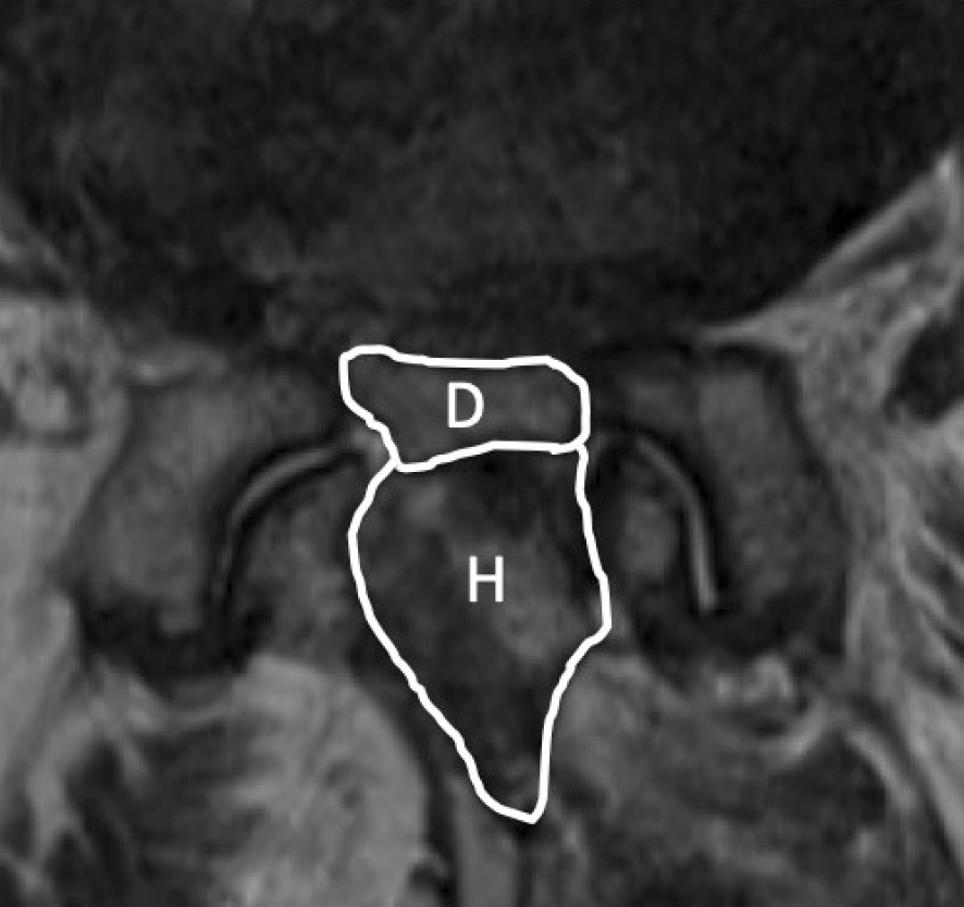
    Methods: We ultimately included 88 patients (35 males, 53 females) who underwent one level of MEL for lumbar spinal canal stenosis from April 2016 to March 2022. Patients who did not undergo anticoagulation therapy were classified into Group A (65 patients), those who stopped anticoagulation therapy at the perioperative periods were classified into Group B (9 patients), and those who continued oral administration of LDA throughout the perioperative periods were classified into Group C (14 patients). Surgery time, intraoperative estimate blood loss (EBL), differences between hemoglobin (Hb) and platelet (Plt) before and after surgery, perioperative complications, and cross-sectional area of hematoma and dural sac on MRI taken within 1 week and at 6 months or more after surgery were assessed between three groups. The EuroQol-5 dimensions (EQ-5D), Oswestry Disability Index (ODI), and Japanese Orthopaedic Association Back Pain Evaluation Questionnaire (JOABPEQ) were also evaluated as the clinical outcomes.

    Results: There was no statistically significant difference between the three groups in operation time, intraoperative EBL, differences between Hb and Plt before and after surgery, and cross-sectional area of hematoma and dural sac on MRI. A case of hematoma removal was confirmed in Group A. There was also no statistically significant difference between the three groups in EQ-5D, ODI, and each domain of JOABPEQ.

    Conclusions: The continuation of LDA throughout the perioperative periods did not affect perioperative complications and clinical outcomes of one-level MEL. In MEL, it might be possible to continue oral administration of LDA throughout the perioperative periods.

    Download PDF (355K)
  • Shuhei Iwata, Toshiaki Kotani, Tsuyoshi Sakuma, Yasushi Iijima, Shun O ...
    2023 Volume 7 Issue 4 Pages 356-362
    Published: July 27, 2023
    Released on J-STAGE: July 27, 2023
    Advance online publication: February 13, 2023
    JOURNAL OPEN ACCESS

    Introduction: This study aims to investigate risk factors for cage subsidence following minimally invasive lateral corpectomy for osteoporotic vertebral fractures.

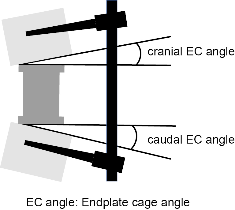
    Methods: Eight males and 13 females (77.2±6.0 years old) with osteoporotic vertebral fractures who underwent single corpectomy using a wide-footprint expandable cage with at least a 1-year follow-up were retrospectively included. The endplate cage (EC) angle was defined as the angle between the vertebral body's endplate and the cage's base on the cranial and caudal sides. A sagittal computed tomography scan was performed immediately after surgery and at the final follow-up, with cage subsidence defined as subsidence of ≥2 mm on the cranial or caudal side. Risk factors were analyzed by dividing cases into groups with (n=6) and without (n=15) cage subsidence.

    Results: No significant differences were noted in age, bone mineral density, number of fixed vertebrae, sagittal parameters, preoperative and final kyphosis angle, amount of kyphosis angle correction, bone union, screw loosening, and number of other vertebral fractures preoperatively and 1-year postoperatively between the two groups. No difference was noted in cranial EC angle, but a significant difference was noted in caudal EC angle in the group with (10.7±4.1°) and without (4.7±4.2°) subsidence (P=0.008). Logistic regression analysis with the dependent variable as presence or absence of subsidence showed that caudal EC angle (>7.5°) was a significant factor (odds ratio: 20, 95% confidence interval: 1.655-241.7, P=0.018).

    Conclusions: In minimally invasive lateral corpectomy for osteoporotic vertebral fractures, a cage tilted more than 7.5° to the caudal vertebral endplate is a risk factor for cage subsidence. The cage should be placed as perpendicular to the endplate as possible, especially to the caudal vertebral body, to avoid cage subsidence.

    Download PDF (360K)
  • Yujiro Kagami, Hiroaki Nakashima, Naoki Segi, Ryuichi Shinjo, Shiro Im ...
    2023 Volume 7 Issue 4 Pages 363-370
    Published: July 27, 2023
    Released on J-STAGE: July 27, 2023
    Advance online publication: March 13, 2023
    JOURNAL OPEN ACCESS

    Introduction: This study aimed to evaluate the efficacy of condoliase injection therapy (CIT) for lateral lumbar disc herniation (LLDH).

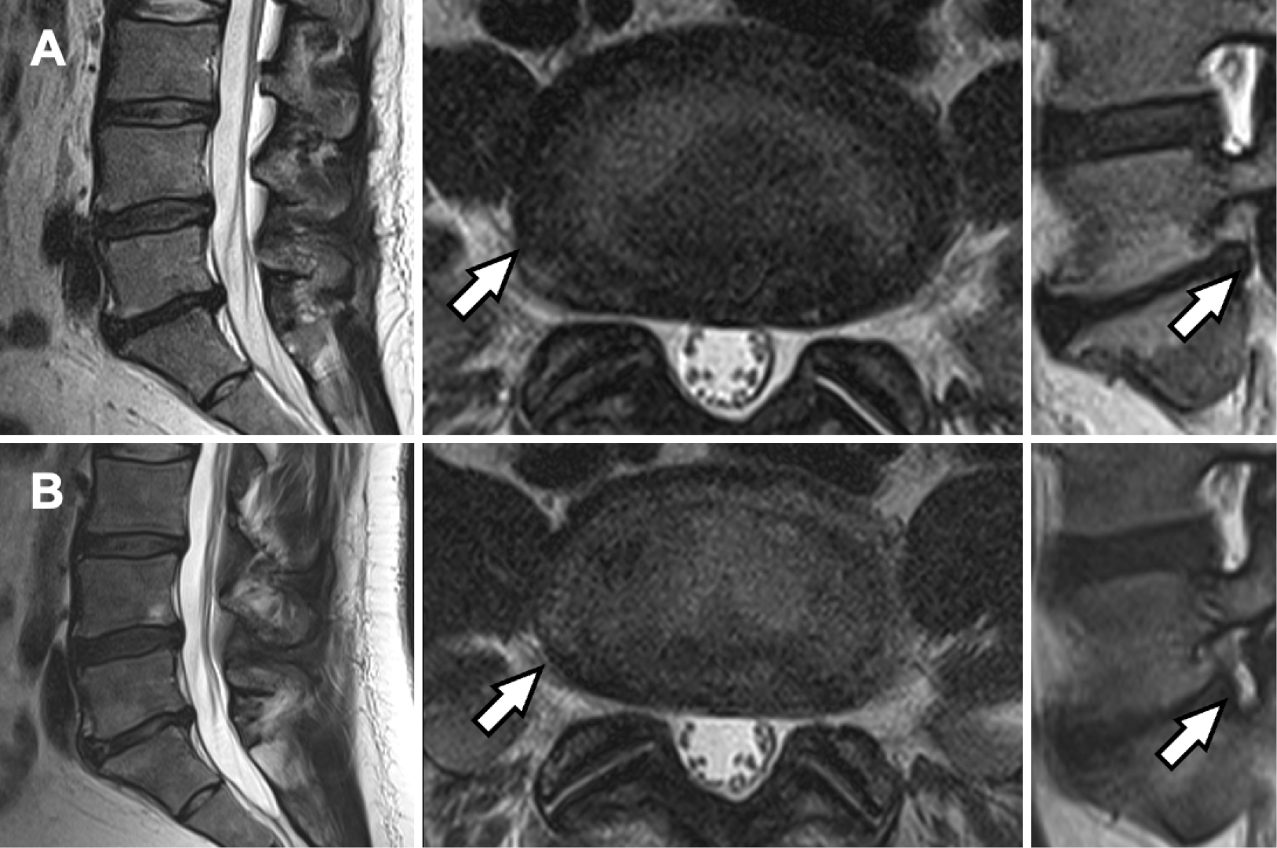
    Methods: This retrospective study included 157 of 180 enrolled patients (70 males, 87 females; mean age: 52.6±16.9 years). These patients were divided into two groups (group L: LLDH, group M: medial LDH [subligamentous and transligamentous]). From baseline to 1 year after injection (final follow-up), leg pain was assessed using the visual analog scale (VAS) and the Japanese Orthopedic Association (JOA) scoring for CIT's clinical efficacy of CIT. Radiography and magnetic resonance imaging conducted before and 3 months after the injection were assessed. Patients with a VAS improvement of ≥50% at the final follow-up were defined as responders. The responder and nonresponder LLDH groups were also compared.

    Results: Groups L and M showed comparable responder rates (75.0% and 77.4%, respectively) (P=0.80). VAS and JOA scores at 1 year showed no significant differences between the groups (P=0.82 and 0.80, respectively). VAS score at 1 month after injection reduced considerably in the responder group compared with that in the nonresponder group (19.7 vs. 66.0, P<0.01) and continued to decrease at the last follow-up (3.5 vs. 52.0, P<0.001). Nonresponders had significantly lower disc heights after 3 months. However, intervertebral instability, alignment, and disc degeneration did not differ between the responders and nonresponders.

    Conclusions: The response rate of CIT for LLDH was comparable to that for medial LDH. Therefore, CIT is an effective treatment for LLDH.

    Download PDF (628K)
  • Yosuke Horiuchi, Mitsuru Yagi, Satoshi Suzuki, Yohei Takahashi, Satosh ...
    2023 Volume 7 Issue 4 Pages 371-376
    Published: July 27, 2023
    Released on J-STAGE: July 27, 2023
    Advance online publication: March 13, 2023
    JOURNAL OPEN ACCESS

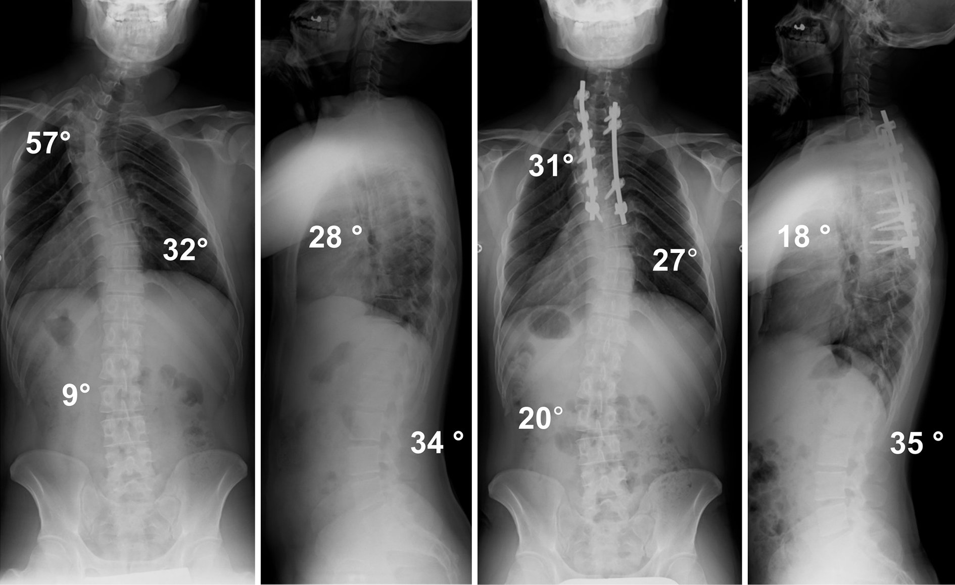
    Introduction: Adolescent idiopathic scoliosis (AIS) with a major curve at the main thoracic (MT) area is classified as Lenke type 1, 2, or 3 depending on the flexibility of the proximal thoracic (PT) curve and lumbar curve. No definite classification has been established for a major curve at the PT spine. The purpose of this study is to investigate the radiographic characteristics before and after correction surgery for AIS with a major curve at the PT area.

    Methods: This is a retrospective cohort study at a single academic institution. Twelve patients with a major curve at the PT spine participated in our study and followed for at least two years after surgery. We evaluated the pre- and postoperative Cobb angles of the curve, curve range, location of the apex, sagittal parameters, and shoulder balance-related parameters. All patients were treated by posterior correction and fusion surgery using pedicle screw constructs.

    Results: The patients were classified as having a double-curve (DC) type, in which the MT curve was structural, or a single-curve (SC) type, in which the MT curve was corrected to less than 25° on supine side-bending films. The mean correction rates for the PT curve were favorable in both groups (DC, 65.7%±9.6%; SC, 39.2%±4.9%). The mean Cobb angle of the lumbar curve improved in the DC group (preoperative, 17.1°±4.0°; postoperative, 5.0°±4.2°) but deteriorated in the SC group (preoperative, 7.1°±1.2°; postoperative, 12.4°±4.4°) after surgery.

    Conclusions: We illustrated the postoperative radiographical changes of 12 consecutive patients with the major curve at the PT curve. Although posterior correction and fusion surgery corrected the PT curve satisfactorily in both DC and SC patients, the Cobb angle of the lumbar curve deteriorated after surgery in all SC patients. Surgeons need to pay attention to the fusion area, especially LIV, when operating the SC curve type.

    Download PDF (861K)
  • Tomoyuki Asada, Toshiaki Kotani, Tsuyoshi Sakuma, Yasushi Iijima, Keit ...
    2023 Volume 7 Issue 4 Pages 377-384
    Published: July 27, 2023
    Released on J-STAGE: July 27, 2023
    Advance online publication: March 13, 2023
    JOURNAL OPEN ACCESS

    Introduction: This study aimed to investigate the brace compliance and clinical background of patients with adolescent idiopathic scoliosis (AIS) who demonstrate different degrees of psychological brace-related stress.

    Methods: Forty-five patients initiating brace treatment with a Cobb angle between 25° and 45° were included. Patients receiving brace treatment for AIS were administered a questionnaire for brace-related stress (i.e., the Japanese version of the Bad Sobernheim Stress Questionnaire-Brace [JBSSQ-brace]). Based on their scores, we allocated the patients into two stress groups: mild-stress (≥16 points) and below-moderate-stress (<16 points). We investigated the character of brace compliance and brace-related psychological stress in all patients and compared the demographics and brace compliance between both groups.

    Results: Forty-one of 45 patients completed the study. The mean JBSSQ-brace scores were 18.7±5.1, 19.1±5.2, and 18.7±5.0 points at the 1-month, 4-month, and 1-year follow-ups, respectively. There was no significant change in JBSSQ-brace scores over one year after the brace prescription (P=0.332). There was no difference in-brace compliance between seasons during the first month of brace prescription (P=0.252). Both groups' overall brace compliance was comparable (below-moderate: 17.1±7.1 h/day vs. mild: 20.4±3.0 h/day; P=0.078). The mild-stress group showed better compliance than the below-moderate-stress group on weekdays (below-moderate: 17.0±6.9 h/day vs. mild: 20.5±2.8 h/day; P=0.048) and at nighttime (below-moderate: 82.3%±27.0%/nighttime vs. mild: 93.8%±12.4%/nighttime; P=0.008).

    Conclusions: Overall, brace compliance was comparable among patients with different brace-related stress, but brace compliance during weekdays and nighttime was significantly better in the mild-stress group.

    Download PDF (235K)
  • Kathryn M. DeFoe, Jeremiah Atkinson, Jean Stansbury, Angela Sinner, Wa ...
    2023 Volume 7 Issue 4 Pages 385-389
    Published: July 27, 2023
    Released on J-STAGE: July 27, 2023
    Advance online publication: March 13, 2023
    JOURNAL OPEN ACCESS

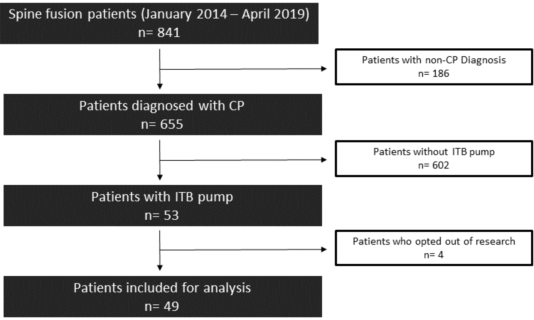
    Introduction: Patients with cerebral palsy (CP) may receive intrathecal baclofen (ITB) to reduce muscle spasticity and dystonia. It can be challenging to identify the proper dose of ITB, and anecdotally these dosing needs may change after spinal fusion surgery. This study aimed to evaluate the need for changes in ITB dosing following a spinal fusion in pediatric neuromuscular scoliosis (NMS) patients and identify predisposing factors for those changes.

    Methods: This was a retrospective case-control study of NMS patients with an ITB pump who later received a spinal fusion surgery. Dosing changes and the indications for the changes were postoperatively noted. Demographics, preoperative factors, and surgical factors were evaluated for correlation with dosing changes.

    Results: A total of 49 patients were included in this study. Most had no change in ITB dose (71.4%), and others required a change that averaged about 10%. Male patients, those with larger pumps, and those that had a longer hospital stay were more likely to require a decrease in dose. Complications were similar between groups. Three catheters were revised during surgery: two continued on the same dose and one required an increase in dose after surgery.

    Conclusions: Spinal fusion after ITB pump placement is feasible and safe. Most patients did not require dosing changes after spine fusion; however, careful evaluation postoperatively remains prudent.

    Download PDF (103K)
  • Hisanori Gamada, Masaki Tatsumura, Shun Okuwaki, Toru Funayama, Masash ...
    2023 Volume 7 Issue 4 Pages 390-395
    Published: July 27, 2023
    Released on J-STAGE: July 27, 2023
    Advance online publication: March 13, 2023
    JOURNAL OPEN ACCESS

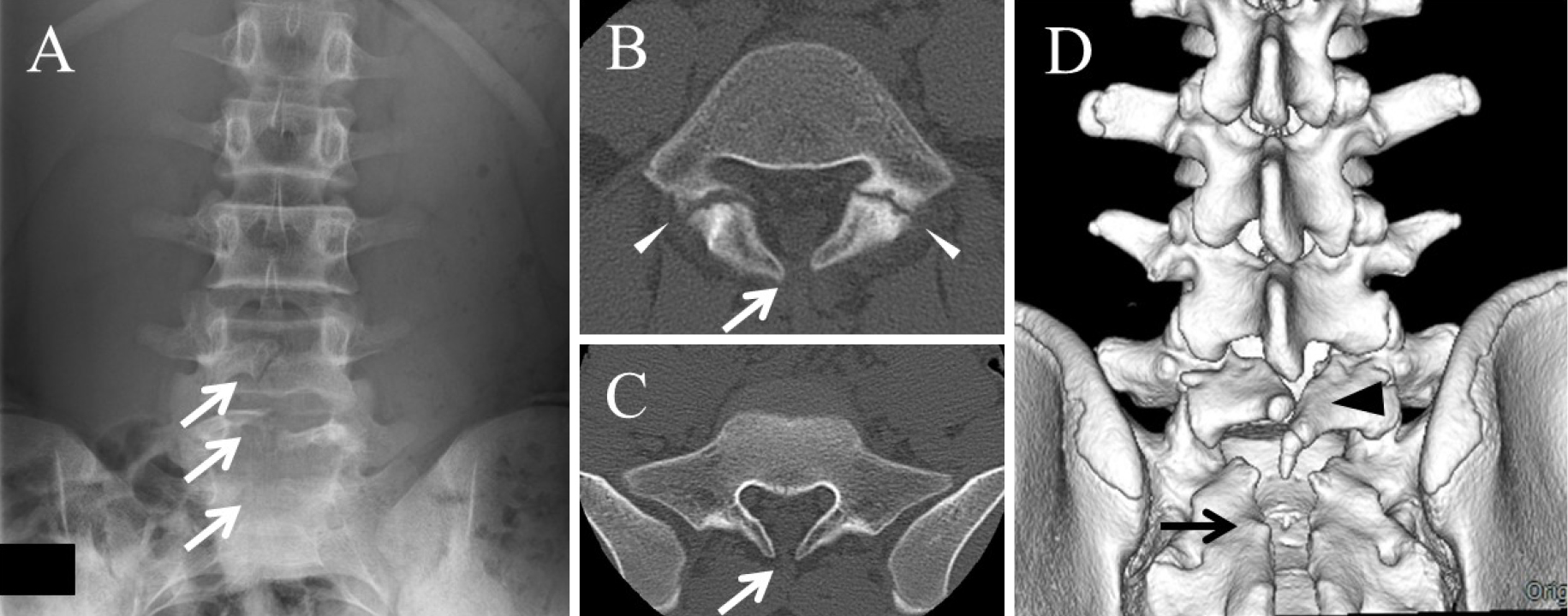
    Introduction: The effect of spina bifida occulta (SBO) on bone union in lumbar spondylolysis is controversial. The study aim was to assess the effect of SBO on bone union after conservative treatment of L5 spondylolysis, the most common level.

    Methods: The study included 191 lesions in 145 patients (mean age, 14.0 years) with conservatively treated L5 spondylolysis. We examined the relationships between bone union after conservative treatment and the number of SBOs, levels, and single or multilevel status. Fisher's exact probability test, chi-square test, and Welch's T test were performed.

    Results: The SBO incidence was 53%, with at least one SBO at any vertebral level. SBO at S1 (p=0.034) or S2 (p=0.0003), two SBOs (p=0.0018), and three SBOs (p=0.011) were associated with a lower bone union rate. The bone union rate was significantly lower for lesions with SBOs at both S1 and S2 than without (42% vs. 79%; p<0.0001).

    Conclusions: The SBO incidence in L5 spondylolysis was 53%. SBO at S1 or S2 and a higher number of SBOs were associated with lower bone union rates. In particular, the bone union rate of lesions with SBOs at both S1 and S2 was <50%.

    Download PDF (333K)
TECHNICAL NOTE
  • Masaki Tatsumura, Shun Okuwaki, Hisarnori Gamada, Reo Asai, Fumihiko E ...
    2023 Volume 7 Issue 4 Pages 396-401
    Published: July 27, 2023
    Released on J-STAGE: July 27, 2023
    Advance online publication: June 09, 2023
    JOURNAL OPEN ACCESS

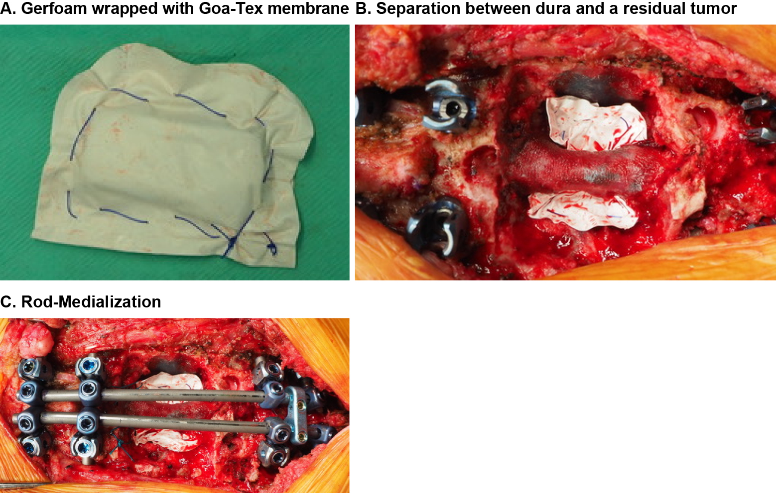
    Introduction: Lumbar spondylolysis is a common fatigue fracture of the pars interarticularis of the lamina of the lumbar spine in adolescent athletes presenting with pars clefts. Some pseudarthrotic lumbar spondylolysis causes low back pain or radiculopathy. This study presents a case of pseudarthrotic lumbar spondylolysis that was successfully treated using a modified smiley face rod technique.

    Technical Note: We developed a modified smiley face rod technique, which places pedicle screws in the lateral edge of the pedicle to preserve the erector spinae muscles and inserts a U-shaped rod between the spinous processes to preserve the supraspinous ligament. When a U-shaped rod penetrates the interspinous ligament subcutaneously, the resection of the supraspinous ligaments can be avoided. When the screw head is positioned more anterolaterally, a compression force is applied perpendicular to the surface of the pars cleft by rod clamping. This intrasegmental fusion technique preserves the mobile segment and simultaneously repairs the pars cleft. It is less invasive and more appropriate than interbody fusion for young athletes to avoid the possibility of future adjacent segment disorders.

    Conclusions: This is a minimally invasive procedure that can easily achieve bone fusion and should be introduced for patients who are suffering from the symptoms of pseudarthrotic lumbar spondylolysis.

    Download PDF (1789K)
CLINICAL CORRESPONDENCE
feedback
Top