Abstract
Gastric acid secretion levels are an important determinant of the manifestation of acid-related upper GI diseases such as gastroesophageal reflux disease. We recently reported that gastric acid secretion levels did not change from the 1990s to 2010s in H. pylori-negative asymptomatic Japanese outpatients with a mean age of 63 years old. However, because young people have a quite different lifestyle, including dietary pattern, from elderly people in Japan, it is worth investigating any chronologic changes in gastric acid secretion in younger generations. The aim of this analysis is to investigate the potential changes in gastric acid secretion from 1995 to 2014 in young Japanese healthy volunteers. Eighty-eight young Japanese healthy volunteers under the age of 40 with a mean age of 26 were extracted from a database accumulated from 1995 to 2014 for the present analysis. Their gastrin-stimulated gastric acid level was determined with the endoscopic gastrin test. In 76 H. pylori-negative subjects, gastric acid secretion levels showed a significant positive correlation with the calendar year when the test was performed (r = 0.27, p < 0.01). A similar trend was observed in 12 H. pylori-positive subjects. A chronological 5-year time period showed a significant positive association with gastric acid secretion in univariate and multivariate analyses (p < 0.01 and p = 0.01, respectively). Gastric acid secretion levels have been increasing in young Japanese healthy volunteers during the last 20 years. We need to monitor future trends in the prevalence of acid-related diseases such as gastro-esophageal reflux disease in Japan.
Introduction
Gastro-esophageal reflux disease (GERD) is caused by exposure of the esophageal epithelium to the refluxed gastric acid, and the GERD spectrum comprises from reflux erosive esophagitis, then Barrett’s esophagus, and finally to esophageal adenocarcinoma. The prevalence of esophageal adenocarcinoma have been remarkably increased over the last 3 decades in Western countries, while there has no such apparent increase in Asian countries at the present (Thrift and Whiteman 2012; Arnold et al. 2015). Nonetheless, the prevalence of reflux esophagitis has been increasing over the last 2 decades in Asian countries (Wong and Kinoshita 2006); consequently, whether complicated GERD such as Barrett’s esophagus and esophageal adenocarcinoma will also increase in this geographic area is a major concern from the Asian perspective. Gastric acid secretion levels are an important determinant of the manifestation of acid-related upper GI diseases such as GERD (Collen et al. 1990; Cadiot et al. 1997). Hence, potential chronological changes in gastric acid secretion levels in Japanese people could be important when predicting the future trend of complicated GERD in Japan.
Previously, Kinoshita et al. (1997) reported that gastric acid secretion levels increase in Japanese healthy volunteers from the 1970s to 1990s in both H. pylori-negative and -positive subjects. Meanwhile, we recently reported that gastric acid secretion levels were unchanged from the 1990s to 2010s in H. pylori-negative asymptomatic Japanese outpatient subjects with a mean age of 63 years old (Iijima et al. 2015). However, because the lifestyle of young people in Japan is quite different from that of elderly people in areas such as dietary pattern, it is worth investigating chronologic changes in gastric acid secretion in the young generation. Although Ishimura et al. (2015) also recently demonstrated that gastric acid secretion did not change during the last 20 years in Japanese people, they did not compare chronological changes in gastric acid secretion in young people.
In this study, using our accumulating database, we investigated potential changes in gastric acid secretion from 1995 to 2014 in young Japanese healthy volunteers under the age of 40 years old.
Methods
In a series of our previous studies (Iijima et al. 1998, 2009, 2013; Iwabuchi et al. 2013; Shinkai et al. 2014), we enrolled 95 healthy male volunteers to investigate their gastric acid secretion level with the endoscopic gastrin test (EGT) described below over the 20-year time period from 1995 to 2014. These healthy volunteers had been recruited mainly from Tohoku university students or post-graduate students, and they were not included in our preceding report comprising hospital outpatients (Iijima et al. 2015). Among them, 88 young subjects under the age of 40 with a mean age of 26 (5) were extracted for the present analysis. None of them had any medication or abdominal symptoms, and additionally, they turned out to be endoscopically normal. The age, body weight, and smoking status of each subject on the study day were recorded. Smoking status was classified as either current smoker or current non-smoker.
The original studies were implemented in accord with the Helsinki Declaration, and each subject provided written informed consent prior to study entry. The current retrospective study was approved by the Tohoku University School of Medicine Ethics Committee (2014-1-414).
Endoscopic gastrin test
During a routine endoscopic procedure, the gastrin-stimulated gastric acid secretory response was estimated for each subject using the endoscopic gastrin test (EGT), which is a modified method of the conventional gastrin-stimulated maximal acid output test (Iijima et al. 1998). The details of the EGT were described previously. Briefly, the subjects were injected intramuscularly with pentagastrin at a dose of 6 µg/kg (Sigma, St. Louis, Mo, USA) 15 min prior to undergoing endoscopy. After entering the stomach with the endoscope, pooled gastric fluid was aspirated and discarded. Gastric fluid secreted between 20 and 30 min after pentagastrin injection was aspirated and collected under direct visualization during routine endoscopic procedure of the stomach and duodenal bulb. The volume of fluid sample collected over the 10-min period was recorded, and the H+ concentration was measured by titration. The acid output in the 10-min period was calculated by multiplying the sample volume by H+ concentration, and the EGT result was expressed as H+ mEq/10 min. We previously reported that EGT values correlate highly with the maximal acid output (MAO) and peak acid output (PAO) measured by conventional methods (correlation coefficient: 0.90 and 0.92, respectively) and show high reproducibility (coefficient of variation, 5.6) (Iijima et al. 1998).
H. pylori determination
H. pylori infection status was evaluated by a rapid urease test (Helicocheck, Otsuka, Tokyo, Japan), histological assessment (Giemsa staining) with gastric biopsies from the antrum and the body, and a serum IgG antibody to H. pylori test (E Plate ‘‘Eiken’’ H. pylori antibody; Eiken Chemical Co., Ltd., Tokyo, Japan). The subjects were judged to be H. pylori-negative if all three tests were negative and H. pylori-positive if one or more the tests was positive (Kikuchi et al. 2011).
Statistics
Data for categorical valuables were described as the actual number (percentage), and data for continuous variables were described as the means (standard deviation [SD]) unless otherwise stated. Comparisons between H. pylori-negative and H. pylori-positive subjects were completed using the χ2 test for categorical valuables and the unpaired t test for continuous variables.
To investigate chronological changes of gastric acid secretion, the EGT values were initially plotted against the calendar year (continuous variables) when the test was performed, and the correlation between the 2 parameters was investigated by a linear regression analysis. Then, the chronological periods of the test were divided into 4 groups within a 5-year period: 1995-1999, 2000-2004, 2005-2009, and 2010-2014, and the correlation between the chronological period and the EGT values was estimated with Spearman’s rank test. Further, using the 4 chronological groups (1995-1999, 1; 2000-2004, 2; 2005-2009, 3; 2010-2014, 4) as an independent variable and gastric acid secretion as a dependent variable, unadjusted and adjusted linear regression analyses were performed. In the adjusted analysis, age (years), body weight (kg), H. pylori infection, and current smoking status (non-smokers, 0; smokers, 1) were included in the model because these parameters have been shown to affect gastric acid secretion (Iijima et al. 2004). All analyses were performed with SPSS statistics ver. 20 (IBM), and a p value of less than 0.05 was considered to be statistically significant.
Results
Of 88 healthy male volunteers enrolled in this analysis, 12 were judged to be H. pylori-positive, and the remaining 76 were H. pylori-negative. A comparison of various parameters between the H. pylori-negative and H. pylori-positive subjects is shown in Table 1. The mean age was 25.8 (4.9) years old and 26.8 (4.6) among H. pylori-negative and H. pylori-positive subjects, respectively, and it was not significantly different between the 2 groups. Similarly, the mean body weight and prevalence of current smokers were not significantly different between the groups. Meanwhile, the mean gastric acid secretion was significantly lower in the H. pylori-positive subjects than in the H. pylori-negative subjects (2.9 (2.2) mEq/10 min vs. 4.1 (1.1), p < 0.01), which is consistent with our previous report (Iijima et al. 2004).
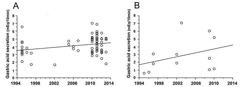
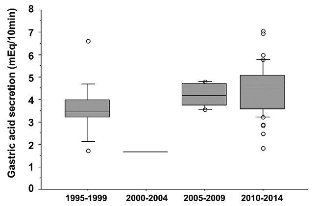
Gastric acid secretion levels were plotted against the calendar year when the test was performed according to the H. pylori infection status, which is shown in Fig. 1. In H. pylori-negative subjects, gastric acid secretion levels showed a significant positive correlation with the calendar year (Fig. 1A, r = 0.27, p = 0.01). A box-and-whisker diagram in which the chronological periods of the study were divided into 4 groups within a 5-year period indicates that gastric acid secretion levels significantly increased (r = 0.32, p < 0.01) in H. pylori-negative subjects, with a 20% increase in the last 5-year period (2010-2014) compared with the first 5-year period (1995-1999) (Fig. 2). In H. pylori-positive subjects, gastric acid secretion levels were positively correlated with the calendar year as well, although it was not significantly different due to the small number of subjects (Fig. 1B).
Further regression analysis to investigate the association between chronological time and gastric acid secretion was performed in the H. pylori-negative subjects (Table 2). A univariate regression analysis revealed that chronological time period showed a significant positive association with gastric acid secretion (p < 0.01), and the association was also evident in a multivariate analysis adjusted by age, body weight, and smoking status (p = 0.01). Meanwhile, body weight showed a significant positive association with gastric acid secretion in the multivariate analysis (p = 0.04).
Discussion
In this study, we found a weak, but significant increase in gastric acid secretion during the 20-year period between 1995 and 2014 in H. pylori-negative young Japanese healthy volunteers with a mean age of 26 years. This finding contrasts with our recent report in which gastric acid secretion did not change during the same period in H. pylori-negative asymptomatic outpatients with a mean age of 63 years (Iijima et al. 2015). Moreover, in that study, an additional subgroup analysis in 47 H. pylori-negative outpatients over the age of 65 reveals that there is no change in gastric acid secretion between 1995 and 2014 (r = 0.03, p = 0.83). Thus, chronological changes in gastric acid secretion in H. pylori-negative Japanese subjects seem to differ between the young and elderly generations.
The rationale for the different chronological behavior in gastric acid secretion between young and elderly Japanese subjects is unclear at the moment. Body size is a significant contributor determining gastric acid secretion (Iijima et al. 2004; Derakhshan et al. 2006), as verified in the present analysis showing a significant positive association between body weight and gastric acid secretion in H. pylori-negative subjects. However, the emerging positive association between chronological time period and gastric acid secretion in H. pylori-negative subjects was independent of body weight, and in addition, body weight did not significantly change in the subjects enrolled in this analysis during the observation period (the first 5-year period: 66.7 (9.3) kg vs. the last one: 64.8 (6.1), p = 0.3)). Hence, body size could not explain the significant increase in gastric acid secretion in H. pylori-negative young subjects observed in this analysis.
Alternatively, diet pattern may be important. There is a considerable difference in the dietary pattern between young and elderly Japanese people. For example, a very recent nationwide survey conducted in 2013 indicated that 47% of Japanese men with an age of 20-29 have a modern westernized dietary pattern consisting of meat, non-Japanese noodles, and sugar sweetened beverages, whereas only 2.9% of those with an age of 60-69 had such a dietary pattern; instead, men aged 60-69 have a more traditional Japanese dietary pattern consisting of rice and vegetables (Katagiri et al. 2015). Because dietary fat intake is known to stimulate gastric acid secretion (Saqui-Salces et al. 2012), such differences in dietary pattern among Japanese generations could be at least partly responsible for the different chronological changes in gastric acid secretion between young and elderly Japanese people.
Whether the escalating level of gastric acid secretion observed in young Japanese people persists for their lifetime is uncertain at present. This could be an important clinical issue because the persistence of elevated gastric acid secretion could lead to more prevalent acid-related diseases such as GERD with advancing age through aging-related motility disorders of the esophagus (Lee et al. 2007). On the other hand, changes in individual dietary patterns with aging might alleviate gastric acid secretion levels.
A major limitation of this study is imbalance in the number of enrolled subjects among 4 chronological periods, that is, the majority of the subjects derived from the last period. In this study, we applied the same definition of the chronological periods as in our preceding study (Iijima et al. 2015) so as to help compare the difference between the two studies.
Increased gastric acid secretion in Japanese young adults observed in this analysis could lead to increased prevalence in reflux esophagitis in that age-group. A recent study reported the prevalence of reflux esophagitis in healthy Japanese people under the age of 40 years old is 14.6% (Chiba et al. 2012), however, there have no reports which directly compared chronological change in prevalence of reflux esophagitis in various age-groups in a defined population. A further study to investigate chronological change in the prevalence of reflux esophagitis among Japanese young adults is warranted.
In conclusion, gastric acid secretion levels have increased in young Japanese healthy volunteers during the last 20 years. Whether this increase in gastric acid secretion in the young generation will persist for their lifetime is uncertain, thus, we need to monitor future trends in the prevalence of acid-related diseases such as GERD in Japan.
Conflict of Interest
The authors declare no conflict of interest.
References
-
Arnold,
M.,
Soerjomataram,
I.,
Ferlay,
J. &
Forman,
D.
(2015) Global incidence of oesophageal cancer by histological subtype in 2012. Gut, 64, 381-387.
-
Cadiot,
G.,
Bruhat,
A.,
Rigaud,
D.,
Coste,
T.,
Vuagnat,
A.,
Benyedder,
Y.,
Vallot,
T.,
Le Guludec,
D. &
Mignon,
M.
(1997) Multivariate analysis of pathophysiological factors in reflux oesophagitis. Gut, 40, 167-174.
-
Chiba,
H.,
Gunji,
T.,
Sato,
H.,
Iijima,
K.,
Fujibayashi,
K.,
Okumura,
M.,
Sasabe,
N.,
Matsuhashi,
N. &
Nakajima,
A.
(2012) A cross-sectional study on the risk factors for erosive esophagitis in young adults. Intern. Med., 51, 1293-1299.
-
Collen,
M.J.,
Lewis,
J.H. &
Benjamin,
S.B.
(1990) Gastric acid hypersecretion in refractory gastroesophageal reflux disease. Gastroenterology, 98, 654-661.
-
Derakhshan,
M.H.,
El-Omar,
E.,
Oien,
K.,
Gillen,
D.,
Fyfe,
V.,
Crabtree,
J.E. &
McColl,
K.E.
(2006) Gastric histology, serological markers and age as predictors of gastric acid secretion in patients infected with Helicobacter pylori. J. Clin. Pathol., 59, 1293-1299.
-
Iijima,
K.,
Ichikawa,
T.,
Okada,
S.,
Ogawa,
M.,
Koike,
T.,
Ohara,
S. &
Shimosegawa,
T.
(2009) Rebamipide, a cytoprotective drug, increases gastric mucus secretion in human: evaluations with endoscopic gastrin test. Dig. Dis. Sci., 54, 1500-1507.
-
Iijima,
K.,
Iwabuchi,
T.,
Ara,
N.,
Koike,
T.,
Shinkai,
H.,
Kamata,
Y.,
Ichikawa,
T.,
Ishihara,
K. &
Shimosegawa,
T.
(2013) Reactive increase in gastric mucus secretion is an adaptive defense mechanism against low-dose aspirin-induced gastropathy. Dig. Dis. Sci., 58, 2266-2274
-
Iijima,
K.,
Koike,
T.,
Abe,
Y.,
Ohara,
S.,
Nakaya,
N. &
Shimosegawa,
T.
(2015) Time series analysis of gastric acid secretion over a 20-year period in normal Japanese men. J. Gastroenterol., 50, 853-861.
-
Iijima,
K.,
Ohara,
S.,
Koike,
T.,
Sekine,
H. &
Shimosegawa,
T.
(2004) Gastric acid secretion of normal Japanese subjects in relation to Helicobacter pylori infection, aging, and gender. Scand. J. Gastroenterol., 39, 709-716.
-
Iijima,
K.,
Ohara,
S.,
Sekine,
H.,
Koike,
T.,
Kubota,
Y.,
Kato,
K.,
Asaki,
S. &
Toyota,
T.
(1998) A new endoscopic method of gastric acid secretory testing. Am. J. Gastroenterol., 93, 2113-2118.
-
Ishimura,
N.,
Owada,
Y.,
Aimi,
M.,
Oshima,
T.,
Kamada,
T.,
Inoue,
K.,
Mikami,
H.,
Takeuchi,
T.,
Miwa,
H.,
Higuchi,
K. &
Kinoshita,
Y.
(2015) No increase in gastric acid secretion in healthy Japanese over the past two decades. J. Gastroenterol., 50, 844-852.
-
Iwabuchi,
T.,
Iijima,
K.,
Ara,
N.,
Koike,
T.,
Shinkai,
H.,
Ichikawa,
T.,
Kamata,
Y.,
Ishihara,
K. &
Shimosegawa,
T.
(2013) Increased gastric mucus secretion alleviates non-steroidal anti-inflammatory drug-induced abdominal pain. Tohoku J. Exp. Med., 231, 29-36.
-
Katagiri,
R.,
Asakura,
K.,
Uechi,
K.,
Masayasu,
S. &
Sasaki,
S.
(2015) Adequacy of iodine intake in three different Japanese adult dietary patterns: a nationwide study. Nutr. J., 14, 129.
-
Kikuchi,
R.,
Abe,
Y.,
Iijima,
K.,
Koike,
T.,
Ara,
N.,
Uno,
K.,
Asanuma,
K.,
Asano,
N.,
Imatani,
A. &
Shimosegawa,
T.
(2011) Low serum levels of pepsinogen and gastrin 17 are predictive of extensive gastric atrophy with high-risk of early gastric cancer. Tohoku J. Exp. Med., 223, 35-44.
-
Kinoshita,
Y.,
Kawanami,
C.,
Kishi,
K.,
Nakata,
H.,
Seino,
Y. &
Chiba,
T.
(1997) Helicobacter pylori independent chronological change in gastric acid secretion in the Japanese. Gut, 41, 452-458.
-
Lee,
J.,
Anggiansah,
A.,
Anggiansah,
R.,
Young,
A.,
Wong,
T. &
Fox,
M.
(2007) Effects of age on the gastroesophageal junction, esophageal motility, and reflux disease. Clin. Gastroenterol. Hepatol., 5, 1392-1398.
-
Saqui-Salces,
M.,
Dowdle,
W.E.,
Reiter,
J.F. &
Merchant,
J.L.
(2012) A high-fat diet regulates gastrin and acid secretion through primary cilia. FASEB. J., 26, 3127-3139.
-
Shinkai,
H.,
Iijima,
K.,
Koike,
T.,
Nakagawa,
K.,
Maejima,
R.,
Endo,
H.,
Ara,
N.,
Asano,
N.,
Imatani,
A.,
Ohara,
S. &
Shimosegawa,
T.
(2014) Calcium carbonate breath test for non-invasive estimation of gastric acid secretion. Tohoku J. Exp. Med., 232, 255-261.
-
Thrift,
A.P. &
Whiteman,
D.C.
(2012) The incidence of esophageal adenocarcinoma continues to rise: analysis of period and birth cohort effects on recent trends. Ann. Oncol., 23, 3155-3162.
-
Wong,
B.C. &
Kinoshita,
Y.
(2006) Systematic review on epidemiology of gastroesophageal reflux disease in Asia. Clin. Gastroenterol. Hepatol., 4, 398-407.