Abstract
The Holliday Junction-Recognition Protein (HJURP) was upregulated in several tumors, which was associated with poor outcome. This study investigated the effects of the HJURP-mediated c-Jun N-terminal kinase (JNK)/ signal transducer and activator of transcription 3 (STAT3) pathway on bladder urothelial carcinoma (BLUC). Online databases were used to analyze HJURP expression in BLUC and the correlation of HJURP to JNK1 [mitogen-activated protein kinase 8 (MAPK8)], JNK2 (MAPK9), STAT3, marker of proliferation Ki-67 (MKI67), proliferating cell nuclear antigen (PCNA), cyclin dependent kinase 2 (CDK2), CDK4 and CDK6. HJURP expression was detected in BLUC cells and human normal primary bladder epithelial cells (BdECs). BLUC cells were treated with HJURP lentivirus activation /shRNA lentivirus particles or JNK inhibitor SP600125. HJURP was upregulated in BLUC tissues and correlated with poor prognosis of patients (all P < 0.05). HJURP in tumor positively correlated with MAPK8 (R = 0.30), MAPK9 (R = 0.30), STAT3 (R = 0.15), MKI67 (R = 0.60), PCNA (R = 0.46), CDK2 (R = 0.39), CDK4 (R = 0.24) and CDK6 (R = 0.21). The JNK inhibitor SP600125 decreased p-JNK/JNK and p-STAT3/STAT3 in BLUC cells, which was reversed by HJURP overexpression (P < 0.05). The HJURP-mediated JNK/STAT3 pathway promoted BLUC cell proliferation and inhibited cell apoptosis (P < 0.05). HJURP reversed the arrested G0/G1 phase of BLUC cells by SP600125. HJURP acted as an oncogene to regulate BLUC cell proliferation, apoptosis and the cell cycle by mediating the JNK/STAT3 pathway. Therefore, HJURP targeting might be an attractive novel therapeutic target for early diagnosis and treatment in BLUC.
Introduction
Bladder cancer (BCa) is the most common malignancy in the bladder mucosa of the urinary tract and one of the most prevalent cancers worldwide (Dobruch and Oszczudlowski 2021). According to estimates in 2020 (worldwide, both sexes, all ages), the incidence and the mortality of BCa were 573,278 and 212,536, respectively, which ranked 10th for malignant cancer incidence and 13th in malignancy-caused deaths worldwide. However, the incidence and the mortality of BCa were 5.9% and 2.7%, respectively, in China (http://gco.iarc.fr/today/home). BCa pathologically includes bladder urothelial carcinoma (BLUC, also known as transitional cell carcinoma), squamous cell carcinoma and adenocarcinoma, small cell carcinoma, mixed carcinoma, carcinosarcoma and metastatic carcinoma, and BLUC is the most common type, accounting for up to 95%. BLUC generally places a heavy financial burden on patients. BLUC originates from the urothelial cells on the inside of the bladder (Hyldgaard and Jensen 2021; Martinez-Rojo et al. 2021). Most BCa patients are males aged 65 years and older, and female patients are less common. The major risk factors for BCa include smoking, exposure to chemicals, radiotherapy and chemotherapy (Martinez-Rojo et al. 2021). The treatment methods of BCa are limited to surgery and immunotherapy or chemotherapy (Siracusano et al. 2020). Recent extensive analyses of molecular alterations have created the possibility of some novel therapeutic approaches for BCa (Rozanec and Secin 2020).
Holliday junction-recognition protein (HJURP), also known as FAKTS, URLC9, or HFLEG1, is located at 2q37.1, and it encodes a protein consisting of 748 amino acids. It is expressed in thymus, placenta, small intestine, liver, skeletal muscle, bone marrow and colon tissues (http://www.genecards.org/cgi-bin/carddisp.pl?gene=HJURP). HJURP is abnormally expressed in many diseases, including several cancers. For example, Tsevegjav et al. (2022) revealed that HJURP was highly expressed in most oral cancer cell lines and tissues compared to normal oral epithelial cells. Milioli et al. (2017) observed the overexpression of HJURP in basal-like breast cancer, which was linked to abnormal proliferation, cell invasion and metastasis in breast cancer and is consistent with the definition of basal-like breast carcinoma (Lehmann et al. 2011). Valente et al. (2013) reported that HJURP was overexpressed in 40 astrocytoma samples of different grades, and tumors with high HJURP expression correlated with poor survival prognosis, which indicated that HJURP overexpression was an independent prognostic factor for the risk of death of astrocytoma patients. Based on the online databases UALCAN and GEPIA, we found the upregulation of HJURP in BLUC and its positive correlation to c-Jun NH(2)-terminal kinase 1 (JNK1, also known as mitogen-activated protein kinase 8, MAPK8) (Hess et al. 2002), JNK2 (MAPK9) (Jaeschke et al. 2005), and STAT3 in BLUC. The JNK/STAT3 signaling pathway is abnormally activated in BCa, and many genes inhibit this pathway to play a potential therapeutic role in BCa (Lee et al. 2020; Mirzaei et al. 2021). Therefore, we hypothesized that HJURP mediated the JNK/STAT3 signaling pathway to exert a regulatory role in BLUC. The present study detected and compared the expression of HJURP in different BLUC cell lines and human normal primary bladder epithelial cells (BdECs). BLUC cell lines were treated with HJURP lentivirus activation particles/shRNA lentivirus particles or the JNK inhibitor SP600125, and we detected cell proliferation, apoptosis and cell cycle distribution to verify our hypothesis. The full names of abbreviations were listed in Supplementary Table S1.
Materials and Methods
HJURP expression in BLUC and its effect on BLUC prognosis were analyzed using TCGA
The online TCGA databases UALCAN (http://ualcan.path.uab.edu/analysis.html) and GEPIA (http://gepia.cancer-pku.cn/) were used for analyses with HJURP as the keyword. HJURP expression in BLUC tumor tissues and normal tissues was analyzed and compared. Disease-free survival (DFS, also called relapse-free survival and RFS) analysis was performed using the log-rank test for hypothesis testing.
Cell culture
Human normal primary bladder epithelial cells (BdECs) were purchased from American Type Culture Collection (PCS-420-010, ATCC, Manassas, VA, USA) and cultured in basic culture medium for bladder epithelial cells (PCS-420-032, ATCC) supplemented with a bladder epithelial growth kit (PCS-420-042, ATCC). SW780 cells (Grade I, CRL-2169, ATCC) were cultured in Leibovitz’s L-15 medium with 10% fetal bovine serum (FBS). The 5637 cells (Grade II) were cultured in RPMI-1640 medium with 10% FBS. T24 cells (HTB-4, Grade III) were cultured in McCoy’s 5A medium with 10% FBS. TCCSUP cells (Grade IV) were cultured in minimum essential medium (Eagle) in Earle’s Balanced Salt Solution (BSS) with nonessential amino acids (30-2003, ATCC) containing 1 mM sodium pyruvate (90%) and 10% FBS. The conditions for cell culture included 37°C, 95% air, and 5% CO2.
Cell treatment
T24 and TCCSUP cells were transfected with control (sc-437282)/HJURP (sc-405278-LAC) lentivirus activation particles or control (sc-437282)/HJURP (sc-94530-V) shRNA lentivirus particles (all purchased from Santa Cruz Biotechnology, Inc., Santa Cruz, CA, USA) to investigate the effect of HJURP on the JNK/STAT3 signaling pathway. T24 and TCCSUP cells were treated with both HJURP lentivirus activation particles and the JNK inhibitor SP600125 (ab120065, Abcam, Cambridge, MA, USA) to examine whether HJURP overexpression reversed the inhibitory effect of SP600125 on the JNK pathway. To further analyze whether HJURP regulated the JNK/STAT3 pathway to affect the proliferation, apoptosis and cell cycle of BLUC cells, T24 and TCCSUP cells were divided into a Mock group, Control activation group, HJURP activation group, SP600125 group and HJURP activation + SP600125 group. When cell confluence reached 70-90%, transfection was performed with Lipofectamine 3000 Transfection Reagent (L3000150, Thermo Fisher Scientific Co., Ltd., Shanghai, China) in accordance with the manufacturer’s instructions. Subsequent experiments were performed 48 h after transfection.
Quantitative reverse-transcription polymerase chain reaction (qRT-PCR)
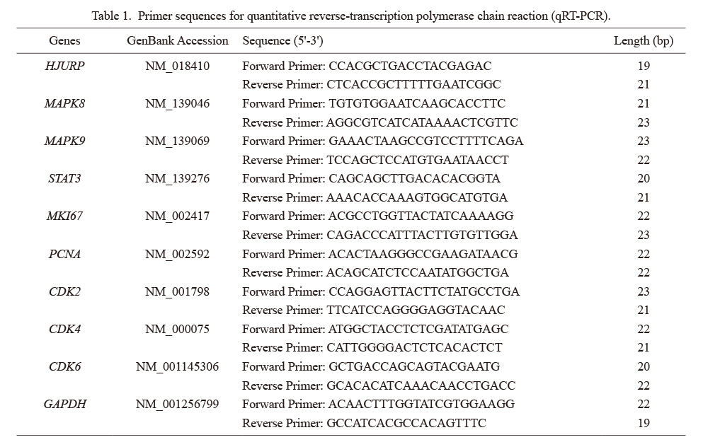
TRIzol reagent (15596026, Thermo Fisher Scientific Co., Ltd.) was used to extract total RNA from cells. A spectrophotometer was used to determine the purity and concentration of RNA. The extracted RNA was reverse transcribed into cDNA using a reverse transcription kit. A Maxima SYBR Green qPCR (K0253, Thermo Fisher Scientific Co., Ltd.) was used to analyze gene expression levels. Glyceraldehyde-3-phosphate dehydrogenase (GAPDH) was used as an internal reference. The 2-ΔΔCt method was used to calculate the relative expression levels of mRNA (Table 1).
Western blotting
Cells were collected for protein extraction with radioimmunoprecipitation assay (RIPA) Lysis Buffer (sc-24948, Santa Cruz Biotechnology, Inc.), and the protein concentration was determined using a BCA Protein Assay Kit (sc-202389, Santa Cruz Biotechnology, Inc.). Sodium dodecyl sulfate-polyacrylamide gel electrophoresis (SDS-PAGE) was used to separate proteins, which were transferred to polyvinylidene fluoride (PVDF) membranes. The PVDF membrane was blocked in Tris-buffered saline (TBS) buffer with 5% defatted milk powder for 1 h. Rabbit monoclonal antibodies were added overnight at 4°C, including an anti-HJURP antibody (ab233541, Abcam) at a 1:1,000 dilution, anti-β-actin antibody (ab8227) at a 1 µg/ml dilution, anti-JNK antibody (ab179461) at a 1:20,000 dilution, anti-JNK (phospho) antibody (ab124956) at a 1:1,000 dilution (purified), anti-STAT3 (phospho) antibody (ab76315) at a 1:20,000 dilution (purified), and anti-STAT3 antibody (ab68153) at a 1:1,000 dilution. After washing, goat anti-rabbit IgG H&L (HRP) (ab97051) at a 1:100,000 dilution was added for incubation at room temperature for 2 h. Enhanced chemiluminescence (ECL) was used for the development and visualization of protein bands. QUANTITY ONE software (Bio-Rad Laboratories, Hercules, CA, USA) was used to analyze the density of protein bands, and the experiment was repeated three times.
MTT (3-[4,5-dimethylthiazol-2-yl]-2,5 diphenyl tetrazolium bromide) assay to detect cell proliferation
T24 and TCCSUP cells were collected at the logarithmic growth phase and inoculated into 96-well plates at a density of 5 × 10⁴ cells/well to culture for 24 h, 36 h, 48 h, 72 h and 96 h. Each well received 20 μL MTT (5 mg/mL) for 4 h of continued culture. The culture medium was discarded, and 150 μL dimethyl sulfoxide (DMSO) was added to each well for full dissolution and 10 min of oscillation prior to the development and measurement of absorbance at a wavelength of 490 nm.
Flow cytometry detection of cell cycle distribution
Cells (5 × 10⁵ cells/well) were seeded in 6-well plates for 48 h of incubation, fixed in 80% ethanol, and centrifuged at 200 × g for 5 min before disposal of the culture medium. The cells were washed twice with 1 ml of phosphate-buffered saline (PBS), centrifuged and suspended in 100 μl of PBS. The cells were incubated in 80% ethanol for 2 h (4°C) and preserved at −20°C. The cells were stained with propidium iodide (PI), centrifuged at 200 × g for 5 min, washed twice with 1 ml PBS, and centrifuged again. The cell precipitate was incubated for 30 min (37°C) in 100 μl PBS with 1% FBS, PI (5 μg) and RNaseA (20 μg). A FACScalibur flow cytometer (Becton Dickinson, CA, USA) was used to detect the fluorescence of PI, and FlowJo software was used to analyze the data.
Annexin V-FITC/PI staining detection of cell apoptosis
After centrifugation, cells were suspended in 200 µL PBS, followed by the addition of annexin V-FITC 10 µL and PI 5 µL for 15 min at room temperature in the dark. Binding buffer (30 µL) was added, and cell apoptosis was detected within 0.5 h. Annexin V+/PI− stained cells were counted as early apoptotic cells, and annexin V+/PI+ stained cells were late apoptotic/necrotic cells (Yu et al. 2019).
Statistical methods
GraphPad Prism 6.0 (GraphPad Software, CA, USA) was used for graphical analyses, and SPS 22.0 was used for statistical analyses. All data are presented as the means ± standard deviation (SD). One-way ANOVA was used to test the significance of mean values, and Tukey’s test was used to compare intergroup differences. P < 0.05 indicated a statistically significant difference.
Results
HJURP was upregulated in BLUC tissues and cell lines
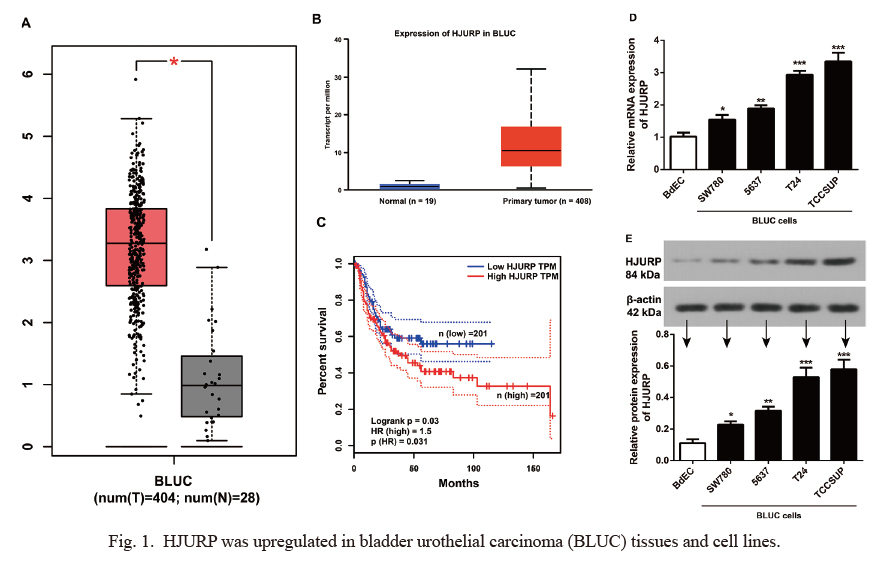
To detect the expression of HJURP in BLUC, we used the UALCAN database and found that HJURP was one of the 25 overexpressed genes in BLUC (Supplementary Fig. S1). HJURP was significantly upregulated in cancerous tissues of BLUC patients (all P < 0.01, Fig. 1A, B), and it was associated with the disease-free survival of BLUC patients (log-rank P = 0.030, HR = 1.5, Fig. 1C). Patients with high HJURP expression (n = 201) had appreciably shorter DFS compared to patients with low HJURP expression (n = 201). HJURP expression in BLUC cell lines (SW780, 5637, T24 and TCCSUP) was apparently higher than BdECs (all P < 0.05, Fig. 1D, E).
HJURP regulated the JNK/STAT3 signaling pathway in BLUC
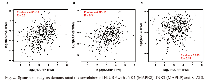
Spearman analysis of the GEPIA database revealed that HJURP expression significantly positively correlated with JNK1 (MAPK8, R = 0.30, P = 4.5E-10, Fig. 2A), JNK2 (MAPK9, R = 0.30, P = 4.9E-10, Fig. 2B) and STAT3 (R = 0.15, P = 0.003, Fig. 2C). QRT-PCR and Western blotting were used to detect the transfection efficiency of T24 and TCCSUP cells transfected with HJURP lentivirus activation particles and HJURP shRNA lentivirus particles. HJURP gene expression was apparently upregulated and downregulated, respectively (all P < 0.05, Fig. 3A). Moreover, the gene expressions of MAPK8 (Fig. 3B), MAPK9 (Fig. 3C) and STAT3 (Fig. 3D) were positively regulated by HJURP lentivirus particles (all P < 0.05). HJURP overexpression activated the JNK/STAT3 signaling pathway with the enhanced expressions of p-JNK/total-JNK and p-STAT3/total-STAT3, and HJURP shRNA exerted the opposite effect (all P < 0.05, Fig. 3E-H). A subsequent experiment found that the JNK inhibitor SP600125 had no obvious effect on HJURP expression in BLUC cells (all P > 0.05, Fig. 3I, J), but HJURP activation particles reversed the inhibitory effect of SP600125 on p-JNK/total-JNK and p-STAT3/total-STAT3 expression in T24 and TCCSUP cells (all P < 0.05, Fig. 3K, L).
Spearman analysis of the GEPIA database revealed a significant positive correlation between HJURP and the proliferation-related genes MKI67 (R = 0.60) and PCNA (R = 0.46) (Fig. 4A). The MTT assay found that HJURP upregulation led to significantly increased proliferation of BLUC cells, and SP600125 apparently inhibited the proliferation of BLUC cells. Compared to the SP600125 group, the HJURP activation + SP600125 group showed increased cell proliferation and a significant upregulation of MKI67 and PCNA expression (all P < 0.05, Fig. 4B, C).
HJURP mediates the JNK/STAT3 signaling pathway to affect BLUC cell cycle distribution
As shown in Fig. 5A, B, SP600125 arrested T24 and TCCSUP cells at the G0/G1 phase and reduced the proportion of S phase cells, which was reversed by HJURP overexpression particles. Analysis of the GEPIA database found that HJURP significantly positively correlated with the G1 phase cycle-related genes CDK2 (R = 0.39, P = 4.4E-16), CDK4 (R = 0.24, P = 1.5E-06) and CDK6 (R = 0.21, P = 1.5E-05) (Fig. 5C). HJURP overexpression promoted the expression of CDK2, CDK4 and CDK6 in T24 and TCCSUP cells (all P < 0.05), and SP600125 reduced the expression of CDK2, CDK4 and CDK6 in BLUC cells (all P < 0.05). Compared to the SP600125 group, T24 and TCCSUP cells in the HJURP activation + SP600125 group showed markedly increased expression of CDK2 and CDK4 (all P < 0.05, Fig. 5D).
HJURP mediates the JNK/STAT3 signaling pathway to inhibit BLUC cell apoptosis
Annexin V-FITC/PI staining was performed to detect cell apoptosis (Fig. 6). Compared to the Mock group, HJURP significantly reduced the apoptosis rate of BLUC cells (T24 and TCCSUP), and SP600125 appreciably increased the cell apoptosis rate (all P < 0.05). HJURP overexpression effectively reversed the proapoptotic effect of SP600125 on BLUC cells, which was demonstrated by the dramatic decrease in the number of apoptotic cells in the HJURP activation + SP600125 group compared to the SP600125 group (all P < 0.05).
Discussion
HJURP was upregulated in BLUC tissues, which was correlated with the poor prognosis of patients and the expression MAPK8, MAPK9 and STAT3 by online database. This was the first study which revealed that decreased p-JNK/JNK and p-STAT3/STAT3 by a JNK inhibitor SP600125 in BLUC cells was reversed after transfection with HJURP activation lentivirus particles. The HJURP-mediated JNK/STAT3 pathway could affect BLUC cell proliferation, cell apoptosis, and cell cycle.
HJURP, a centromeric histone chaperone involved in de novo histone H3 variant CenH3 (CENP-A) recruitment (Niikura et al. 2015), was one of the 25 overexpressed genes in BLUC based on biological information analyses, and HJURP expression was significantly higher in BLUC cell lines (SW780, 5637, T24 and TCCSUP) than BdECs. Similar expression of HJURP was also observed in many other cancers (Dou et al. 2022; Jia et al. 2022; Tsevegjav et al. 2022). Notably, the mRNA and protein levels of HJURP were significantly upregulated in BCa tissues in a transcriptome and in vivo study (Cao et al. 2017), which is consistent with the findings in the current study. The overexpression of HJURP in hepatocellular carcinoma (HCC) tissues primarily resulted from the hypomethylation of the HJURP promoter region (Li et al. 2021). However, whether the upregulation of HJURP in BLUC was also affected by methylation was not known, and further investigations are needed. As a DNA repair protein, HJURP is associated with the proliferative competence, drug resistance and poor prognosis of cancer cells (Sousa et al. 2019). High HJURP expression predicts a higher risk of metastatic relapse and poor outcome in breast cancer patients (Montes de Oca et al. 2015). Zhang et al. (2021) also revealed that the prognostic factor HJURP influenced the prognosis of non-small-cell lung cancer (NSCLC) patients. BLUC patients with high HJURP expression had apparently shorter disease-free survival than patients with low HJURP expression, which suggests that HJURP acts as an oncogene in bladder cancer and affects the prognostic outcome of patients.
Based on the Spearman analysis of the GEPIA database, HJURP significantly positively correlated with JNK1 (MAPK8), JNK2 (MAPK9) and STAT3. A previous study demonstrated that JNK activation was essential for tobacco smoke-induced lung cancer, chemical carcinogen-induced gastric cancer, hepatocellular carcinoma (Liu et al. 2012), and BLUC (Shen et al. 2016) in animal models. STAT3 mediates many cell processes, including the metabolism, survival, host defense, growth and differentiation, and it is unremittingly activated in cancer cells compared to normal cells (Saleem et al. 2020). STAT3 activation upregulates the expression of genes related to angiogenesis, metastasis and proliferation (Cui et al. 2020), and inhibition of STAT3 activation may be a novel approach to inhibit tumor growth (Saleem et al. 2020). The carcinogenic effect of the JNK/STAT3 signaling pathway was found in many cancers, such as epithelial ovarian cancer, lung cancer, and oral squamous cell carcinoma (OSCC) (Gkouveris et al. 2017; Cui et al. 2020; Wang et al. 2020). A previous study reported that ectopic HJURP expression promoted the proliferation of prostate cancer cells in vitro and in vivo via the JNK signaling pathway (Lai et al. 2021). Based on the online analysis, we found that HJURP expression in BLUC tissues was in a positive correlation with the expression of MAPK8, MAPK9 and STAT3. In addition, HJURP knockdown decreased gene expressions of MAPK8, MAPK9 and STAT3 in BLUC cells with decreased ratio of p-JNK/JNK and p-STAT3/STAT3, while the HJURP overexpression caused opposite effect. All mentioned above indicated that HJURP may target the transcription of JNK/STAT3, thus inducing protein degradation. We treated BLUC cells with the JNK pathway inhibitor SP600125 (Bennett et al. 2001; Pereira et al. 2012) and found that SP600125 had no obvious effect on HJURP expression. However, HJURP overexpression counteracted the inhibitory effect of SP600125 on the JNK pathway, which indicates that HJURP may play a regulatory role in BLUC by mediating the JNK signaling pathway.
The histone chaperone HJURP mediates the deposition of CENP-A by colocalizing with CENP-A at the centromere in early G1 phase (Andronov et al. 2019). Online database analyses found a significant positive correlation between HJURP and the G1 phase cycle-related genes CDK2, CDK4 and CDK6. A previous study showed that low HJURP expression arrested cancer cells at the G1 phase (Filipescu et al. 2017), including serous ovarian cancer cells (Dou et al. 2022). U343MG cells with conspicuous senescence were significantly arrested at the G1 phase with a decreased proportion of S phase cells on the fifth day after HJURP suppression (Serafim et al. 2020). SP600125 inhibited cell growth by arresting cells at the G1 or G2/M phase in multiple myeloma (MM) cell lines (Hideshima et al. 2003). The inhibitory effect of SP600125 on human mast cell proliferation was also related to cell cycle arrest at the G1 phase (Wang et al. 2006). SP600125 arrested BLUC cells at the G0/G1 phase and reduced the expression of CDK2, CDK4 and CDK6 in the present study, which was reversed by HJURP overexpression. Inhibition of HJURP expression reduced the proliferative competence of four colorectal cancer (CRC) cell lines in vitro (Kang et al. 2020). HJURP knockdown downregulated clonogenic capacity and severely damaged the survival of five distinct glioblastoma (GBM) cell lines (Serafim et al. 2020). We also found that HJURP overexpression reversed the inhibitory effect of SP600125 on the proliferation of BLUC cells, which was accompanied by increased cell apoptosis and cell cycle alterations. We performed Spearman analysis using the GEPIA database and found a significant positive correlation between HJURP and cell proliferation markers (Helpap et al. 2003), including MKI67 and PCNA. A similar trend was reported in breast cancer, where the HJURP mRNA level significantly correlated with the proliferation index MKI67 (Hu et al. 2010). HJURP overexpression also upregulated the expression of MKI67 and PCNA in BLUC cells in our study, and SP600125 reduced the expression of MKI67 and PCNA. These results suggest that HJURP mediates the JNK/STAT3 signaling pathway to affect the proliferation, apoptosis and cell cycle distribution of BLUC cells.
There are some potential limitations of our study. First, this was an in vitro cell study, therefore results shown here do not necessarily replicate the effects of human tumor microenvironment. Secondly, we did not evaluate the underlying molecular mechanisms of HJURP-mediated JNK/STAT3 signaling pathway in BLUC in vivo. Thirdly we tested only 2 cell lines and it is not known how other BLUC cell lines would respond to SP600125.
In summary, HJURP was upregulated in BLUC tissues and cell lines and acted as an oncogene to promote proliferation, inhibit apoptosis, and regulate the cell cycle distribution of BLUC cells via regulation of the JNK/STAT3 signaling pathway. Findings of our study theorize that HJURP is a novel protein which has an important role in progression of BLUC possibly associated with JNK/STAT3 signaling pathway, and it might be an attractive novel therapeutic target for its early diagnosis and treatment.
Acknowledgments
This work was supported by the Zhejiang University Cooperation Project of Lishui (No. 2018ZDHZ12), Zhejiang Public Welfare Technology Application Research Project (No.LGF20H050003), Zhejiang Medical and Health Science and Technology Plan Project (No. 2020382934), Traditional Chinese medicine of Zhejiang province science and technology plan project (No. 2020ZB309), Lishui Public Welfare Technology Application Research Project (No. 2019SJZC46), and Lishui Public Welfare Technology Application Research Project (No. 2020GYX22).
Conflict of Interest
The authors declare no conflict of interest.
References
- Andronov, L., Ouararhni, K., Stoll, I., Klaholz, B.P. & Hamiche, A. (2019) CENP-A nucleosome clusters form rosette-like structures around HJURP during G1. Nat. Commun., 10, 4436.
- Bennett, B.L., Sasaki, D.T., Murray, B.W., O Leary, E.C., Sakata, S.T., Xu, W., Leisten, J.C., Motiwala, A., Pierce, S., Satoh, Y., Bhagwat, S.S., Manning, A.M. & Anderson, D.W. (2001) SP600125, an anthrapyrazolone inhibitor of Jun N-terminal kinase. Proc. Natl. Acad. Sci. U. S. A., 98, 13681-13686.
- Cao, R., Wang, G., Qian, K., Chen, L., Qian, G., Xie, C., Dan, H.C., Jiang, W., Wu, M., Wu, C.L., Xiao, Y. & Wang, X. (2017) Silencing of HJURP induces dysregulation of cell cycle and ROS metabolism in bladder cancer cells via PPARgamma-SIRT1 feedback loop. J. Cancer, 8, 2282-2295.
- Cui, L., Yang, G., Ye, J., Yao, Y., Lu, G., Chen, J., Fang, L., Lu, S. & Zhou, J. (2020) Dioscin elicits anti-tumour immunity by inhibiting macrophage M2 polarization via JNK and STAT3 pathways in lung cancer. J. Cell. Mol. Med., 24, 9217-9230.
- Dobruch, J. & Oszczudlowski, M. (2021) Bladder cancer: current challenges and future directions. Medicina (Kaunas), 57, 749.
- Dou, Z., Qiu, C., Zhang, X., Yao, S., Zhao, C., Wang, Z., Chu, R., Chen, J., Chen, Z., Li, R., Wang, K., Liu, P., Liu, C., Song, K. & Kong, B. (2022) HJURP promotes malignant progression and mediates sensitivity to cisplatin and WEE1-inhibitor in serous ovarian cancer. Int. J. Biol. Sci., 18, 1188-1210.
- Filipescu, D., Naughtin, M., Podsypanina, K., Lejour, V., Wilson, L., Gurard-Levin, Z.A., Orsi, G.A., Simeonova, I., Toufektchan, E., Attardi, L.D., Toledo, F. & Almouzni, G. (2017) Essential role for centromeric factors following p53 loss and oncogenic transformation. Genes Dev., 31, 463-480.
- Gkouveris, I., Nikitakis, N., Avgoustidis, D., Karanikou, M., Rassidakis, G. & Sklavounou, A. (2017) ERK1/2, JNK and STAT3 activation and correlation with tumor differentiation in oral SCC. Histol. Histopathol., 32, 1065-1076.
- Helpap, B., Schmitz-Drager, B.J., Hamilton, P.W., Muzzonigro, G., Galosi, A.B., Kurth, K.H., Lubaroff, D., Waters, D.J. & Droller, M.J. (2003) Molecular pathology of non-invasive urothelial carcinomas (part I). Virchows Arch., 442, 309-316.
- Hess, P., Pihan, G., Sawyers, C.L., Flavell, R.A. & Davis, R.J. (2002) Survival signaling mediated by c-Jun NH(2)-terminal kinase in transformed B lymphoblasts. Nat. Genet., 32, 201-205.
- Hideshima, T., Hayashi, T., Chauhan, D., Akiyama, M., Richardson, P. & Anderson, K. (2003) Biologic sequelae of c-Jun NH(2)-terminal kinase (JNK) activation in multiple myeloma cell lines. Oncogene, 22, 8797-8801.
- Hu, Z., Huang, G., Sadanandam, A., Gu, S., Lenburg, M.E., Pai, M., Bayani, N., Blakely, E.A., Gray, J.W. & Mao, J.H. (2010) The expression level of HJURP has an independent prognostic impact and predicts the sensitivity to radiotherapy in breast cancer. Breast Cancer Res., 12, R18.
- Hyldgaard, J.M. & Jensen, J.B. (2021) The inequality of females in bladder cancer. APMIS, 129, 694-699.
- Jaeschke, A., Rincon, M., Doran, B., Reilly, J., Neuberg, D., Greiner, D.L., Shultz, L.D., Rossini, A.A., Flavell, R.A. & Davis, R.J. (2005) Disruption of the Jnk2 (Mapk9) gene reduces destructive insulitis and diabetes in a mouse model of type I diabetes. Proc. Natl. Acad. Sci. U. S. A., 102, 6931-6935.
- Jia, Y., Zhou, J., Tan, T.K., Chung, T.H., Chen, Y., Chooi, J.Y., Sanda, T., Fullwood, M.J., Xiong, S., Toh, S.H.M., Balan, K., Wong, R.W.J., Lim, J.S.L., Zhang, E., Cai, Z., et al. (2022) Super enhancer-mediated upregulation of HJURP promotes growth and survival of t(4;14)-positive multiple myeloma. Cancer Res., 82, 406-418.
- Kang, D.H., Woo, J., Kim, H., Kim, S.Y., Ji, S., Jaygal, G., Ahn, T.S., Kim, H.J., Kwak, H.J., Kim, C.J., Baek, M.J. & Jeong, D. (2020) Prognostic relevance of HJURP expression in patients with surgically resected colorectal cancer. Int. J. Mol. Sci., 21, 7928.
- Lai, W., Zhu, W., Xiao, C., Li, X., Wang, Y., Han, Y., Zheng, J., Li, Y., Li, M. & Wen, X. (2021) HJURP promotes proliferation in prostate cancer cells through increasing CDKN1A degradation via the GSK3beta/JNK signaling pathway. Cell Death Dis., 12, 583.
- Lee, E.H., Chun, S.Y., Kim, B., Yoon, B.H., Lee, J.N., Kim, B.S., Yoo, E.S., Lee, S., Song, P.H., Kwon, T.G. & Ha, Y.S. (2020) Knockdown of TRPM7 prevents tumor growth, migration, and invasion through the Src, Akt, and JNK pathway in bladder cancer. BMC Urol., 20, 145.
- Lehmann, B.D., Bauer, J.A., Chen, X., Sanders, M.E., Chakravarthy, A.B., Shyr, Y. & Pietenpol, J.A. (2011) Identification of human triple-negative breast cancer subtypes and preclinical models for selection of targeted therapies. J. Clin. Invest., 121, 2750-2767.
- Li, Y., Yi, Q., Liao, X., Han, C., Zheng, L., Li, H., Yu, Q., Yan, X., Chen, X., Zhu, H., Zhao, B., Lin, Q., Liang, L., Wang, L., Qin, F., et al. (2021) Hypomethylation-driven overexpression of HJURP promotes progression of hepatocellular carcinoma and is associated with poor prognosis. Biochem. Biophys. Res. Commun., 566, 67-74.
- Liu, J., Chen, B., Lu, Y., Guan, Y. & Chen, F. (2012) JNK-dependent Stat3 phosphorylation contributes to Akt activation in response to arsenic exposure. Toxicol. Sci., 129, 363-371.
- Martinez-Rojo, E., Berumen, L.C., Garcia-Alcocer, G. & Escobar-Cabrera, J. (2021) The role of androgens and androgen receptor in human bladder cancer. Biomolecules, 11, 594.
- Milioli, H.H., Tishchenko, I., Riveros, C., Berretta, R. & Moscato, P. (2017) Basal-like breast cancer: molecular profiles, clinical features and survival outcomes. BMC Med. Genomics, 10, 19.
- Mirzaei, S., Gholami, M.H., Mahabady, M.K., Nabavi, N., Zabolian, A., Banihashemi, S.M., Haddadi, A., Entezari, M., Hushmandi, K., Makvandi, P., Samarghandian, S., Zarrabi, A., Ashrafizadeh, M. & Khan, H. (2021) Pre-clinical investigation of STAT3 pathway in bladder cancer: paving the way for clinical translation. Biomed. Pharmacother., 133, 111077.
- Montes de Oca, R., Gurard-Levin, Z.A., Berger, F., Rehman, H., Martel, E., Corpet, A., de Koning, L., Vassias, I., Wilson, L.O., Meseure, D., Reyal, F., Savignoni, A., Asselain, B., Sastre-Garau, X. & Almouzni, G. (2015) The histone chaperone HJURP is a new independent prognostic marker for luminal A breast carcinoma. Mol. Oncol., 9, 657-674.
- Niikura, Y., Kitagawa, R., Ogi, H., Abdulle, R., Pagala, V. & Kitagawa, K. (2015) CENP-A K124 ubiquitylation is required for CENP-A deposition at the centromere. Dev. Cell, 32, 589-603.
- Pereira, A.C., Soares-Martins, J.A., Leite, F.G., Da Cruz, A.F., Torres, A.A., Souto-Padron, T., Kroon, E.G., Ferreira, P.C. & Bonjardim, C.A. (2012) SP600125 inhibits Orthopoxviruses replication in a JNK1/2 -independent manner: implication as a potential antipoxviral. Antiviral Res., 93, 69-77.
- Rozanec, J.J. & Secin, F.P. (2020) Epidemiology, etiology and prevention of bladder cancer. Arch. Esp. Urol., 73, 872-878.
- Saleem, M.Z., Alshwmi, M., Zhang, H., Din, S.R.U., Nisar, M.A., Khan, M., Alam, S., Alam, G., Jin, L. & Ma, T. (2020) Inhibition of JNK-mediated autophagy promotes proscillaridin A- induced apoptosis via ROS generation, intracellular Ca(+2) oscillation and inhibiting STAT3 signaling in breast cancer cells. Front. Pharmacol., 11, 01055.
- Serafim, R.B., Cardoso, C., Di Cristofaro, L.F.M., Pienna Soares, C., Araujo Silva, W. Jr., Espreafico, E.M., Paco-Larson, M.L., Price, B.D. & Valente, V. (2020) HJURP knockdown disrupts clonogenic capacity and increases radiation-induced cell death of glioblastoma cells. Cancer Gene Ther., 27, 319-329.
- Shen, K.H., Li, C.F., Chien, L.H., Huang, C.H., Su, C.C., Liao, A.C. & Wu, T.F. (2016) Role of galectin-1 in urinary bladder urothelial carcinoma cell invasion through the JNK pathway. Cancer Sci., 107, 1390-1398.
- Siracusano, S., Rizzetto, R. & Porcaro, A.B. (2020) Bladder cancer genomics. Urologia, 87, 49-56.
- Sousa, J.F., Serafim, R.B., Freitas, L.M., Fontana, C.R. & Valente, V. (2019) DNA repair genes in astrocytoma tumorigenesis, progression and therapy resistance. Genet. Mol. Biol., 43, e20190066.
- Tsevegjav, B., Takano, A., Zhu, M., Yoshitake, Y., Shinohara, M. & Daigo, Y. (2022) Holliday junction recognition protein as a prognostic biomarker and therapeutic target for oral cancer. Int. J. Oncol., 60, 26.
- Valente, V., Serafim, R.B., de Oliveira, L.C., Adorni, F.S., Torrieri, R., Tirapelli, D.P., Espreafico, E.M., Oba-Shinjo, S.M., Marie, S.K., Paco-Larson, M.L. & Carlotti, C.G. Jr. (2013) Modulation of HJURP (Holliday Junction-Recognizing Protein) levels is correlated with glioblastoma cells survival. PLoS One, 8, e62200.
- Wang, B., Tsukada, J., Higashi, T., Mizobe, T., Matsuura, A., Mouri, F., Sawamukai, N., Ra, C. & Tanaka, Y. (2006) Growth suppression of human mast cells expressing constitutively active c-kit receptors by JNK inhibitor SP600125. Genes Cells, 11, 983-992.
- Wang, C.X., Xiong, H.F., Wang, S., Wang, J., Nie, X., Guo, Q., Li, X., Qi, Y., Liu, J.J. & Lin, B. (2020) Overexpression of TEM8 promotes ovarian cancer progression via Rac1/Cdc42/JNK and MEK/ERK/STAT3 signaling pathways. Am. J. Transl. Res., 12, 3557-3576.
- Yu, H., Wu, C.L., Wang, X., Ban, Q., Quan, C., Liu, M., Dong, H., Li, J., Kim, G.Y., Choi, Y.H., Wang, Z. & Jin, C.Y. (2019) SP600125 enhances C-2-induced cell death by the switch from autophagy to apoptosis in bladder cancer cells. J. Exp. Clin. Cancer Res., 38, 448.
- Zhang, Y.Q., Yuan, Y., Zhang, J., Lin, C.Y., Guo, J.L., Liu, H.S. & Guo, Q. (2021) Evaluation of the roles and regulatory mechanisms of PD-1 target molecules in NSCLC progression. Ann. Transl. Med., 9, 1168.