International Journal of Surgical Wound Care
Online ISSN : 2435-2128
4 巻, 3 号
選択された号の論文の7件中1~7を表示しています
Original Article
  • Kosuke Morioka, Ami Higashiyama, Airi Tazaki, Sawa Ikarimoto, Takashi ...
    2023 年 4 巻 3 号 p. 81-91
    発行日: 2023年
    公開日: 2023/09/01
    ジャーナル フリー
    Background: Osteoradionecrosis significantly reduces quality of life. In this study, we reviewed our experience of surgically managing complicated cases of osteonecrosis and discussed the perioperative problems encountered.
    Methods: We retrospectively evaluated 15 patients with osteoradionecrosis of the craniofacial or trunk skeleton who underwent flap reconstruction surgery at our institution between January 2002 and December 2021. In all cases, the irradiated tissues were excised, followed by coverage with well-vascularized tissue. Surgical methods and postoperative complications were evaluated as outcomes.
    Results: The 15 patients included six men and nine women, with a mean age of 67.8 ± 17.0 years (range 20–83 years). The affected bone was the skull in one case, mandible in five cases, clavicle in one case, ribs in five cases, sacrum in two cases, and pubic bone in one case. In seven cases, a postoperative fistula formed in the craniofacial or pelvic region. The fistulas in the craniofacial region were closed by the removal of the infected plate or excision of the sequestrum.
    Conclusions: Surgical treatment of osteoradionecrosis should include excision of the irradiated tissues followed by coverage with well-vascularized tissue. Well-vascularized transplanted tissues can be effective for wound healing in complicated cases.
    A 75-year-old man with osteoradionecrosis of the skull (case 1). Fullsize Image
    (a) Preoperative photograph showing necrotic parietal bone. (b) Sagittal preoperative computed tomography (CT) showing epidural abscess and brain edema. (c) Sagittal T1-weighted image showing an area of low intensity in the parietal bone. (d) Fresh bleeding after wide excision of the devitalized tissue. (e) The latissimus dorsi musculocutaneous flap and serratus anterior muscle with the eighth and ninth ribs designed on the right chest wall. (f) Ribs fixed with absorbable stitches across the parietal bone defect and the serratus anterior muscle covering the central portion of the defect. (g) CT image taken 14 months after surgery with a red arrow denoting the progression of necrosis of the marginal bone. (h) Photograph taken 16 months after surgery showing closure of the fistula.
Case Reports
  • Takuya Seike, Kensuke Sasaki
    2023 年 4 巻 3 号 p. 92-98
    発行日: 2023年
    公開日: 2023/09/01
    ジャーナル フリー
    電子付録
    Lymphorrhea occasionally occurs as a complication of surgical manipulation and is often difficult to treat. This report describes two cases of inguinal lymphorrhea diagnosed after lymph node biopsy and arterial puncture and treated with lymph embolization and transcutaneous sclerotherapy using OK-432.
    In both cases, intranodal lymphatic embolization was performed with n-butyl-2-cyanoacrylate after lymphangiography of the lymph nodes around the wound, followed by debridement, sclerotherapy with OK-432, and wound closure.
    No recurrence of lymphorrhea was observed.
    Conservative treatment for lymphorrhea comprises compression, fat restriction, and negative-pressure wound therapy, including identification and ligation of the lymphatic vessels, closure with a musculocutaneous flap, lymphaticovenular and lymphaticolymphatic anastomosis. The combination of intranodal lymphatic embolization, sclerotherapy with OK-432, and wound closure performed in this study is a minimally invasive method that can achieve wound closure in a short period.
    Sclerotherapy with OK-432 and closed wound. Fullsize Image
    (a) Intraoperative design. (b) After debridement. (c) OK-432 injection in progress. (d) Immediately after the operation. The release wound was debrided under local anesthesia, and the wound OK-432:5KE was injected into the wound. A continuous suction drain was placed, and the wound was sutured closed.
  • Eri Ichijo, Masahiro Kuwabara, Saori Kawakami, Alan Kawarai Lefor, Nao ...
    2023 年 4 巻 3 号 p. 99-103
    発行日: 2023年
    公開日: 2023/09/01
    ジャーナル フリー
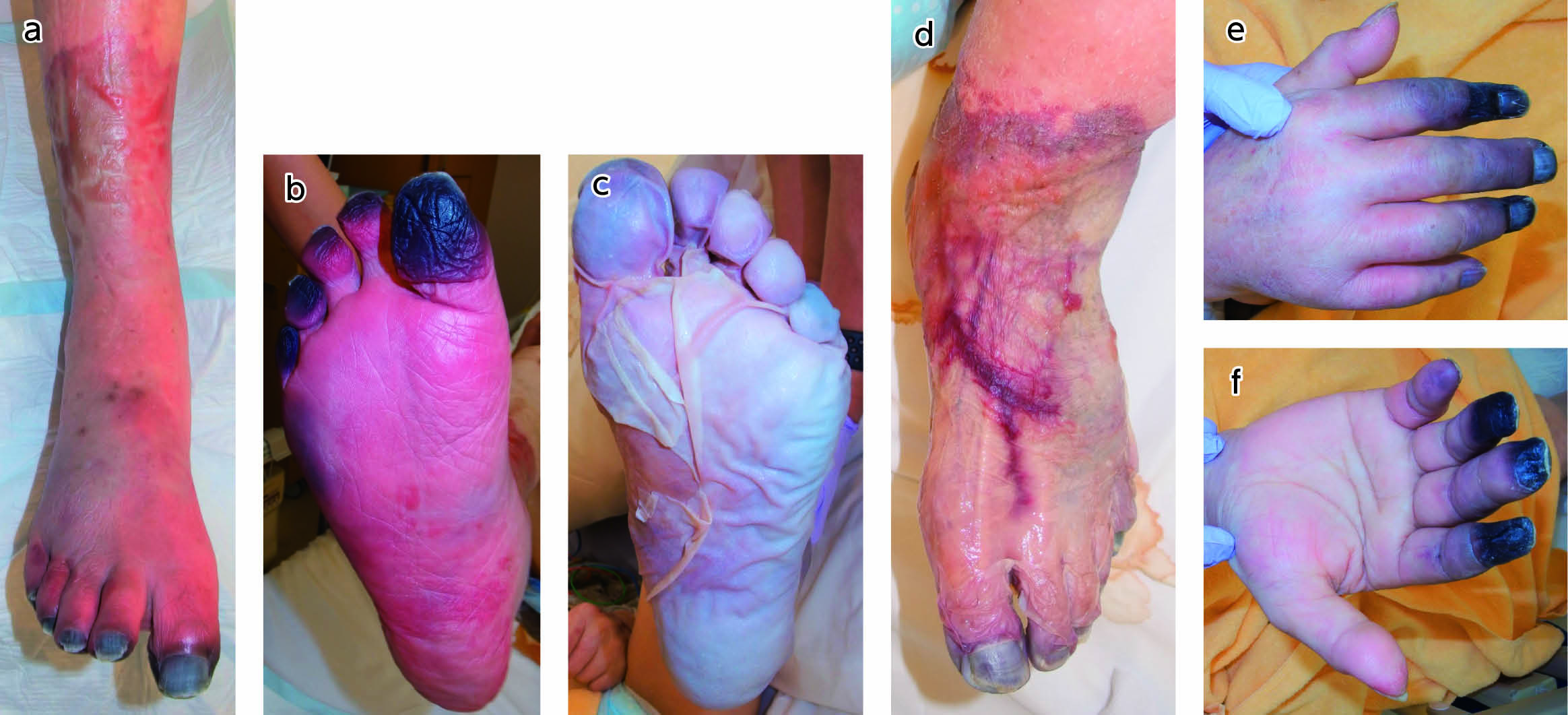
    Severe acute respiratory syndrome coronavirus 2, the causative virus of coronavirus disease 2019, causes thrombosis via direct effects on vascular endothelial cells. Necrosis of the extremities due to arterial occlusion has been reported in patients with coronavirus disease 2019. Here, we report a case of multiple arterial occlusions associated with coronavirus disease 2019. Although the extent to which severe acute respiratory syndrome coronavirus 2 infection is responsible for the development of necrosis of the extremities in this patient is uncertain, surgeons who routinely treat patients with necrosis of the extremities should consider severe acute respiratory syndrome coronavirus 2 infection as a possible cause of necrosis of the extremities, which can present with rapid progression in multiple extremities.
    Day 26 after being tested positive for coronavirus disease 2019. Fullsize Image
    (a), (b) Necrosis of the right first, second, third, fourth, and fifth toes. (c), (d) Severe cyanosis of the left foot from the ankle to toes. (e), (f) Necrosis of the right second, third, fourth, and fifth fingers.
  • A Case Report
    Hirohiko Aosaki, Shoichi Ishikawa, Takeshi Kurihara, Yohei Ishikawa, S ...
    2023 年 4 巻 3 号 p. 104-108
    発行日: 2023年
    公開日: 2023/09/01
    ジャーナル フリー
    This case report details our experience with lower-extremity ischemia due to extracorporeal membrane oxygenation. The patient presented with massive myonecrosis caused by ischemia of the thighs and lower legs. Biphasic wound closure was performed using ultrasound debridement and negative-pressure wound therapy to prepare the wound bed after knee disarticulation and above-knee amputation.
    Necrosis mainly observed on the posterior aspect of the right lower leg. Fullsize Image
  • A Case Report
    Ryosuke Oseki, Noboru Shiroshita, Hirokazu Sukegawa, Takayuki Sugimoto ...
    2023 年 4 巻 3 号 p. 109-113
    発行日: 2023年
    公開日: 2023/09/01
    ジャーナル フリー
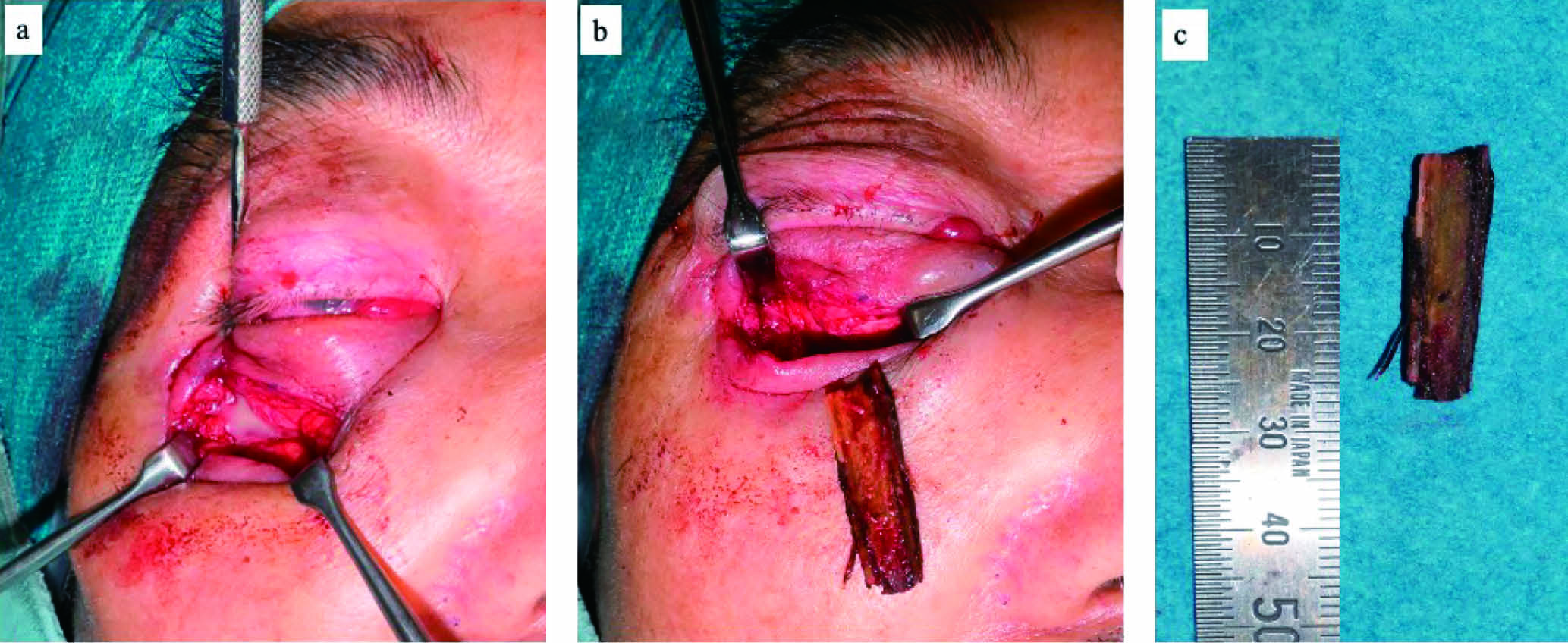
    Intra-orbital wooden foreign bodies pose a significant challenge for clinicians as they are uncommon and difficult to diagnose due to their unique characteristics. Surgery is usually recommended as the presence of a foreign body can potentially induce severe complications, such as diplopia, proptosis, and loss of vision. However, the specific symptoms associated with these foreign bodies are not yet well understood and may, in some cases, take several years to manifest. Moreover, imaging studies have a high likelihood of failing to detect the presence of a foreign body. In this case, a 74-year-old man presented with worsening swelling and redness of the right lower eyelid. He had fell in his garden 6 months prior and underwent computed tomography, which did not reveal any evidence of foreign bodies in the orbit. Subsequently, he was treated for an optic canal fracture by an ophthalmologist. Based on the patient’s symptoms and a secondary computed tomography scan, we performed surgery addressing the posttraumatic granuloma, abscess, and foreign body. During the procedure, a piece of wood was successfully extracted from his orbit. In conclusion, when patients experience orbital trauma and present with chronic symptoms, the possibility of a foreign body retained in the orbit should be considered.
    Intraoperative findings of the orbit. Fullsize Image
    (a) Abscesses are drained from the subperiosteal space. (b) A piece of the wooden chip is extracted from the orbital apex. (c) The size of the wooden chip is 25 mm.
  • Ryo Kitai, Koji Kanayama, Masamitsu Suhara, Yasuaki Mochizuki, Shuji Y ...
    2023 年 4 巻 3 号 p. 114-120
    発行日: 2023年
    公開日: 2023/09/01
    ジャーナル フリー
    Buerger disease or thromboangiitis obliterans is a non-atherosclerotic, segmental, inflammatory, peripheral arterial occlusive disease in young male smokers. Revascularization via endovascular treatment and/or bypass surgery is less common and is technically challenging in patients with Buerger disease. Herein, we report the case of a 30-year-old male patient who underwent endovascular treatment followed by bypass surgery, which successfully salvaged his critically ischemic limbs. This case demonstrates that a revascularization strategy for critical limb ischemia due to Buerger disease could be beneficial for limb salvage. To achieve effective limb salvage, a collaborative approach among endovascular specialists, vascular surgeons, and reconstructive surgeons is recommended.
    Postoperative findings. Fullsize Image
    After the bypass surgery, the ischemic necrosis site gradually became clear (A), and patchy skin necrosis was found over a wide area of the left planta (B). (C) The skin color of the foot improved daily, and most of the necrotic tissue at the proximal level of the metatarsophalangeal joint was peeled off.
  • Masashi Hayakawa, Chizuki Kondo, Tsuneo Yasumura, Toyonori Tsuzuki, Hi ...
    2023 年 4 巻 3 号 p. 121-127
    発行日: 2023年
    公開日: 2023/09/01
    ジャーナル フリー
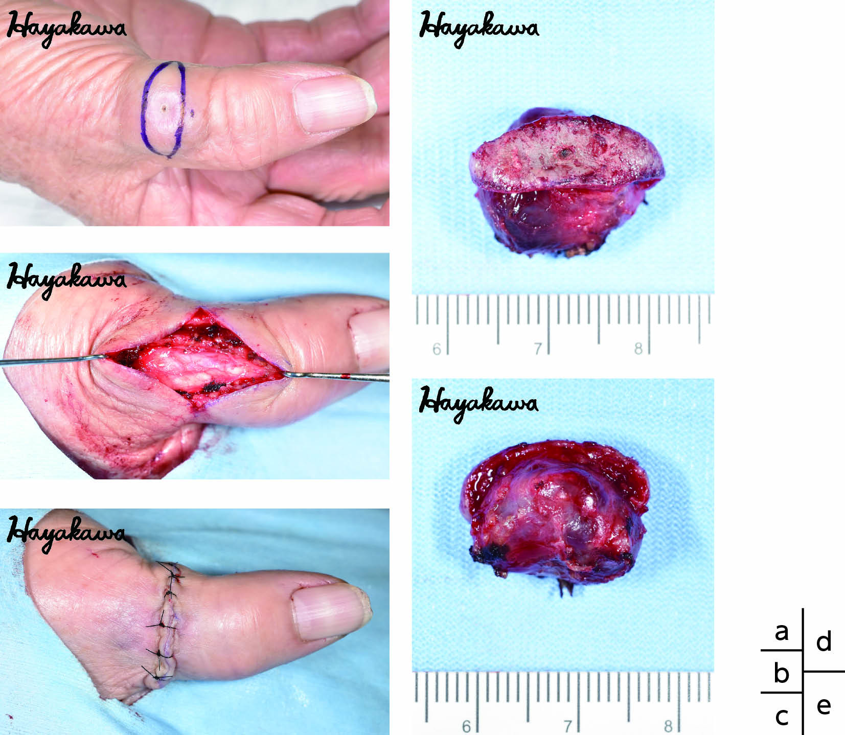
    To the best of our knowledge, only four patients with chronic expanding hematoma (CEH) of the digits have been reported. Herein, along with other literature reviews, we describe a case with CEH of the thumb. An 81-year-old woman presented to our hospital with an enlarged subcutaneous mass in the thumb and underwent en bloc resection. Based on the clinical and histopathological findings, the patient was diagnosed with CEH. She had no remarkable history of trauma, but was taking antiplatelet drugs. These drugs may contribute to the pathogenesis of CEH of the thumb. We examined the clinical characteristics of this condition by reviewing previously reported cases and found no clear bias, including age of onset, male-to-female ratio, and left–right differences. The treatment strategy and recurrence rates of this disease remain controversial; however, in this case, the patient’s condition resolved without any recurrence after the mass was surgically resected en bloc.
    Intraoperative findings and resected specimen. Fullsize Image
    (a) To excise skin that is expanded and includes a punch biopsy scar, a spindle-shaped incision line is designed on the skin. (b) Macroscopic features of the resected specimen. (c) Macroscopic features of the resected specimen immediately after surgery: the wound is temporally sutured and closed. (d) Superficial layer of the skin. (e) Margin.
feedback
Top