Journal of Plastic and Reconstructive Surgery
Online ISSN : 2436-259X
Volume 2, Issue 1
Displaying 1-7 of 7 articles from this issue
Original Research
  • Yuichi Ichikawa, Miho Tobita, Rina Takahashi, Tomoyuki Ito, Daiki Send ...
    2023 Volume 2 Issue 1 Pages 1-8
    Published: January 27, 2023
    Released on J-STAGE: January 27, 2023
    Advance online publication: June 17, 2022
    JOURNAL OPEN ACCESS

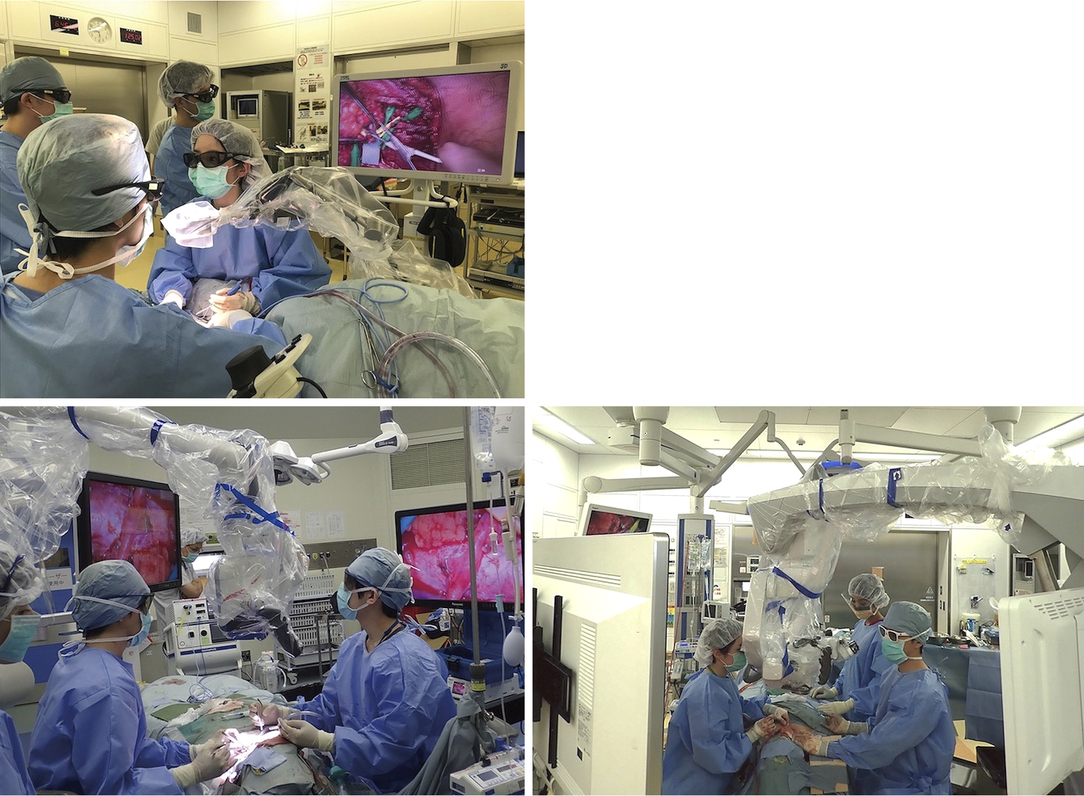
    Objectives: The use of three-dimensional monitors and digital microscopes for microsurgery is becoming prevalent and has great expectations of providing ergonomic advantages for surgeons. However, it remains unclear if this approach does provide ergonomic benefits, and whether transitional difficulties will be encountered when using it in place of a conventional optical microscope. Therefore, the purpose of this study was to clarify both the subjective and objective ergonomic advantages and the learning curve associated with the use of a digital microscope.

    Methods: Seventeen cases of head and neck reconstruction using a digital microscope were compared with those using a conventional optical microscope with respect to the time required for vascular anastomoses, microvascular complications, and ergonomics of the surgeon. The surgeons' learning curve was evaluated by comparing the time required for the transitions during the vascular anastomoses in each series. An objective ergonomics study was conducted by evaluating muscle fatigue using electromyography (EMG) during simulated vascular anastomosis.

    Results: The time required for vascular anastomosis transitions using a digital microscope gradually decreased in a linear fashion. In the objective study to check muscle fatigue by using EMG, a digital microscope was found to provide an ergonomic advantage for surgeons in the lower trapezius muscle part as compared to a conventional optical microscope.

    Conclusions: At present, a digital microscope provides modest ergonomic benefits to surgeons and requires a certain amount of time to learn.

    Download PDF (861K)
  • Munetomo Nagao, Takehiko Ohura, Makoto Takahashi, Masayuki Osawa
    2023 Volume 2 Issue 1 Pages 9-16
    Published: January 27, 2023
    Released on J-STAGE: January 27, 2023
    Advance online publication: June 17, 2022
    JOURNAL OPEN ACCESS

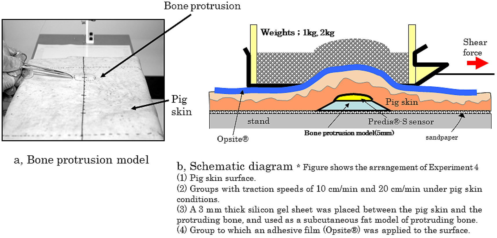
    Objectives: Subcutaneous pressure and subcutaneous shear force were investigated using a bone protrusion model to investigate a polyurethane film dressing (hereinafter referred to as adhesive film) for preventing shear force. Our hypothesis was that when pressure or shear force was applied to the body surface, the adhesive film would 1) decrease the shear force on the skin surface and 2) decrease the effect of the external force on the subcutaneous layer.

    Methods: We built a bone protrusion model using pig skin equipped with a plastic stand and sensors that can simultaneously measure subcutaneous pressure and shear force. In this experimental model, changes in shear force and pressure were measured using a surface friction-measuring device and a subcutaneous sensor.

    Results: In the bone protrusion model, the effect of body weight on the surface shear force was significant. Contrarily, in the subcutaneous fat model, the effect of the body weight on the subcutaneous shear force was particularly small. When the adhesive film was applied, the surface friction, subcutaneous pressure, and subcutaneous shear force all significantly decreased.

    Conclusions: A bone protrusion model was created, and the surface friction, subcutaneous pressure, and subcutaneous shear force were continuously measured and found to decrease. The pressure and shear force were reduced when the adhesive film was used. These results will be of great help in the treatment of bedsores.

    Download PDF (694K)
Case Report
  • Yasushi Mochizuki, Akihiko Oyama, Akiyoshi Kajikawa
    2023 Volume 2 Issue 1 Pages 17-19
    Published: January 27, 2023
    Released on J-STAGE: January 27, 2023
    Advance online publication: June 17, 2022
    JOURNAL OPEN ACCESS

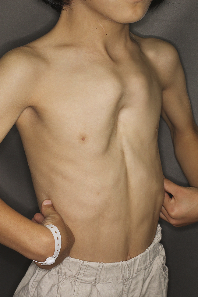
    Surgical correction of pouter pigeon chest deformity traditionally requires invasive procedures including total removal of the affected costal cartilages and major dissection of the sternum. We present a novel minimally invasive two-stage technique that was applied to an 8-year-old girl. First, the depressed lower chest was elevated using a plate with the Nuss procedure. Then, after 30 months, simultaneously with plate removal, correction of the remaining projection of the upper chest was also performed. This involved excision of only two pairs of deformed costal cartilage and a local osteotomy at the manubriosternal joint. Intraoperative blood loss was 20 mL, and the patient was discharged 7 days postoperatively. Because both the primary and secondary stages involve manual bending of the costal cartilages, our procedure may be suitably indicated only for patients in their early teenage years.

    Download PDF (528K)
  • Ken Yamashita, Takatoshi Yotsuyanagi, Yusuke Hamamoto, Ayako Gonda, Ar ...
    2023 Volume 2 Issue 1 Pages 20-24
    Published: January 27, 2023
    Released on J-STAGE: January 27, 2023
    Advance online publication: June 17, 2022
    JOURNAL OPEN ACCESS

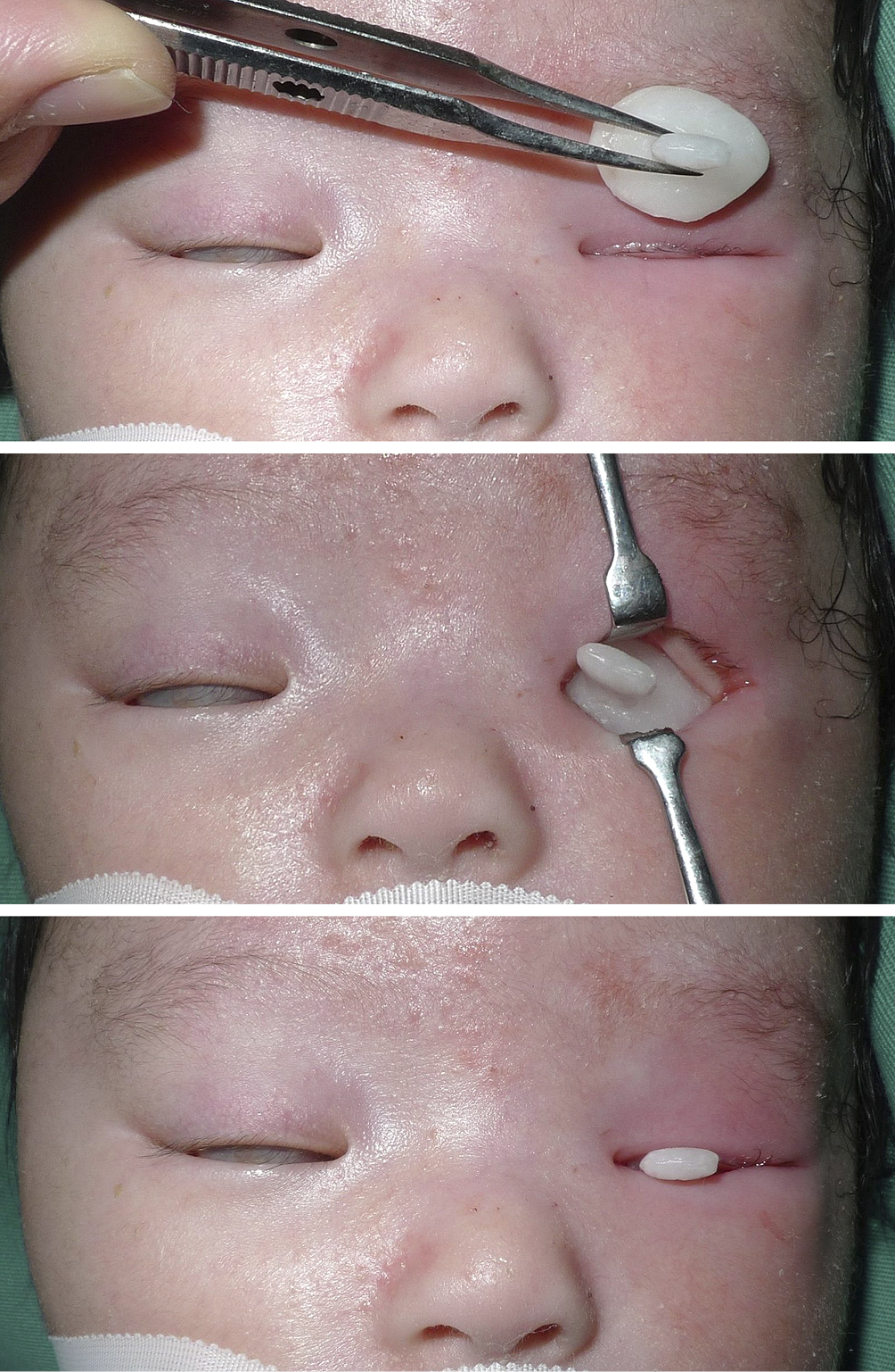
    Clinical congenital anophthalmia causes facial asymmetry and difficulty in wearing ocular prostheses due to underdevelopment of the orbits. Orbital expansion must be started early after birth for treatment, but consensus regarding the treatment for clinical congenital anophthalmia has not been established. We report a successful orbital expansion using a thermoplastic splint. A one-month-old boy had left clinical congenital anophthalmia (ophthalmatrophia), right congenital glaucoma, and right anterior segment ocular dysgenesis. Two months after birth, left orbital expansion was initiated using a custom-made thermoplastic splint. The splint was replaced with larger ones in steps as the infant grew, and the orbit was successfully expanded. There is no appreciable size difference between the left and right orbit after 5 years of follow up. Treatment using a splint for the conjunctival sac is a simple and effective procedure that can be performed on an outpatient basis. Safer and simpler material should be widely used for the treatment.

    Download PDF (1009K)
  • Nadia Hui Shan Sim, Yang Chong Chua, Wei Hao Liang, Agasthian Thirugna ...
    2023 Volume 2 Issue 1 Pages 25-28
    Published: January 27, 2023
    Released on J-STAGE: January 27, 2023
    Advance online publication: June 17, 2022
    JOURNAL OPEN ACCESS

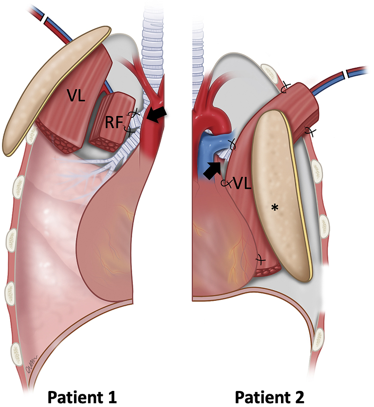
    A bronchial stump blow-out is a serious complication following pneumonectomy/lobectomy. A blown-out stump requires well-vascularized tissue for healing and successful closure. We illustrate the use of the free anterolateral thigh flap in two patients. For patient 1, a chimeric anterolateral thigh flap was used to seal the bronchial stump following an upper lobectomy for tuberculous bronchiectasis. For patient 2, an anterolateral thigh flap was used to seal the main bronchial stump that had blown-out following pneumonectomy for actinomyces lung abscess. The latissimus dorsi muscle was unavailable in both cases due to a previous thoracotomy. Patient 1 had a persistent bronchial leak post-operatively, which resolved after anti-tuberculous treatment. Patient 2 recovered uneventfully and was discharged 14-days post-operatively. Their follow-ups were 13 years and 4 months respectively. In conclusion, the free anterolateral thigh flap is an alternative option to seal a blown-out bronchial stump in the absence of the latissimus dorsi muscle flap.

    Download PDF (726K)
  • Yoshinori Suzuki, Kenjiro Hasegawa, Shogo Ebisudani
    2023 Volume 2 Issue 1 Pages 29-33
    Published: January 27, 2023
    Released on J-STAGE: January 27, 2023
    Advance online publication: September 05, 2022
    JOURNAL OPEN ACCESS

    We present a rare case of a forearm radiation ulcer. Recently, technological advances in intravascular treatments involving X-ray radiography have enabled the application of this treatment in more patients. This has caused more frequent problems due to radiation ulcers. Here we report the case of a 54-year-old man who developed an ulcer on his left forearm six months after placing a stent graft to treat an abdominal aortic aneurysm. A radiation ulcer was diagnosed because of the distance between the position of the ulcer and pigment deposition in the lateral abdominal region and because of the following pathological findings: coagulative necrosis and fibrinoid degeneration and narrowing of the lumen in the small blood vessels. The ulcer was treated by debridement (skin defect size, 22 × 10 cm), followed by reconstruction with an anterolateral thigh flap (23 × 12 cm). Cosmetic and functional improvements were observed, with the latter indicated by an increased range of motion.

    Levels of evidence: V

    Download PDF (1222K)
Technical Note
feedback
Top