Journal of Plastic and Reconstructive Surgery
Online ISSN : 2436-259X
Volume 3, Issue 4
Displaying 1-7 of 7 articles from this issue
Original Research
  • Kazunobu Hashikawa, Toshihiko Satake, Yuko Asano, Tsutomu Kashimura, S ...
    2024 Volume 3 Issue 4 Pages 138-141
    Published: October 27, 2024
    Released on J-STAGE: October 27, 2024
    Advance online publication: April 06, 2024
    JOURNAL OPEN ACCESS

    Objectives: No detailed country-specific cross-sectional studies have been reported on endoscopic plastic surgery. For the first time, the Japan Society of Plastic and Reconstructive Surgery conducted a nationwide survey to ascertain the status of endoscopic procedures in plastic surgery in Japan. This article discusses the current situation based on this survey findings.

    Methods: A questionnaire survey was conducted among 566 Japan Society of Plastic and Reconstructive Surgery-certified training facilities regarding the status of endoscopic plastic surgery procedures completed between January 2020 and December 2021.

    Results: Of the 143 institutions, 33 conducted endoscopic plastic surgery operations during the period, for a total of 753 procedures. Two hundred sixty-two procedures were conducted in the craniomaxillofacial region at 19 institutions and 491 in the extremities and trunk region at 19 institutions. Five intraoperative accidents were recorded, but no cases were moved to traditional surgery based on intraoperative choices. Sixteen postoperative problems were reported, but there were no reports of fatalities.

    Conclusions: The number of endoscopic plastic surgery cases is anticipated to rise, and this nationwide survey will contribute to this by offering useful data and insights.

    Download PDF (312K)
  • Isabella L. Lipkin, Renxi Li, Bharat G. Ranganath
    2024 Volume 3 Issue 4 Pages 142-150
    Published: October 27, 2024
    Released on J-STAGE: October 27, 2024
    Advance online publication: May 10, 2024
    JOURNAL OPEN ACCESS

    Objectives: Comorbidities that impair wound healing, increase infection risk, and compromise tissue viability influence rates of hospital readmission after autologous reconstruction and implant-based reconstruction. This study aimed to evaluate patient factors that increase risk for 30-day hospital readmission after autologous reconstruction and implant-based reconstruction and identify differences in the comorbidities that affect readmission risk after each method.

    Methods: Patients from 2005 to 2021 were selected by autologous reconstruction and implant-based reconstruction current procedural terminology codes from the American College of Surgeons National Surgical Quality Improvement Program database. A multivariable regression model identified the significant predictors of unplanned readmission.

    Results: Comorbidities that increase risk for readmission after autologous reconstruction but not implant-based reconstruction include dialysis (OR 3.87, p = 0.042) and malnutrition (OR 3.20, p = 0.003). Risk factors for readmission after implant-based reconstruction but not autologous reconstruction include bleeding disorder (OR 2.62, p < 0.0001), previous infection (OR 1.49, p = 0.045), recent sepsis (OR 2.16 p = 0.0003), anemia (OR 1.13, p = 0.0018), and hypoalbuminemia (OR 1.35, p = 0.0213). Predictors of unplanned readmission after both methods include chronic obstructive pulmonary disorder, obesity, inpatient status prior to procedure, Black or White race, chronic steroid use, smoking, diabetes, and hypertension.

    Conclusions: These findings may be used to individualize preoperative discussions and help guide optimization of risk factors. In addition, while autologous reconstruction and implant-based reconstruction are often combined into one category for discussion of factors that increase complication risk, our study suggests that the types of reconstruction differ with regard to the comorbidities that increase risk for hospital readmission.

    Download PDF (693K)
  • Zakarya Alami Hassani, Nawfal Fejjal
    2024 Volume 3 Issue 4 Pages 151-156
    Published: October 27, 2024
    Released on J-STAGE: October 27, 2024
    Advance online publication: August 23, 2024
    JOURNAL OPEN ACCESS

    Objectives: This study aimed to discuss the risks and complications associated with skin expansion in the pediatric population, a common plastic surgery technique.

    Methods: Fifty-one patients who underwent skin expansion between 2013 and 2022 at the Plastic Surgery and Burns Unit of the Children's Hospital in Rabat were included in this retrospective study. This study aimed to report all results, analyze them, and compare them with existing literature to determine predictive elements of complications in the pediatric population.

    Results: The study used 96 expanders of three different brands in 70 procedures. Only one expander was often used, but up to seven expanders were used in some cases. Expansion sessions began after an average of 15 days of expander placement, with an average total expansion rate of 105.5%.

    Complications were encountered in 16 of 96 expanders (16.6%), with infection being the most serious complication, found in four cases. Other complications included skin complications, expander malfunctions, skin necrosis, loose sutures, exposure of the valve, and hair loss with stretch marks. The use of multiple expanders was the only statistically relevant factor toward complications.

    Conclusions: The study concluded that skin expansion is not without risks and reported complication rate range from 9.6% to 30% in the pediatric population. Multicenter studies specific to each variable will allow better identification of the risk factors and standardization of the indications. The study provides valuable insights into the risks and complications associated with skin expansion in the pediatric population, which could help guide medical professionals in making informed decisions when considering this procedure.

    Download PDF (573K)
  • Satomi Furukawa, Rie Hirano, Ai Sugawara, Satoshi Fujimura, Rica Tanak ...
    2024 Volume 3 Issue 4 Pages 157-164
    Published: October 27, 2024
    Released on J-STAGE: October 27, 2024
    Advance online publication: May 10, 2024
    JOURNAL OPEN ACCESS

    Objectives: The number and quality of endothelial progenitor cells decrease in patients with connective tissue diseases. This limits the efficacy of mononuclear cell therapy for ischemic ulcers associated with connective tissue diseases. To overcome these problems, we developed a serum-free quality and quantity control culture method that potentially improves the function of endothelial progenitor cells and expands their numbers. Here, we show the effect of quality and quantity control culture on mononuclear cells from patients with connective tissue diseases.

    Methods: Peripheral blood mononuclear cells were isolated from C57BL/6JJmsSlc-lpr/lpr mice with systemic lupus erythematosus, patients with connective tissue diseases, and healthy volunteers. Mononuclear cells were cultured using the quality and quantity control culture method, and the number of endothelial progenitor cells was analyzed using flow cytometry, an endothelial progenitor cell culture assay, and an endothelial progenitor cell colony-forming assay. Flow cytometry was also used to examine mononuclear cell subpopulations. A human umbilical vein endothelial cell tube-forming assay was used to examine the function of quality and quantity control cultured mononuclear cells.

    Results: Mice with systemic lupus erythematosus showed a significantly lower number of endothelial progenitor cells, which increased to the same levels as those of the control mice after quality and quantity control culture. In humans, the numbers of endothelial progenitor cells and M2 macrophages were significantly increased and the number of proinflammatory cells was decreased after quality and quantity control culture in both healthy volunteers and patients with connective tissue diseases. The human umbilical vein endothelial cell tube formation assay showed higher angiogenic potential in quality and quantity control cultured mononuclear cells from patients with connective tissue diseases than that in quality and quantity control cultured mononuclear cells from healthy controls.

    Conclusions: Our study suggests that the quality and quantity control culture method is effective in recovering the angiogenic ability of mononuclear cells from patients with connective tissue diseases.

    Download PDF (600K)
Case Report
  • Sean Neo, Hui-Chai Fong, Boon Hui Chan, Chee Liam Foo
    2024 Volume 3 Issue 4 Pages 165-167
    Published: October 27, 2024
    Released on J-STAGE: October 27, 2024
    Advance online publication: July 05, 2024
    JOURNAL OPEN ACCESS

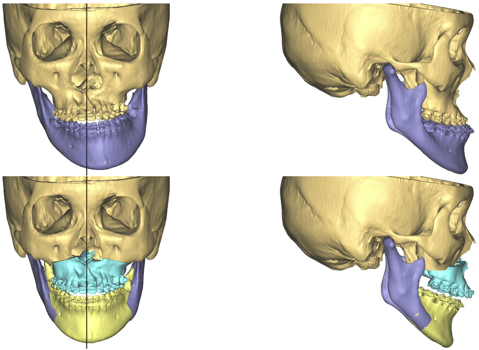
    While rare, complications following Le Fort I maxillary osteotomy includes hemorrhage, ischemia, infection, and vascular abnormalities. This study aimed to present an uncommon case of delayed presentation of a middle meningeal pseudoaneurysm formation after Le Fort I osteotomy. The patient initially presented with pulsatile tinnitus, and the pseudoaneurysm was subsequently treated successfully with image-guided embolization.

    Download PDF (520K)
  • Atsuki Yamada, Ken Yukawa, Tomoaki Eguchi
    2024 Volume 3 Issue 4 Pages 168-174
    Published: October 27, 2024
    Released on J-STAGE: October 27, 2024
    Advance online publication: April 06, 2024
    JOURNAL OPEN ACCESS

    Mycobacterium abscessus (M. abscessus) is a nontuberculous mycobacterium that causes skin and soft tissue infections. Treatment methods for these infections, including the type and duration of antimicrobial agents, have not been established. The function of surgery is also unknown. We report a case of skin and soft tissue infection of the left upper arm in a healthy 47-year-old Japanese man who was treated with complete surgical excision and antimicrobial therapy as feasible on an outpatient basis, which resulted in relapse <1 month later. Given the possibility of developing antibiotic-resistant bacteria, insufficient antimicrobial treatment should not be administered without careful consideration. There are currently few effective oral antimicrobial agents against M. abscessus, so careful antimicrobial therapy along with hospitalization (at least during the early treatment phase) is preferred, regardless of the use/nonuse of surgical treatment.

    Download PDF (930K)
  • Kahoko Yamada, Nobuyuki Mitsukawa
    2024 Volume 3 Issue 4 Pages 175-178
    Published: October 27, 2024
    Released on J-STAGE: October 27, 2024
    Advance online publication: June 07, 2024
    JOURNAL OPEN ACCESS

    A 54-year-old woman presented with an anterior neck subcutaneous tumor that had appeared one month prior. Mild tenderness was noted. As a diagnosis was difficult to make based on physical examination and ultrasonography, a magnetic resonance imaging (MRI) scan was performed. Both examinations revealed a 1-cm subcutaneous mass with well-defined margins; the MRI scan was hypointense on T1-weighted images and slightly hyperintense with low point foci on T2-weighted images. Subsequently, an excisional biopsy was performed, and the pathologic diagnosis of glomus tumor was obtained. Glomus tumors usually present as a painful subcutaneous mass beneath the nail bed but may be painless or occur in areas other than the fingers. Because glomus tumors in the neck resemble a variety of diseases, their diagnosis may be delayed. This case highlights the importance of considering glomus tumors as a potential cause of neck subcutaneous tumors.

    Download PDF (797K)
feedback
Top