Journal of Plastic and Reconstructive Surgery
Online ISSN : 2436-259X
Volume 2, Issue 4
Displaying 1-8 of 8 articles from this issue
Original Research
  • Ikkei Tamada, Marika Otaki, Riho Ishikawa
    2023 Volume 2 Issue 4 Pages 123-128
    Published: October 27, 2023
    Released on J-STAGE: October 27, 2023
    Advance online publication: April 14, 2023
    JOURNAL OPEN ACCESS

    Objectives: The secondary alveolar bone graft (ABG) is one of the key components in the treatment of cleft lip and palate. However, an ABG is commonly accompanied by postoperative facial edema. Although appropriate assessment of facial edema can facilitate the early detection of major postoperative complications, the usual course of swelling post-ABG is not yet fully understood. The present, prospective cohort study was conducted to clarify the typical course of facial swelling post-ABG.

    Methods: Nineteen operations in 17 patients who underwent an ABG between September 2019 and August 2020 were enrolled. Postoperatively, facial photographs of each patient were taken on four consecutive days; then, the degree of swelling visible in the photographs was assessed by three plastic surgeons. This assessment was repeated one month later.

    Results: Both the first and the second assessments demonstrated an inter-rater agreement of 63.2%. Cohen's kappa values for the three possible rater combinations ranged 0.377-0.755 and 0.453-0.574 in the first and the second assessment, respectively. The intra-rater reliability was 77.2%, with a Cohen's kappa value of 0.543, indicating moderate agreement.

    The swelling peaked on postoperative day 2 and began improving from postoperative day 3.

    Conclusions: These findings will hopefully help physicians treating alveolar clefts detect postoperative complications earlier and enable them to explain the prognosis of swelling to patients and their families with greater certainty.

    Download PDF (544K)
  • Norihiro Ohba, Goichi Haraoka, Hirotsuna Oiwa, Kozue Toyoda, Mayuko Oh ...
    2023 Volume 2 Issue 4 Pages 129-136
    Published: October 27, 2023
    Released on J-STAGE: October 27, 2023
    Advance online publication: March 15, 2023
    JOURNAL OPEN ACCESS

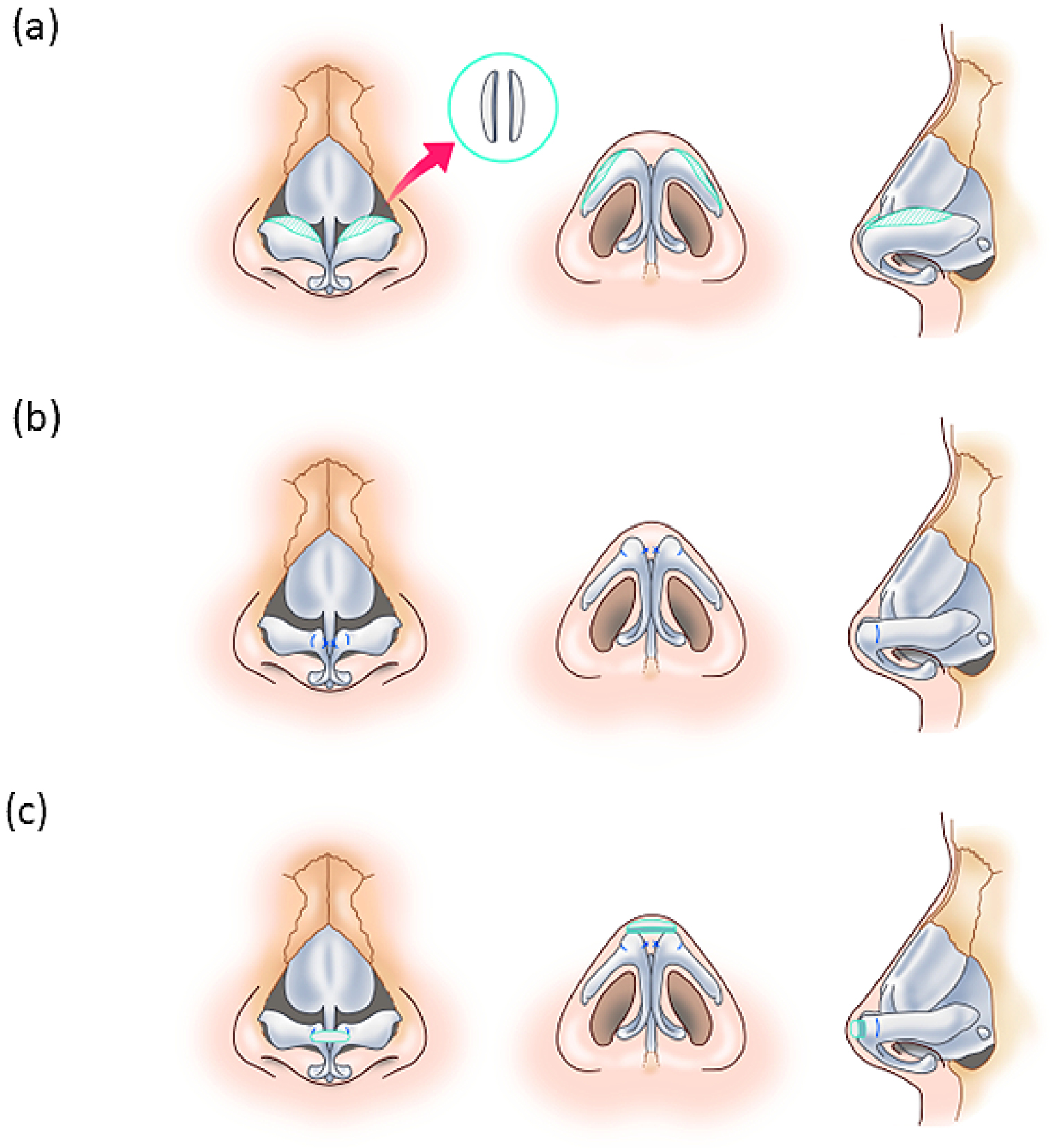
    Objectives: Many Asian patients with nasal aesthetic surgery desire a sharper, more defined nasal tip. In many cases, their nasal cartilage is not abundant enough, necessitating cartilage transplantation from an extranasal location. We investigated which patients would qualify for a method of reshaping the nasal tip without needing donor cartilage by reshaping the lower cartilage using the transdomal suture technique and local cartilage grafting.

    Methods: We performed a single-center retrospective cohort study of patients undergoing nasal tip surgery. We divided patients into two groups. Group 1 comprised 13 patients with nasal tips with thin skin and soft tissue and relatively abundant cartilage. Group 2 included 31 patients with nasal tips with thicker, firmer skin and soft tissue and a smaller amount of cartilage. No patients had prior nasal surgery, and all patients were followed up for at least 3 months. Patients in both groups underwent the transdomal suture technique and local cartilage grafting. Five patients in Group 2 underwent alar reduction. Preoperative and postoperative lateral view photographs were superimposed and analyzed to measure changes in tip projection.

    Results: Patients in Group 1 showed a mean increase in tip projection of 3.9%. Patients in Group 2 demonstrated a mean decrease in tip projection of −1.5% without alar reduction and −5.6% with alar reduction.

    Conclusions: If the nasal tip is predominantly cartilaginous, patients may undergo surgery that does not involve donor cartilage from another site with good results. However, most patients, who have a relatively small amount of nasal tip cartilage, are not good candidates for this procedure.

    Download PDF (1515K)
Case Report
  • Maki Fujii, Masayuki Okochi, Yuzo Komuro
    2023 Volume 2 Issue 4 Pages 137-141
    Published: October 27, 2023
    Released on J-STAGE: October 27, 2023
    Advance online publication: March 15, 2023
    JOURNAL OPEN ACCESS

    The reconstruction of soft tissue defects in the peritoneal region after malignant tumor resection is challenging. To reconstruct large defects, primary closure or local flaps are inadequate, and they do not provide cosmetically and functionally good results. The gracilis, gluteal, and rectus abdominis flaps are workhorse flaps for reconstructing large peritoneal defects. Surgical procedures should be carefully chosen on the basis of the size and location of the defect. In this report, we describe two cases of bilateral large defect reconstruction after malignant tumor resection using a V-shaped deep inferior epigastric artery perforator flap. Our procedure is useful as it shows that single flaps can be used to reconstruct large and bilateral peritoneal defects.

    Download PDF (814K)
  • Haruna Yoshiike, Kenichiro Kawai, Soh Nishimoto, Yoshihiro Ide, Hiroya ...
    2023 Volume 2 Issue 4 Pages 142-146
    Published: October 27, 2023
    Released on J-STAGE: October 27, 2023
    Advance online publication: March 15, 2023
    JOURNAL OPEN ACCESS

    A "poroid cell neoplasm" is a general term used to describe nodular tumors that consist of poroid cells and cuticular cells. Eccrine poroma, the most common type of poroid cell neolplasm, is a tumor continuous with the epidermis and extends into the dermis in a cord-like fashion. This tumor is almost always seen as a single lesion, with cases of multiple eccrine poromas (eccrine poromatosis) being exceedingly rare. In fact, there have only been about 20 reported cases of multiple eccrine poromas worldwide; all of which were associated with some immunosuppressive state. Here we report another case of eccrine poromatosis; this time in a patient who received an allogeneic peripheral blood stem cell transplant for acute lymphocytic leukemia 18 years prior. Our case of eccrine pormatosis is particularly rare that it involved a truncal tumor distribution, whereas the other reported cases were generally isolated to the extremities.

    Download PDF (697K)
  • Teruyo Yamashita, Jun Arata, Shuko Kaito
    2023 Volume 2 Issue 4 Pages 147-149
    Published: October 27, 2023
    Released on J-STAGE: October 27, 2023
    Advance online publication: March 15, 2023
    JOURNAL OPEN ACCESS

    The complications of insulin self-injection include subcutaneous bleeding, nerve damage, and subcutaneous induration due to repeated injection at the same site. However, considering the short length and thinness of the needle, it is unlikely that these complications will ever become serious. Here, we report a case of a 50-year-old man with abdominal giant hematoma due to insulin self-injection, which requires surgical treatment. The patient had obesity and took anticoagulants and steroids for 12 years. These drugs seemed to have affected the vessel wall and led to its compromise or destruction and subsequent hemorrhagic events.

    Download PDF (865K)
  • Toru Miyanaga, Takayoshi Kaneko, Mikio Yagishita, Miyuki Kishibe, Masa ...
    2023 Volume 2 Issue 4 Pages 150-155
    Published: October 27, 2023
    Released on J-STAGE: October 27, 2023
    Advance online publication: June 14, 2023
    JOURNAL OPEN ACCESS
    Supplementary material

    Calcaneal osteomyelitis with pseudarthrosis after calcaneal fracture is rare, and its treatment is challenging. We describe the use of a free fibular osteocutaneous flap to treat a 52-year-old man who presented with calcaneal osteomyelitis and pseudarthrosis after an operation for a closed comminuted fracture. The patient was previously treated with multiple surgeries and antibiotics for 8 months, but he was not cured. We performed radical osteomyelitis bone debridement and reconstruction with a free fibular osteocutaneous flap and autotrabecular bone grafting from the iliac crest. The skin flap survived, and rehabilitation was initiated early. At 2 years postoperatively, his pain and ambulatory function had markedly improved, and he could walk without problems in daily life. Free fibular osteocutaneous flap and autogenous cancellous bone grafting are considered practical options for the treatment of calcaneal osteomyelitis with pseudarthrosis.

    Download PDF (1000K)
  • Sayaka Nakamura, Susumu Saito, Yoshihiro Ishida, Yo Kaku, Ken Natsuga, ...
    2023 Volume 2 Issue 4 Pages 156-162
    Published: October 27, 2023
    Released on J-STAGE: October 27, 2023
    Advance online publication: June 14, 2023
    JOURNAL OPEN ACCESS

    Epidermolysis bullosa is a group of inherited skin fragility disorders with blister formation in the basement membrane zone. Chronic scarring after repeated blistering of the hands causes narrowing of the first web, flexion contractures of the digits, and pseudosyndactyly. Junctional epidermolysis bullosa is a type of epidermolysis bullosa that is characterized by blister formation in the lamina lucida. This condition is associated with survival into adulthood. In adult survivors, hand function might be required in order to participate in normal social activities. However, there is a lack of literature on the management of hand surgery for adult patients with junctional epidermolysis bullosa. We herein describe an adult patient with junctional epidermolysis bullosa who had pseudosyndactyly and flexion contractures in both hands. Five surgeries were performed. Contractures were released and reconstructed using split- or full-thickness skin grafts. Delayed wound healing was always observed due to epidermal necrosis in the graft. After epithelialization, a satisfactory functional outcome was obtained.

    Download PDF (1808K)
Brief Report
  • Paolo Gennaro, Glauco Chisci, Flavia Cascino, Michael Aboud Gasser, Gu ...
    2023 Volume 2 Issue 4 Pages 163-171
    Published: October 27, 2023
    Released on J-STAGE: October 27, 2023
    Advance online publication: April 14, 2023
    JOURNAL OPEN ACCESS

    Objective: Lymphatic fistula is a complication that may occur in all cases where lymphostasis arise, due to a damage to the lymphatic drainage system. The current study evaluates the effectiveness of supramicrosurgical lymphatico-venular anastomosis (s-LVA) for treating lymphatic fistula.

    Patients and Methods: This is a retrospective study that aims to evaluate the effectiveness of s-LVA in treating lymphatic fistula. The inclusion criterion was the presence of persistent chronic lymphatic fistula; the exclusion criteria were refuse of the patient, terminal patients.

    All patients presented with lymphatic fistula. To evaluate lymphatic patency, indocyanine green (ICG) lymphography was performed in preoperative, immediately afterward and 1 month after s-LVA. Postoperative follow-up was performed for 1 year. The site for skin incision was selected on the basis of the ICG lymphography and the venous mapping. s-LVA was performed with 11-0 sutures using an operating microscope. Afterward, the skin incision was sutured with 6-0 absorbable sutures.

    Results: Eight patients (five males and three females) were enrolled in this study. Lymphatic vessels identified during s-LVA resulted ectasic. ICG lymphography demostrated patency of the anastomosis in all patients since the first postoperative day except one case, where the anastomosis resulted patent 1 month after s-LVA. No clinical complications were reported during the postoperative follow-up: no cellulitis and no recurrence of the fistula occurred in the year after s-LVA. Total recovery of fistula was observed and confirmed for the year after the intervention.

    Conclusions: This study evidenced positive results with lymphorrhea interruption and recovery of the fistula after the intervention.

    Download PDF (1209K)
feedback
Top