Abstract
Low-dose bupivacaine can limit the spinal block level with minimal hemodynamic effects and yield a rapid recovery, but sometimes it may not provide adequate anesthesia for surgery. Dexmedetomidine, a selective α2-adrenoreceptor agonist, was shown to be a potent antinociceptive agent when given intrathecally in animals and humans. The purpose of this study was to evaluate the adjuvant effects of intrathecal dexmedetomidine in elderly patients undergoing transurethral prostate surgery with low-dose bupivacaine spinal anesthesia. Fifty-four patients undergoing transurethral prostate surgery were randomized into two groups receiving either dexmedetomidine 3 µg (n=27) or normal saline (n=27) intrathecally with 6 mg of 0.5% hyperbaric bupivacaine. The characteristics of the spinal block and postoperative analgesic effects were evaluated. The peak block level was similar for the two groups. However, the dexmedetomidine group demonstrated a faster onset time to the peak block and longer duration of spinal block than the saline group (p<0.01). The motor block scales at the time of peak sensory block and regression of 2-sensory dermatomes were higher in the dexmedetomidine group than in the saline group (p<0.001). There was less analgesic request and the time to the first analgesic request was longer in the dexmedetomidine group than in the saline group (each 487, 345 min, p<0.05). Dexmedetomidine 3 µg when added to intrathecal bupivacaine 6 mg produced fast onset and a prolonged duration of sensory block and postoperative analgesia in elderly patients for transurethral surgery. However, recovery of motor block could be delayed in dexmedetomidine-added patients.
Spinal block is a common anesthetic technique for patients undergoing transurethral resection of prostate (TURP). Most patients indicated for TURP are elderly and frequently associated with cardiopulmonary, endocrine, or other comorbidities.1) Thus, it is important to limit the block level to minimize hemodynamic changes during the spinal anesthesia in such patients.2,3) Low-dose local anesthetics can limit the block level and induce rapid recovery from anesthesia, but sometimes these low-dose local anesthetics may not provide an adequate anesthetic level for surgery. Intrathecal opioids or clonidine are frequently co-administered with local anesthetics to improve the anesthetic quality and postoperative analgesia2–8); the synergistic action of clonidine with local anesthetics is well-established.6–8) Unlike opioids, intrathecal clonidine does not produce respiratory depression or pruritus, but usual dose of clonidine (15–150 µg) may be associated with bradycardia, hypotension, or sedation.6–8)
Dexmedetomidine is an S-enantiomer of medetomidine with a higher specificity for α2-adrenoreceptor (α2 : α1, 1620 : 1) compared to clonidine (α2 : α1, 220 : 1). It was first introduced into practical use as intravenous sedative after the approval of U.S. Food and Drug Administration in 1999. Since then it has been investigated as the anxiolytic, sympatholytic, and analgesic properties related to α2-adrenoceptor binding, and it is now being used as a co-analgesic drug.9) As adjuvant, neuraxial administration is the appropriate route to dexmedetomidin, because the analgesic effect of α2-agonists mostly occurs at spinal level, and dexmedetomidin’s high lipophilicity facilitates rapid absorption into the cerebrospinal fluid and binding to the spinal cord α2-adrenoreceptor. Intrathecally-administered dexmedetomidine has been shown to exert potent antinociceptive effects in animals.9–11) To date, a few studies have reported on the effects of intrathecal dexmedetomidine combined with local anesthetics in humans.12–19) In those studies, using 10–15 mg of bupivacaine, dexmedetomidine (3–15 µg) prolonged the block duration of local anesthetics with a low-rate of side effects. Those studies, however, showed too high a block level (even up to T2) followed by a prolonged regression time. To the best of our knowledge, the influence of intrathecally-administered dexmedetomidine on spinal block using low-dose bupivacaine is unknown.
We hypothesized that a small-dose of dexmedetomidine (3 µg) added to low-dose bupivacaine (6 mg) would produce an appropriate sensory block for TURP, rapid recovery from the limited motor block, and effective postoperative analgesia.
The aim of this study was to compare the characteristics of spinal block, hemodynamic changes, and postoperative analgesia, following administration of intrathecal 3 µg dexmedetomidine combined with low-dose bupivacaine in elderly patients undergoing TURP.
MATERIALS AND METHODS
Clinical SettingThis randomized, double blind, and controlled study was approved by the Institutional Ethics Committee of Yonsei University Health System (4-2010-0513) and registered at www.ClinicalTrials.gov (NCT 01342562) in April 2011. Written informed consent was obtained from all patients. Patients with a history of back surgery or the presence of an infectious focus on the back, coagulopathy, hypersensitivity to local anesthetics or dexmedetomidine, cooperation difficulty, heart block/dysrrhythmia, or neurological disorders were excluded from the study. Also the patients with severe hepatic failure were excluded because the clearance of dexmedetomidine is diminished to 32% of normal value.
Fifty four elderly patients undergoing TURP were included in this study. Using a random number sequence, patients were enrolled in one of two groups: Group D receiving 3 µg of dexmedetomidine (Precedex® 100 µg/mL, Hospira, Inc., IL, U.S.A.) combined with 6 mg of 0.5% hyperbaric bupivacaine (Marcaine® Spinal Heavy; Astra, Sodertalje, Sweden) and Group S receiving the same amount of normal saline and bupivacaine. Dexmedetomidine 100 µg/mL was mixed with preservative-free normal saline to 10 µg/mL. The 0.3 mL of dilute dexmedetomidine was added to the bupivacaine in group D. Also, 0.3 mL of preservative-free normal saline was added to the bupivacaine in groups S. The total volume of drug solutions was 1.5 mL of 0.4% bupivacaine.
An independent investigator prepared the drug solutions and provided the coded drug to the anesthetic administrator before the start of the anesthesia. The anesthetic administrator, patients, outcome assessors, and data analysts were blinded to the allocation.
Spinal AnesthesiaPatients were hydrated with 300 mL of 0.9% sodium chloride solution prior to anesthesia. The fluid was minimally infused during the surgery to avoid overloading associated with the systemic absorption of irrigating fluid. Spinal puncture was performed at L3-4 or L4-5 with a midline approach using a 25 G Quincke needle in the lateral decubitus position. After confirmation of free flow and clear cerebrospinal fluid, the drug was administered and the patients were then placed in the supine neutral position.
AssessmentThe primary end-point of this study was the time to regression of 2-sensory dermatomes from peak sensory block level. The secondary end-points were the motor block scales at peak sensory block and regression of 2-sensory dermatomes, as well as the postoperative analgesic requirement.
The block levels were checked on the bilateral mid-thoracic line with an ice cube and pin-prick every 2 min from the drug injection. The cold peak block level was assessed by an ice cube and the sensory peak block level was assessed by pin-prick. The peak block level was defined as the same block level that persisted for four consecutive tests. For cases with a discrepancy in the dermatomal level between the right and left sides, the higher level was selected. The degree of motor block was scored using the modified Bromage scale (0=able to move the hip, knee, and ankle; 1=unable to move the hip, but able to move the knee and ankle; 2=unable to move the hip and knee, but able to move the ankle; 3=unable to move the hip, knee, and ankle). The sedation score was assessed every 15 min for 1 h using the Ramsay sedation scale (1=anxious and agitated; 2=cooperative and tranquil; 3=drowsy but responds to command; 4=asleep but responds to tactile stimulation; and 5=asleep and no response). Mean arterial pressure (MAP) and heart rate (HR) were measured every 2 min for 20 min. When the MAP or HR was decreased by 20% from the baseline, 4 mg of ephedrine or 0.5 mg of atropine was intravenously injected. The peak block level, time to reach the peak block, motor block scale at peak sensory block, and supplemental analgesic requirement (fentanyl 50 µg) were recorded. In the post anesthetic care unit (PACU), the time to the sensory regression of 2-dermatomes and motor block scales were recorded. After discharge from the PACU, the time to the first analgesic request (meperidine or tramadol) was recorded. An independent investigator assessed the pain scales using the visual analog scale (VAS, 0–10) at the PACU (arrival and 30 min) and at 6 h, 24 h, and 36 h after discharge from the PACU.
Although several studies reported that intrathecal dexmedetomidine did not result in any neurological deficits in animals20–24) and human,12–19) follow-up investigation was required to confirm neurological safety in this study. As the Institutional Review Board (IRB) of our institution recommended that we confirm the safety of patients for possible neurological effects, a follow-up evaluation was done for one month. An independent coordinator interviewed all the patients when the patient visited the out-patient department for follow-up a week after discharge. Patients were checked for any newly developed pain or paresthesia in the back, buttocks, or legs. After one month, the coordinator had a telephone-interview with the patients and confirmed that they were free of any newly developed unusual sensation on back, buttock, or legs possibly related to spinal anesthesia.
Statistical AnalysisSample size estimation was made based on a previous study.17) The primary end-point of this study was the time to the regression of 2-dermatomes from the peak sensory block level. In a previous study, the mean time (S.D.) to the sensory regression of 2-dermatomes was 122 (37) min and 80 (28) min in the dexmedetomidine group and plain group. When their results were extrapolated to our study, the mean time to the sensory regression of 2-dermatomes was calculated as 61.2 and 40.2 min. The difference of the means was 21 min and S.D. was estimated as 23.5 min. For an α=0.05 and a power of 80%, 23 patients were required per group to detect a 20-min difference in the mean time. We decided to include 27 patients per group to allow for possible drop-out.
Statistical analysis was performed using PASW Statistics 18™ (SPSS Inc., Chicago, IL, U.S.A.). Data are presented as mean (S.D.), median (IQR), or numbers as appropriate. Patient characteristics (age, weight, height, duration of operation, irrigation volume, prostate volume, time to peak block, time to 2-dermatomes regression, and maximum motor block scale) were analyzed using the independent two sample t-test. Peak sensory block levels were compared using the Mann–Whitney U-test. The number of patients by motor block scales was analyzed with the chi-square test. A Kaplan–Meier survival curve was obtained for the time to the first analgesic requirement. The linear mixed model (post-hoc: Bonferroni correction) was used for comparison of VAS between the two groups.
RESULTS
No patient dropped out from this study (Fig. 1). Patient characteristics were not different between the two groups (Table 1). Thirty-nine of 54 patients had more than one systemic disease, such as hypertension (n=24), diabetes mellitus (n=13), coronary disease (n=8), cerebrovascular accident (n=5), arrhythmia (n=3), liver cirrhosis (n=3), chronic obstructive pulmonary disease (n=3), and chronic renal failure (n=2). The mean values of MAP and HR were comparable between the two groups throughout the intraoperative period (Fig. 2).
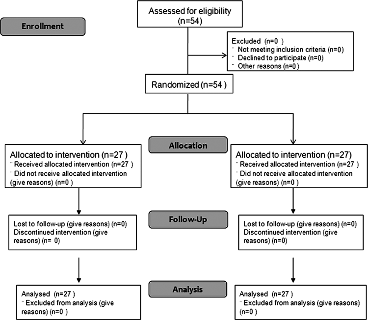
Fig. 1. Consort Flow Diagram for Enrollment
Table 1. Patient Demographics and Intraoperative Data
| Group S (n=27) | Group D (n=27) |
|---|
| Age (years) | 68.8±6.3 | 66.6±6.2 |
| Weight (kg) | 66.0±7.7 | 68.7±10.2 |
| Height (cm) | 165.5±5.9 | 167.0±12.9 |
| Duration of operation (min) | 27.9±12.3 | 30.0±16.7 |
| Irrigation amount (L) | 7.4±3.0 | 8.0±3.9 |
| Prostate volume (g) | 21.4±17.3 | 22.7±15.6 |
Values are presented as the mean±S.D. Group S: saline group, Group D: dexmedetomidine group.
The characteristics of the spinal block and postoperative data are presented in Table 2. Group D demonstrated a shorter time to reach the peak sympathetic and sensory block level, a longer time to reach the regression of 2-sensory dermatomes, and a lower postoperative analgesics requirement compared to Group S (p<0.01). In motor block scales, Group D showed higher scales both at the peak sensory block and regression of 2-dermatomes than Group S (p<0.001) (Fig. 3). All patients showed a sedation score <2 at every time point. VAS was lower in the dexmedetomidine group than in the saline group at PACU (arrival and 30 min), but no differences were shown at the ward (Fig. 4). In the dexmedetomidine group, postoperative analgesic request was significantly lower at the 7-day follow-up (p<0.01) (Table 2). A Kaplan–Meier survival analysis with the Log Rank test (Mantel-Cox) for the time to first analgesic request showed a significant difference between the two groups (p=0.006) (Fig. 5). At follow-up a week after discharge, there were not any newly developed pain or paresthesia in the back, buttocks, or legs in all the patients. Also, none of the patients complained of any neurological symptoms or signs related to spinal anesthesia upon telephone-interview that was done after one month.


Table 2. Spinal Block and Postoperative Characteristics
| Group S (n=27) | Group D (n=27) | p Value |
|---|
| Peak block level |
| Cold | T10 [T6–T12] | T9 [T6–T11] | 0.166 |
| Sensory | T10 [T7–L1] | T10 [T6–T12] | 0.013 |
| Time to peak block (min)* |
| Cold | 10.2±3.3 | 8.1±2.0 | 0.008 |
| Sensory | 10.1±3.2 | 7.9±1.5 | 0.003 |
| Time to regression of 2-sensory dermatomes (min)* | 78.4±27.3 | 109.0±38.9 | 0.002 |
| Hypotension | 1 | 1 | NS |
| Bradycardia | 1 | 1 | NS |
| Supplemental fentanyl | 3 | 0 | NS |
| Postoperative | | | |
| Rescue analgesics* | 11 | 4 | <0.01 |
| Time to first rescue (min)* | 345 [230–543] | 1,360 [487–2053] | 0.039 |
| Nausea | 1 | 0 | >0.05 |
* p<0.01. Values are presented as number, mean±S.D., or median [range]. Cold peak block level and sensory peak block level were checked by an ice cube and pin-prick respectively. Group S: saline group, Group D: dexmedetomidine group, Time to the regression: time to regression of 2-sensory dermatomes from peak sensory block level, Hypotension=SAP of <90 mmHg or <75% from the baseline value, Bradycardia=HR<45 beats min−1, NS: no significant.
DISCUSSION
The primary end-point of this study was the time to regression of 2-sensory dermatomes from the peak sensory block level. In the current study, 3 µg of intrathecal dexmedetomidine significantly prolonged the duration of sensory block compared to the control group for low-dose bupivacaine spinal anesthesia. The time to reach peak block level was also shorter in the dexmedetomidine group than in the control group. In addition, despite similar peak sensory block levels for the two groups, motor block scales were significantly higher in the dexmedetomidine group than in the saline group. In the dexmedetomidine group, postoperative analgesic requirement was lower and the time to the first-analgesic request was longer compared to the saline group.
Patients undergoing TURP are usually elderly having various co-morbidities.1) Thus, it is important to limit the block level to minimize the hemodynamic instability during spinal anesthesia. Although there are several factors influencing the spinal block level, the block level could be more influenced by total dosage of drug, not volume, concentration, or block position.25–28) Therefore, the dose of intrathecal local anesthetic should be decrease to limit the block level. Most anesthesiologists concern that reduced dose of local anesthetic may provide insufficient spinal block. Thus, there have been many trials to reduce the dose of intrathecal local anesthetics and improve the block quality with co-administration of additives such as opioids or clonidine.29,30) However, combined additive can induce their own side effects such as nausea/vomiting,30,31) pruritus,29,30,32) hypotension/bradycardia, and excessive sedation.5,7,8) In our institute, 6 mg bupivacaine has been used to obtain short duration of block and rapid return of motor function in elderly patients undergoing TURP. Although the results were favorable in those patients, we concerned the prolonged postoperative analgesia without residual spinal effect.
The antinociceptive properties of intrathecal α2-adrenoreceptor agonists are produced by inhibiting the release of C-fiber transmitters, by inhibition of release of substance P, and by hyperpolarizing post-synaptic dorsal horn neurons.5,33–35) Also, the potency of α2-adrenoreceptor agonist has been shown to correlate well with their binding affinity to spinal α2-adrenoreceptors.36,37) Since Coombs et al. first introduced the potent antinociceptive effects of intrathecal α2- adrenoreceptor agonists, clonidine (15–150 µg) has been frequently used in spinal anesthesia to improve the quality of local anesthetics.7,8,38) But, side effect such as hypotension, bradycardia, and sedation is increased as dose of clonidine is increased.39)
Dexmedetomidine is a more potent and selective α2-adrenoreceptor agonist than clonidine. In the experimental studies, the α1-adrenoreceptor activity was shown to counterbalance α2-adrenoreceptor induced analgesia. Therefore, greater α2-adrenoreceptor selectivity of dexmedetomidine may enhance the therapeutic window of α2-adrenoreceptor in the treatment of pain and overcome problematic adverse effects of clonidine.
To date, there have been a few clinical human studies on intrathecal dexmedetomidine.12–19) In those studies, it was found that 3–15 µg of dexmedetomidine co-administered with local anesthetics has a dose-dependent effect on anesthetic onset and duration with hemodynamic stability. In a study by De Koch et al., 15 µg and 45 µg intrathecal clonidine showed similar spinal effect when added to local anesthetic.39) Thus, we thought that 30 µg clonidine could be appropriate for intrathecal dose with 6 mg bupivacaine. Based on animal studies that dexmedetomidine showed a similar effect to clonidine with a 1 : 10 dose ratio in the spinal cord of animals,20,36,37) Kanazi et al.17) also used 3 µg of intrathecal dexmedetomidine in their clinical study. Although the optimal dose of intrathecal dexmedetomidine has not been established, 3 µg of dexmedetomidine seems to be appropriate for potentiating the analgesic efficacy of low-dose spinal anesthesia referred from previous studies,6,39) in which 15–45 µg of clonidine combined with low-dose ropivacaine (8 mg) or bupivacaine (6 mg) provided a prolonged spinal block.
In the current study, 3 µg of dexmedetomidine of diluted bupivacaine somewhat increased the peak sensory block level compared to the control group (Group S, Group D; median T10 [range, T7–L1], median T10 [range, T6–T12]; mean T10.48 [S.D. 1.7], mean T9.33 [S.D. 1.47], respectively), and did prolong the duration of sensory block. However other studies using 10–15 mg as general dose of bupivacaine did not show any significant difference in peak sensory block level between bupivacaine group and bupivacaine adding dexmedetomidine group, and excessively reached the block level of median T5–T6.13,14,16–18)
In our study, the motor block was potentiated in the dexmedetomidine group throughout the spinal anesthesia (Fig. 3). Though we did not evaluate the duration of motor block, this indicates that small dose of dexmedetomidine can potentiate the degree of motor block. As seen in animal and human studies, dexmedetomidine prolongs not only the duration of sensory block, but also the degree and duration of the motor block.11–19) The potentiation mechanism of motor block by dexmedetomidine is not well understood, but is suggested to be an additive or synergistic effect to the local anesthetics,11,40) or related to the interference with neuromuscular activity,11,41) or binding of α2-agonists to motor neurons in the dorsal horn.11,33)
In the current study, the numbers of patients that required postoperative analgesics were not different between two groups. This may be a result of the rapid recovery of low spinal anesthesia in both groups. However, postoperative rescue analgesics requirement was less and the time to the first analgesic request was longer in the dexmedetomidine group than in the control group (Fig. 5).
With the use of intrathecal dexmedetomidine, adverse effects should be considered. A number of studies conducted in rats, rabbits, and sheep reported that intrathecal dexmedetomidine showed no neurological deficit at a dose range of 2.5–100 µg.20–24) In rat model of perinatal excitotoxic brain injury, dexmedetomidine provided the potent neuroprotection mediated via the alpha2A-adrenoceptor. In human studies, 3–15 µg of dexmedetomidine showed a prolonged duration of sensory and motor block without any neurological effects.12–19) In the current study, we used 3 µg of dexmedetomidine referred from previous studies, and no patient showed any newly developed numbness, paresthesia, or pain on the back, buttocks, or legs on postoperative one month follow-up.
TURP for benign prostatic hyperplasia is frequently performed in elderly patients having cardiovascular limitations with various systemic diseases. We found that 72% of patients had more than one systemic disease, which corresponded to that of a previous study.1) Considering this, it is desirable to limit the spinal block level as low as possible to avoid hypotension owing to high sympathetic block. In our study, 6 mg of bupivacaine with 3 µg of dexmedetomidine resulted in a peak sympathetic block level of median T9 [range, T6–T11] and did not produce significant hypotension or bradycardia perioperatively. Even though α2-agonists produce hypotension by acting on the spinal cord, hypotension is common after spinal injection at the thoracic level, where sympathetic preganglionic neurons reside.5,41) A small-dose of dexmedetomidine intrathecally administered at the lumbar level does not seem to cause significant hypotension. Because the sympathetic block is usually near-maximal with the usual doses of local anesthetics,5) a low-dose of local anesthetics is recommended to avoid significant hypotension, especially in elderly patients. While most studies on the use of the intrathecal dexmedetomidine showed comparative hemodynamic responses similar to those of currunt study,11–17,19) one study reported a significant reduction in heart rate and blood pressure with the addition of dexmedetomidine.12) However, one could also conclude that general anesthesia following spinal block have influenced the hemodynamic variables, although there was no mention of types of anesthetics used.41–43)
In addition, high spinal block is not required for TURP. Al-Mustafa et al.15) and Kanazi et al.17) found that 12–12.5 mg of bupivacaine combined with 3–10 µg of dexmedetomidine produced a prolonged sensory and motor block in patients who underwent TURP, but the peak sensory block level was too high in their studies (even up to T2). Considering the sensory innervations to the prostate, a sensory block up to T11 is adequate for TURP. The prostate and bladder are innervated by both the sympathetic (pelvic plexus, hypogastric plexus) and parasympathetic (S3, S4) autonomic divisions. The urethral sphincter should be adequately relaxed for the endoscope to pass freely, and the urethral sphincter is also supplied by the sympathetic division of the pelvic plexus (internal sphincter) and somatic fibers of the pudendal nerve (external sphincter). Beers et al.44) suggested that mid-lumbar sensory block is enough for TURP, but sensory block at T12–L1 at least is recommended to avoid discomfort with bladder distension with irrigation.2) In our study, 3 patients in the control group, who showed a peak sensory block level <L1, required fentanyl supplementation for abdominal discomfort during the operation.
Intrathecal α2-agonists induce a dose-dependent sedative effect in humans.5,45) The dose of dexmedetomidine used in our study was at the end of the dosing spectrum, as in a study by Kanazi et al.17) The sedation score was low (<2) in all patients, as in other studies,13,15,18) demonstrating that 3 µg of intrathecal dexmedetomidine may not produce the sedative effects.
In conclusion, our study showed that 3 µg of dexmedetomidine added to 6 mg of bupivacaine produced a fast onset and long duration of sensory block as well as a prolonged postoperative analgesia compared to bupivacaine alone. Although, the dexmedetomidine group showed higher motor block scales at the time of 2-sensory dermatomal regression as well as at the time of peak sensory block, none of the patients reported discomfort in the lower extremities at the time of discharge from the PACU.
REFERENCES
- 1) Hong JY, Yang SC, Ahn S, Kil HK. Preoperative comorbidities and relationship of comorbidities with postoperative complications in patients undergoing transurethral prostate resection. J. Urol., 185, 1374–1378 (2011).
- 2) Kim SY, Cho JE, Hong JY, Koo BN, Kim JM, Kil HK. Comparison of intrathecal fentanyl and sufentanil in low-dose dilute bupivacaine spinal anaesthesia for transurethral prostatectomy. Br. J. Anaesth., 103, 750–754 (2009).
- 3) Kararmaz A, Kaya S, Turhanoglu S, Ozyilmaz MA. Low-dose bupivacaine-fentanyl spinal anaesthesia for transurethral prostatectomy. Anaesthesia, 58, 526–530 (2003).
- 4) Ben-David B, Solomon E, Levin H, Admoni H, Goldik Z. Intrathecal fentanyl with small-dose dilute bupivacaine: better anesthesia without prolonging recovery. Anesth. Analg., 85, 560–565 (1997).
- 5) Eisenach JC, De Kock M, Klimscha W. Alpha(2)-adrenergic agonists for regional anesthesia. A clinical review of clonidine (1984–1995). Anesthesiology, 85, 655–674 (1996).
- 6) Dobrydnjov I, Axelsson K, Thörn SE, Matthiesen P, Klockhoff H, Holmström B, Gupta A. Clonidine combined with small-dose bupivacaine during spinal anesthesia for inguinal herniorrhaphy: a randomized double-blinded study. Anesth. Analg., 96, 1496–1503 (2003).
- 7) Strebel S, Gurzeler JA, Schneider MC, Aeschbach A, Kindler CH. Small-dose intrathecal clonidine and isobaric bupivacaine for orthopedic surgery: a dose-response study. Anesth. Analg., 99, 1231–1238 (2004).
- 8) Elia N, Culebras X, Mazza C, Schiffer E, Tramèr MR. Clonidine as an adjuvant to intrathecal local anesthetics for surgery: systematic review of randomized trials. Reg. Anesth. Pain Med., 33, 159–167 (2008).
- 9) Virtanen R, Savola JM, Saano V, Nyman L. Characterization of the selectivity, specificity and potency of medetomidine as an alpha 2-adrenoceptor agonist. Eur. J. Pharmacol., 150, 9–14 (1988).
- 10) Ishii H, Kohno T, Yamakura T, Ikoma M, Baba H. Action of dexmedetomidine on the substantia gelatinosa neurons of the rat spinal cord. Eur. J. Neurosci., 27, 3182–3190 (2008).
- 11) Calasans-Maia JA, Zapata-Sudo G, Sudo RT. Dexmedetomidine prolongs spinal anaesthesia induced by levobupivacaine 0.5% in guinea-pigs. J. Pharm. Pharmacol., 57, 1415–1420 (2005).
- 12) Mohamed AA, Fares KM, Mohamed SA. Efficacy of intrathecally administered dexmedetomidine versus dexmedetomidine with fentanyl in patients undergoing major abdominal cancer surgery. Pain Physician, 15, 339–348 (2012).
- 13) Al-Ghanem SM, Massad IM, Al-Mustafa MM, Al-Zaben KR, Qudaisat IY, Qatawneh AM, Abu-Ali HM. Effect of adding dexmedetomidine versus fentanyl to intrathecal bupivacaine on spinal block characteristics in gynecological procedures: A double blind controlled study. Am. J. Appl. Sci., 6, 882–887 (2009).
- 14) Hala EEA, Mohamed SA, Hend Y. Dose-related prolongation of hyperbaric bupivacaine spinal anesthesia by dexmedetomidine. Ain Shams J. Anesthesiol., 4, 83–95 (2011).
- 15) Al-Mustafa MM, Abu-Halaweh SA, Aloweidi AS, Murshidi MM, Ammari BA, Awwad ZM, Al-Edwan GM, Ramsay MA. Effect of dexmedetomidine added to spinal bupivacaine for urological procedures. Saudi Med. J., 30, 365–370 (2009).
- 16) Gupta R, Verma R, Bogra J, Kohli M, Raman R, Kushwaha JK. A comparative study of intrathecal dexmedetomidine and fentanyl as adjuvants to bupivacaine. J. Anaesthesiol. Clin. Pharmacol., 27, 339–343 (2011).
- 17) Kanazi GE, Aouad MT, Jabbour-Khoury SI, Al Jazzar MD, Alameddine MM, Al-Yaman R, Bulbul M, Baraka AS. Effect of low-dose dexmedetomidine or clonidine on the characteristics of bupivacaine spinal block. Acta Anaesthesiol. Scand., 50, 222–227 (2006).
- 18) Gupta R, Bogra J, Verma R, Kohli M, Kushwaha JK, Kumar S. Dexmedetomidine as an intrathecal adjuvant for postoperative analgesia. Indian. J. Anesth., 55, 347–351 (2011).
- 19) Shukla D, Verma A, Agarwal A, Pandey HD, Tyagi C. Comparative study of intrathecal dexmedetomidine with intrathecal magnesium sulfate used as adjuvants to bupivacaine. J. Anaesthesiol. Clin. Pharmacol., 27, 495–499 (2011).
- 20) Kalso EA, Pöyhiä R, Rosenberg PH. Spinal antinociception by dexmedetomidine, a highly selective alpha 2-adrenergic agonist. Pharmacol. Toxicol., 68, 140–143 (1991).
- 21) Talke P 1, Xu M 3, Paloheimo M 2, Kalso E 2. Effects of intrathecally administered dexmedetomidine, MPV-2426 and tizanidine on EMG in rats. Acta Anaesthesiol. Scand., 47, 347–354 (2003).
- 22) Horvath G, Joo G, Dobos I, Klimscha W, Toth G, Benedek G. The synergistic antinociceptive interactions of endomorphin-1 with dexmedetomidine and/or S(+)-ketamine in rats. Anesth. Analg., 93, 1018–1024 (2001).
- 23) Xu M, Kontinen VK, Kalso E. Effects of radolmidine, a novel alpha2-adrenergic agonist compared with dexmedetomidine in different pain models in the rat. Anesthesiology, 93, 473–481 (2000).
- 24) Lo WC, Harris J, Clarke RW. Endogenous opioids support the spinal inhibitory action of an alpha 2-adrenoceptor agonist in the decerebrated, spinalised rabbit. Neurosci. Lett., 340, 95–98 (2003).
- 25) Sheskey MC, Rocco AG, Bizzarri-Schmid M, Francis DM, Edstrom H, Covino BG. A dose-response study of bupivacaine for spinal anesthesia. Anesth. Analg., 62, 931–935 (1983).
- 26) Pitkanen M. Spinal (subarachnoid) blockade. Neural blockade in clinical anesthesia and pain medicine. (Cousins MJ, Carr DB, Horlocker TT, Bridenbaugh PO ed.) Vol 4, Wolters Kluwer Health, Lippincott Williams & Wilkins, Philadelphia, p. 221 (2009).
- 27) Ruppen W, Steiner LA, Drewe J, Hauenstein L, Brugger S, Seeberger MD. Bupivacaine concentrations in the lumbar cerebrospinal fluid of patients during spinal anaesthesia. Br. J. Anaesth., 102, 832–838 (2009).
- 28) Veering BT, Immink-Speet TTM, Burm AGL, Stienstra R, van Kleef JW. Spinal anaesthesia with 0.5% hyperbaric bupivacaine in elderly patients: effects of duration spent in the sitting position. Br. J. Anaesth., 87, 738–742 (2001).
- 29) Akcaboy EY, Akcaboy ZN, Gogus N. Low dose levobupivacaine 0.5% with fentanyl in spinal anaesthesia for transurethral resection of prostate surgery. J. Res. Med. Sci., 16, 68–73 (2011).
- 30) Erdil F, Bulut S, Demirbilek S, Gedik E, Gulhas N, Ersoy MO. The effects of intrathecal levobupivacaine and bupivacaine in the elderly. Anaesthesia, 64, 942–946 (2009).
- 31) Vaghadia H, Neilson G, Lennox PH. Selective spinal anesthesia for outpatient transurethral prostatectomy (TURP): randomized controlled comparison of chloroprocaine with lidocaine. Acta Anaesthesiol. Scand., 56, 217–223 (2012).
- 32) Kuusniemi KS, Pihlajamäki KK, Pitkänen MT, Helenius HY, Kirvelä OA. The use of bupivacaine and fentanyl for spinal anesthesia for urologic surgery. Anesth. Analg., 91, 1452–1456 (2000).
- 33) Smith C, Birnbaum G, Carter JL, Greenstein J, Lublin FD. Tizanidine treatment of spasticity caused by multiple sclerosis: results of a double-blind, placebo-controlled trial. US Tizanidine Study Group. Neurology, 44, S34–S42, discussion S42–S43 (1994).
- 34) Lawhead RG, Blaxall HS, Bylund DB. Alpha-2A is the predominant alpha-2 adrenergic receptor subtype in human spinal cord. Anesthesiology, 77, 983–991 (1992).
- 35) Butterworth JF 5th, Strichartz GR. The alpha 2-adrenergic agonists clonidine and guanfacine produce tonic and phasic block of conduction in rat sciatic nerve fibers. Anesth. Analg., 76, 295–301 (1993).
- 36) Post C, Gordh T Jr, Minor BG, Archer T, Freedman J. Antinociceptive effects and spinal cord tissue concentrations after intrathecal injection of guanfacine or clonidine into rats. Anesth. Analg., 66, 317–324 (1987).
- 37) Asano T, Dohi S, Ohta S, Shimonaka H, Iida H. Antinociception by epidural and systemic alpha(2)-adrenoceptor agonists and their binding affinity in rat spinal cord and brain. Anesth. Analg., 90, 400–407 (2000).
- 38) Coombs DW, Saunders RL, Lachance D, Savage S, Ragnarsson TS, Jensen LE. Intrathecal morphine tolerance: use of intrathecal clonidine, DADLE, and intraventricular morphine. Anesthesiology, 62, 358–363 (1985).
- 39) De Kock M, Gautier P, Fanard L, Hody JL, Lavand’homme P. Intrathecal ropivacaine and clonidine for ambulatory knee arthroscopy: a dose-response study. Anesthesiology, 94, 574–578 (2001).
- 40) Talke PO, Caldwell JE, Richardson CA, Kirkegaard-Nielsen H, Stafford M. The effects of dexmedetomidine on neuromuscular blockade in human volunteers. Anesth. Analg., 88, 633–639 (1999).
- 41) De Kock M. Site of hemodynamic effects of alpha 2-adrenergic agonists. Anesthesiology, 75, 715–716 (1991).
- 42) Garcia-Fernandez J, Parodi E, Garcia P, Matute E, A-Gomez-de-Segura I, Cediel R, Gilsanz F. Clinical actions of subarachnoid sevoflurane administration in vivo: a study in dogs. Br. J. Anaesth., 95, 530–534 (2005).
- 43) Grasshoff C, Antkowiak B. Propofol and sevoflurane depress spinal neurons in vitro via different molecular targets. Anesthesiology, 101, 1167–1176 (2004).
- 44) Beers RA, Kane PB, Nsouli I, Krauss D. Does a mid-lumbar block level provide adequate anaesthesia for transurethral prostatectomy? Can. J. Anaesth., 41, 807–812 (1994).
- 45) Filos KS, Goudas LC, Patroni O, Polyzou V. Hemodynamic and analgesic profile after intrathecal clonidine in humans. A dose-response study. Anesthesiology, 81, 591–601, discussion, 27A–28A (1994).
