MATERIALS AND METHODS
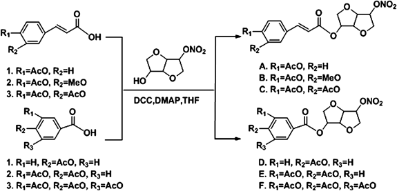
Drugs and ChemicalsThe formulae of NO donor compounds and their general synthesis processes were listed in Fig. 1. Briefly, acetyl acid (compounds 1–6, 10 mmol), isosorbide mononitrate (1.91 g, 10 mmol, CAS No. 16051-77-7, [α]D=168° (c=1.0, ethanol (EtOH))) and dimethylallylpyrophosphate (DMAP) (0.24 g, 1 mmol) were dissolved in 100 mL dry tetrahydrofuran (THF), then dicyclohexylcarbodiimide (DCC) (4.12 g, 10 mmol) was added to the solution at 0°C. The mixture was stirred at room temperature for 5 h. The resulting mixture was filtered and concentrated in vacuo. The crude product was purified by column chromatography over silica gel using ethyl acetate–n-hexane (7 : 3) as eluent. The compounds A–F (83–88% yield) was obtained as white powder. The structures of NO donor compounds were determined by IR, MS, and NMR. Column chromatography was carried out with silica gel 60 Merck for purification, and the purity of NO donor compounds was 99%. Stock solution of NO donor compounds (25 mM) was prepared with dimethyl sulfoxide (DMSO) and stored at −20°C for in vitro test. The stock solution was further diluted with the appropriate assay medium immediately before use. While in vivo test AFIM was prepared with physiological saline before use. ACZ included as a positive control was purchased from Sigma-Aldrich (St. Louis, MO, U.S.A.). The measurement kits for lactate dehydrogenase (LDH), Lactic acid (LAC), bicinchoninic acid (BCA) protein assay, superoxide dismutase (SOD), glutathione peroxidase (GSH-Px), catalase (CAT) and ATPase activities assay kits were obtained from Nanjing Jiancheng Bioengineering Institute (Nanjing, China).
AnimalsAll BALB/c mice (22±2 g) used in this experiment were SPF animals and obtained from the Center for Experimental Animals, Lanzhou Institute of Biological Products (Lanzhou, China). The mice were housed in the Laboratory Animal Care Center of Lanzhou Command General Hospital (elevation 1520 m). Animals were allowed access to food and water ad libitum. The animals were kept on a 12-h day–night cycle. All experimental protocols were reviewed and approved by the Institutional Animal Care and Use Committee at the Lanzhou Command General Hospital. The mice were divided into nine groups with ten animals in each group, (1) Normal control, (2) model, (3) ACZ 200 mg/kg, (4)–(9) compounds A–F 100 mg/kg.
Effect of NO Donors on Survival Time of Mice under Normobaric HypoxiaAirtight anoxia experiment was used to measure the anti-hypoxia effect of NO donors for normobaric hypoxia.21,26) In the normobaric hypoxia test, groups of overnight fasted mice were treated by vena caudalis administration with five NO donors (100 mg/kg), AFIM (50, 75, 100 mg/kg), vehicle (10 mL/kg physiological saline) or ACZ (200 mg/kg). Twenty minutes after administration, each mouse was put into a 250 mL airtight container with 5 g medical soda lime inside. The bottle neck was treated with petroleum jelly for a hermetic condition. Bottle cap was sealed after mouse was put into the bottle. Time from bottle cap sealed to mouse stopped breathing was recorded as survival time. The survival time of oxygen deprivation and prolongation rate (prolongation rate=(survival time of treatment group−survival time of model group)/survival time of model group) were used to compare the anti-hypoxic activity. As soon as the mouse stopped breathing, the thorax was opened, about 0.5 mL blood sample was withdrawn from heart and 0.4 mL was added to the centrifuge tube citrate-stabilized with 3% natrium citricum. The mixture was centrifuged at 2500 g for 5 min. The plasma was collected and used to determine the concentration of lactic acid and lactate dehydrogenase.
Lactic Acid (LD), LD Accumulation Rate and LDH AssessmentTo assess the LD, LD accumulation rate and LDH, blood samples were collected, centrifuged and kept at −20°C until analyses.27) Standard techniques using commercialized assay kits according to the manufacturer’s instructions (Nanjing Jiancheng Bioengineering Institute, China) were performed for analysis. LD accumulation rate were calculated as LD/survival time. LD values were expressed as mmol/L. LD accumulation rates were expressed as µmol/L·min. LDH values were expressed as U/L.
Effect of NO Donors on Heart Rate and Blood Pressure of Rats under Hypobaric Hypoxia TestWe used large low pressure oxygen compartment (Guizhou Fenglei, China) to stimulate high-altitude condition. NO donors and ACZ were administrated as mentioned above. Twenty minutes after being administrated with NO donors or vehicle by vein, rats except normal control group were put into the hypobaric hypoxia compartment and decompressed at a speed of 100 m/min. At last the simulated altitude of 8000 m was obtained. Rats were adapted to this hypobaric hypoxia environment (8% oxygen and 92% nitrogen, 0.035 MPa) for 12 h and then recovered to altitude of 4500 m (100 m/min, 0.06 MPa). Meanwhile the experimenters entered the large low pressure oxygen compartment through a transfer chamber (4500 m) and test the blood pressure and heart rate (HR) of rats by BP-2010 A Series Blood Pressure Meter (Softron, Japan).
Hypobaric Hypoxia TestWe chose the most effective NO donor in normobaric hypoxia test AFIM as the test compound in hypobaric hypoxia test. The method reported by Ma et al. was adjusted and used in this test.21,26) Low Pressure Oxygen compartment (Guizhou Fenglei, China) was used to stimulate high-altitude condition. Sixty BALB/c mice were randomly divided into 6 groups: normal control group, decompression hypoxia model group, ACZ group (200 mg/kg) and AFIM group (50, 75, 100 mg/kg). AFIM and ACZ were administrated as mentioned above. Mice except normal control group were put into a hypobaric hypoxia chamber and decompressed at a speed of 100 m/min, 20 min after being veinly administrated with AFIM or vehicle. When the simulated altitude of 8000 m was obtained, mice were adapted to this hypobaric hypoxia environment (8% oxygen and 92% nitrogen, 0.4 MPa) for 12 h, and then slowly recovered to normal altitude in half an hour. Opened the chamber door, sacrificed the animals by cervical dislocation. Mice hearts and brains were collected and stored at −80°C which were used for morphological analysis, hydrogen peroxide, malonaldehyde and enzymatic activity assays.
Malondialdehyde (MDA) AssessmentThe extent of lipid peroxidation in the mouse blood was estimated by MDA level, which was measured by using the spectrophotometric diagnostic kits (Nanjing Jiancheng Biotechnology Institute, China) as described by Uchiyama and Mihara.28,29)
H2O2 Measurement AssayH2O2 production in tissue homogenate was measured as quantitative index of reactive oxygen species (ROS) generation (indirect indicator of the free radical O2−·) by an H2O2 assay kit (Nanjing Jiancheng Institute, China). H2O2 in mice cerebrum and myocardium was performed on monitoring at the absorbance at 405 nm of the molybdenic acid-peroxide complex. The absorbance values were calibrated to a standard graph generated with known content of H2O2, the unit was defined as 1 mmol of H2O2 per gram fresh protein. Vehicle and AFIM administration mice were treated under hypobaric hypoxia for 12 h. After 12 h of exposure, the brains and hearts were grasped and washed with cold physiological saline once then homogenated and centrifuged in refrigerated centrifuge. The supernatant (100 µL) was added with H2O2 assay solution (100 µL).
Antioxidant Enzyme ActivitiesTo prepare homogenates, the mice cerebral cortex, heart and liver were homogenized with a homogenizer (400 g, 60 s) at 4°C in cold buffer (1/9, tissue/ buffer, w/v) containing 0.01 mol/L Tris–HCl, 0.1 mmol/L ethylenediaminetetraacetic acid (EDTA), 0.01 mol/L saccharose, 0.8% saline.30) The tubes with homogenate were kept in ice water for 30 min and centrifuged at 4°C (2500 g, 10 min), as recommended in the assay kits. The supernatant was separated, and then stored at −80°C. Supernatant was used for assay of various enzymatic activities. Measurement of protein concentration was estimated using commercial BCA assay kits (Nanjing Jiancheng Institute, China). The activities of SOD, GSH-Px, CAT and ATPase were measured using commercial assay kits (Nanjing Jiancheng Institute, China) according to the manufacturer instructions. Briefly, SOD activities were measured following the reduction of nitrite by a xanthine–xanthine oxidase system which was a superoxide anion generator. The activities were expressed as U/mg protein. GSH-Px activities were assayed by the decrease of the GSH, which can be reflected by the alteration of the absorbance at 412 nm. CAT activities were determined by decrease of H2O2 absorption at 405 nm. The activities of SOD, GSH-Px, CAT and ATPase were expressed as U/mg protein, U/g protein, U/mg protein and µmol Pi/mgprot/h, respectively.
Statistical AnalysisAll data were expressed as the mean±standard deviation (S.D.) Data was subjected to ANOVA followed by Student–Newman–Keuls tests. p≤0.05 was considered significant.
RESULTS
To determine the protective capability of NO donor against hypoxia, we administrated mice with three concentrations of AFIM and subjected them to oxidative challenge via normobaric hypoxia and hypobaric hypoxia. Our initial aim was to prove whether NO donor compounds could protect mice against oxidative stress caused by high altitude hypoxia. Our study was to estimate the protective effect of the AFIM in vivo compared to a commonly used anti-hypoxia drug, ACZ, and also to identify an appropriate dose that offers beneficial effects with no toxicity. To test whether AFIM could protect mice through the hypobaric hypoxia progress, we carried out both pathology and biochemistry assay to determine the morphology and physiology change during the test.
Synthesis of NO DonorsThe compound was synthesized according to above method.
Compound A: (1S,4S,5S,8R)-8-Nitrooxy-2,6-dioxabicyclo[3.3.0]octan-4-yl-3-(4-acetoxyphenyl)acrylateYield: 83%; mp: 78–79°C; 1H-NMR: 7.69 (d, 1H, J=15.6 Hz, H-C=C), 7.54 (d, 2H, J=7.8 Hz, Ar-H), 7.13 (d, 2H, J=7.8 Hz, Ar-H), 6.36 (d, 1H, J=16.2 Hz, C=C-H), 5.37 (s, 2H, Cy-H), 5.04 (s, 1H, Cy-H), 4.57 (s, 1H, Cy-H), 4.11–4.12 (m, 1H, Cy-H), 4.06–4.07 (m, 2H, Cy-H), 3.94 (m, 1H, Cy-H), 2.33 (s, 3H, CH3); IR (cm−1): 2935, 1768, 1712, 1631, 1508, 1276, 1251, 1166, 1098, 1098, 911, 851, 750; electrospray ionization (ESI)-MS [M+H]+: 380.1; Anal. Calcd for C17H17NO9: C, 53.83; H, 4.52; N, 3.69. Found: C, 53.89; H, 4.43; N, 3.58%.
Compound B: (1S,4S,5S,8R)-8-Nitrooxy-2,6-dioxabicyclo[3.3.0]octan-4-yl-3-(3-methoxyl-4-acetoxyphenyl)acrylateYield: 85%; mp: 162–163°C; 1H-NMR: 7.66 (d, 1H, J=16.2 Hz, H-C=C), 7.05–7.13 (m, 3H, Ar-H), 6. 37 (d, 1H, J=16.2 Hz, C=C-H), 5.38 (s, 2H, Cy-H), 5.04 (s, 1H, Cy-H), 4.57 (m, 1H, Cy-H), 4.11–4.13 (m, 1H, Cy-H), 4.05–4.07 (m, 2H, Cy-H), 3.94–3.95 (m, 1H, Cy-H), 3.85 (s, 3H, OCH3), 2.17 (s. 3H, CH3); IR (cm−1): 2930, 1760, 1710, 1635, 1625, 1503, 1424, 1270, 1250, 1070, 1040, 915, 855, 750; ESI-MS [M+H]+: 410.1; Anal. Calcd for C18H19NO10: C, 52.81; H, 4.68; N, 3.42. Found: C, 52.91; H, 4.65; N, 3.35%.
Compound C: (1S,4S,5S,8R)-8-Nitrooxy-2,6-dioxabicyclo[3.3.0]octan-4-yl-3-(3,4-diacetoxyphenyl)acrylateYield: 86%; mp: 130–131°C; 1H-NMR: 7.64 (d, 1H, J=16.2 Hz, H-C=C), 7.40 (d, 1H, J=8.4 Hz, Ar-H), 7.36 (s, 1H, Ar-H), 7.23 (d, 1H, J=8.4 Hz, Ar-H), 6.37 (d, 1H, J=16.2 Hz, C=C-H), 5.37 (s, 2H, Cy-H), 5.03 (s, 1H, Cy-H), 4.56 (s, 1H, Cy-H), 4.10–4.12 (m, 1H, Cy-H), 4.04–4.07 (m, 2H, Cy-H), 3.92–3.94 (m, 1H, Cy-H), 2.31 (s, 6H, CH3); IR (cm−1): 2917, 1770, 1718, 1636, 1507, 1424, 1285, 1258, 1170, 1095, 1060, 916, 854, 766; ESI-MS [M+H]+: 438.3; Anal. Calcd for C19H19NO11: C, 52.18; H, 4.38; N, 3.20. Found: C, 52.26; H, 4.33; N, 3.06%.
Compound D: (1S,4S,5S,8R)-8-Nitrooxy-2,6-dioxabicyclo[3.3.0]octan-4-yl-4-acetoxybenzoateYield: 87%; mp: 104–105°C; 1H-NMR: 8.05 (d, 2H, J=7.8 Hz, Ar-H), 7.18 (d, 2H, J=8.4 Hz, Ar-H), 5.47 (s, H, Cy-H), 5.38 (s, H, Cy-H), 5.06 (s, 1H, Cy-H), 4.62 (s, 1H, Cy-H), 4.12–4.16 (m, 1H, Cy-H), 4.06–4.10 (m, 2H, Cy-H), 3.94 (m, 1H, Cy-H), 2.33 (s, 3H, CH3); IR (cm−1): 2952, 1756, 1714, 1639, 1601, 1504, 1415, 1280, 1220, 1198, 1115, 1015, 920, 864, 766; ESI-MS [M+H]+: 354.0; Anal. Calcd for C15H15NO9: C, 51.00; H, 4.28; N, 3.96. Found: C, 51.08; H, 4.33; N, 3.92%.
Compound E: (1S,4S,5S,8R)-8-Nitrooxy-2,6-dioxabicyclo[3.3.0]octan-4-yl-3,4-diacetoxybenzoateYield: 88%; mp: 106–107°C; 1H-NMR: 7.93 (d, 1H, J=7.8 Hz, Ar-H), 7.84 (s, 1H, Ar-H), 7.29 (d, 1H, J=7.8 Hz, Ar-H), 5.47 (s, H, Cy-H), 5.38 (s, H, Cy-H), 5.05 (s, 1H, Cy-H), 4.48 (s, 1H, Cy-H), 4.17–4.17 (m, 1H, Cy-H), 4.09–4.19 (m, 2H, Cy-H), 3.93 (m, 1H, Cy-H), 2.32 (s, 6H, CH3); IR (cm−1): 2941, 1769, 1722, 1646, 1628, 1499, 1425, 1275, 1207, 1163, 1095, 1054, 909, 847, 764; ESI-MS [M+H]+: 412.1; Anal. Calcd for C17H17NO11: C, 49.64; H, 4.17; N, 3.41. Found: C, 49.74; H, 4.23; N, 3.33%.
Compound F: (1S,4S,5S,8R)-8-Nitrooxy-2,6-dioxabicyclo[3.3.0]octan-4-yl-3,4,5-triacetoxybenzoateYield: 86%; mp: 152–153°C; 1H-NMR: 7.77 (s, 2H, Ar-H), 5.47 (s, H, Cy-H), 5.38 (s, H, Cy-H), 5.04 (s, 1H, Cy-H), 4.58 (s, 1H, Cy-H), 4.14–4.16 (m, 1H, Cy-H), 4.08–4.10 (m, 2H, Cy-H), 3.94 (m, 1H, Cy-H), 2.38 (s, 9H, CH3); IR (cm−1): 2936, 1773, 1730, 1646, 1496, 1430, 1322, 1288, 1251, 1205, 1184, 1092, 1054, 909, 851, 770; ESI-MS [M+H]+: 470.2; Anal. Calcd for C19H19NO13: C, 48.62; H, 4.08; N, 2.98. Found: C, 48.71; H, 4.03; N, 3.03%.
NO Donors Prolonged the Survival Time of Mice in Normobaric Hypoxia TestIn the normobaric hypoxia test, the treatment with the NO donors significantly prolonged the survival time of oxygen deprivation mice (Table 1). Data showed that the AFIM had a dose-dependent effect of increasing the mice survival time exposed to hypoxia. The prolongation rates were 19.3% (low dose), 58.8% (middle dose), 64.5% (high dose) and 26.9% (positive control) compare with vehicle, respectively.
Table 1. Effects of NO Donor Compounds on the Survival Time of Mice under Normobaric Hypoxia Condition (
n=10)
| Name | Dose (mg/kg) | Survival time (min) | Prolonged rate (%) |
|---|
| Vehicle | — | 30.1±1.2 | — |
| Acetazolamide | 200 | 38.2±5.1* | 26.9 |
| A | 100 | Death | — |
| B | 50 | 35.9±5.1* | 19.3 |
| 75 | 47.8±9.8** | 58.8 |
| 100 | 49.5±10.5** | 64.5 |
| C | 100 | 37.8±11.0* | 25.6 |
| D | 100 | 37.5±5.4* | 24.6 |
| E | 100 | 34.7±3.2 | 8.3 |
| F | 100 | 48.6±14.6** | 61.5 |
| G | 40 | 44.7±11.5* | 48.5 |
A: (1S,4S,5S,8R)-8-Nitrooxy-2,6-dioxabicyclo[3.3.0]octan-4-yl-3-(4-acetoxyphenyl)acrylate; B: (1S,4S,5S,8R)-8-Nitrooxy-2,6-dioxabicyclo[3.3.0]octan-4-yl-3-(3-methoxyl-4-acetoxyphenyl)acrylate; C: (1S,4S,5S,8R)-8-Nitrooxy-2,6-dioxabicyclo[3.3.0]octan-4-yl-3-(3,4-diacetoxyphenyl)acrylate; D: (1S,4S,5S,8R)-8-Nitrooxy-2,6-dioxabicyclo[3.3.0]octan-4-yl-4-acetoxybenzoate; E: (1S,4S,5S,8R)-8-Nitrooxy-2,6-dioxabicyclo[3.3.0]octan-4-yl-3,4-diacetoxybenzoate; F: (1S,4S,5S,8R)-8-Nitrooxy-2,6-dioxabicyclo[3.3.0]octan-4-yl-3,4,5-triacetoxybenzoate; G: Isosorbide mononitrate. Each group represents the mean±S.D. * p<0.05 vs. Vehicle. ** p<0.01 vs. Vehicle.
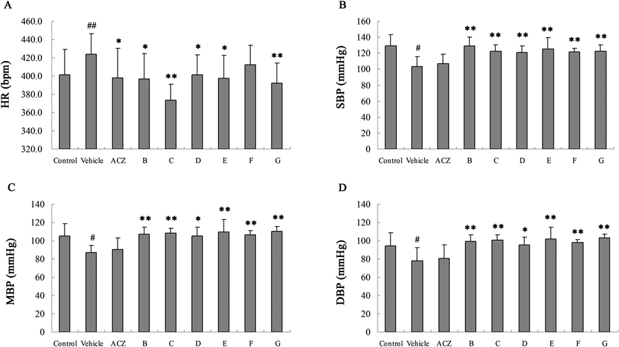
Heart rate accelerate when the body’s need to absorb oxygen and excrete carbon dioxide changes and was expressed as beats per minute (bpm). The heart rate increased significantly in hypobaric hypoxia model group while the blood pressure including systolic blood pressure (SBP), mean artery pressure (MAP) and diastolic blood pressure (DBP) decreased on the contrary. Treatment with NO donors could attenuated these changes compared with model (Fig. 2).
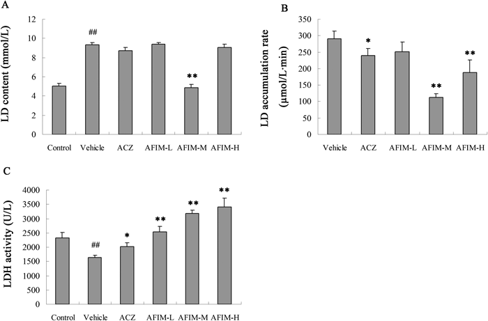
Lactic AcidWe examined the lactic acid level of different groups in the normobaric hypoxia test. There were not significant changes during each group, but the treatment with the AFIM significantly decreased the lactic acid accumulation rate comparing with vehicle31) (Fig. 3). The decrease rates of lactic acid accumulation rate were 13.6% (low dose), 61.2% (middle dose), 35.2% (high dose) and 17.9% (positive control) compare with model, respectively.
Activity of Lactate DehydrogenaseThe activity of lactate dehydrogenase was coincidence with the trend of lactic acid accumulation rate. The treatment with the AFIM significantly decreased the lactate dehydrogenase activity comparing with model (Fig. 3).
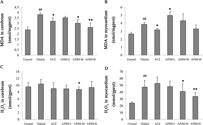
MDAThe level of malonaldehyde was coincidence with the trend of lactic acid accumulation rate. The treatment with the AFIM significantly decreased the MDA comparing with model (Fig. 4).
AFIM Directly Degrade Hypoxia-Induced H2O2 Production in Hypobaric Hypoxia MiceWe determined production of H2O2 as an indication of ROS formation in mice brains and hearts after 6 h hypobaric hypoxia test. Hypoxia induced oxidative stress stimulated cerebrum and myocardium to increase H2O2 production compared to normal control. Treatment with AFIM significantly decreased H2O2 production in myocardium in hypobaric hypoxia mice model (Fig. 4). The degrading rate of H2O2 in AFIM 100 mg/kg group was 29.1% in mice myocardium compare with vehicle group.
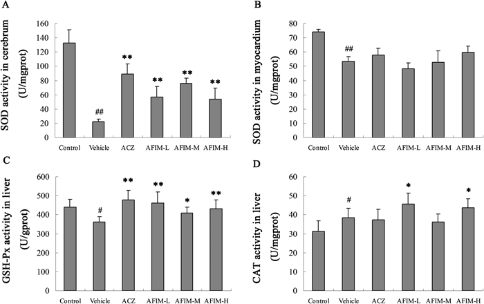
Effect of the AFIM on the Activities of SOD, GSH-Px, CAT and ATP in Hypoxic MiceAs the biomarker of the antioxidant defenses, the activity of SOD in cerebrum and myocardium, GSH and CAT in liver was measured (Fig. 5). The activities of SOD were conspicuous decreased in cerebrum and myocardium in vehicle. Activity of GSH-Px was also descent significantly. AFIM protected SOD activity in mice cerebrum compared with the vehicle group. There is no reduced GSH deficiency observed throughout the trial period at high doses of AFIM and ACZ compared with the vehicle group. The activity of CAT was increased compared with the control group. It was not descent but ascent, which probable because of the positive feedback regulation of H2O2. These results may indirectly indicate that the antioxidant enzymes in AFIM group had good antioxidant activities even after 12 h hypobaric hypoxia test.
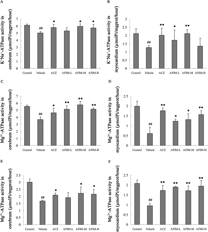
Reduction of Cerebrum and Myocardium ATP Activities in Hypoxic MiceMice cerebrum and myocardium ATP activities were examined using a luciferase assay kit. The biochemical activities of Na+-K+-ATPase, Mg2+-ATPase and Ca2+-ATPase in mice cerebrum and myocardium were significantly lower in vehicle group after 12 h hypoxia treatment compared with normal control group. On the contrary AFIM lessened the decrement of three kinds of ATPase in hypoxic mice cerebrum and myocardium compare to vehicle group. The data showed that AFIM had a dose-dependent effect (Fig. 6).