Regular Articles
Inhibitory Effects of Oligostilbenoids from the Bark of Shorea roxburghii on Malignant Melanoma Cell Growth: Implications for Novel Topical Anticancer Candidates

2016 年 39 巻 10 号 p. 1675-1682
詳細
2016 年 39 巻 10 号 p. 1675-1682
Human malignant melanomas remain associated with dismal prognosis due to their resistance to apoptosis and chemotherapy. There is growing interest in plant oligostilbenoids owing to their pleiotropic biological activities, including anti-inflammatory, antioxidant, and anticancer effects. Recent studies have demonstrated that resveratrol, a well-known stilbenoid from red wine, exhibits cell cycle-disrupting and apoptosis-inducing activities on melanoma cells. The objective of our study was to evaluate the anti-melanoma effect of oligostilbenoids isolated from the bark of Shorea roxburghii. Among the isolates, four resveratrol oligomers, i.e., (−)-hopeaphenol, vaticanol B, hemsleyanol D, and (+)-α-viniferin, possessed more potent antiproliferative action than did resveratrol against SK-MEL-28 melanoma cells. Cell cycle analysis revealed that (−)-hopeaphenol, hemsleyanol D, and (+)-α-viniferin arrested cell division cycle at the G1 phase, whereas vaticanol B had little effect on the cell cycle. In addition, cell proliferation assay also revealed that (+)-α-viniferin induced DNA damage followed by induction of apoptosis in SK-MEL-28 cells, which was confirmed by an increased expression of γ-H2AX and cleaved caspase-3, respectively. The compounds vaticanol B, hemsleyanol D, and resveratrol significantly increased the expression of p21, suggesting that they are able to block cell cycle progression. Moreover, these oligostilbenoids downmodulated cylin D1 expression and extracellular signal-regulated kinase (ERK) activation. Furthermore, hemsleyanol D, (+)-α-viniferin, and resveratrol significantly decreased the expression of cyclin B1, which could also suppress cell cycle progression. The present study thus suggests that these plant oligostilbenoids are effective as therapeutic or chemopreventive agents against melanoma.
Melanoma is known to be the most aggressive form of skin cancer, originating in pigment-producing melanocytes. Although surgical treatment of early melanoma can lead to a cure rate of about 90%, its intrinsic resistance to chemotherapy, aggressive clinical behavior, and tendency to rapidly metastasize may result in unresectable advanced melanoma, which hinders its achieving successful treatment. In addition, the incidence of melanoma continues to increase worldwide.1,2) Hence, development of effective pharmacological or natural chemopreventive and chemotherapeutic agents is imperative.
It is known that cell-cycle arrest is often associated with apoptosis. The tumor protein p53, which responds to various types and levels of stress arising from apoptosis, cell cycle arrest, senescence, DNA repair, and cell metabolism, functions as one of the main regulators of cell survival and apoptosis.3) It is a frequent target for inactivating mutations in various types of human tumor including malignant melanoma, resulting in cell growth and stress resistance to apoptosis.4)
Another signaling pathway involved in cell cycle progression, proliferation, and cell survival is the extracellular signal-regulated kinase 1/2 mitogen-activated protein kinase (ERK1/2 MAPK) pathway. It has been reported that frequent BRAF-activating mutations have been identified in cultured melanoma cell lines and tumor samples, providing evidence that the Ras-Raf-mitogen-activated protein extracellular kinase (MEK)-ERK pathway plays a critical role in melanoma progression.5) When activated, ERK isoforms translocate into the nucleus and transmit mitogenic signals that induce the phosphorylation of specific transcription factors that are required for cell cycle progression.6) A recent study has indicated that the MAPK pathway is the major inducer of cyclin D1 in proliferating cells.7)
A Dipterocarpaceae plant Shorea roxburghii G. DON is widely distributed in Thailand and its neighboring countries. In India, they have been used as folk medicine for the treatment of several diseases including dysentery, diarrhea, and cholera.8) The genus Shorea has been known to be a rich source of oligostilbenoids, which possess important biological activities such as cytotoxic,9) antibacterial,10) and antioxidant properties.11) Resveratrol, a monomeric stilbenoid found in high concentrations in grapes and red wine, has been shown to exhibit chemopreventive activities against a wide variety of cancers, including breast carcinoma, leukemia, colon carcinoma, prostate adenocarcinoma, and melanoma.12–17) Recent studies have demonstrated that its antitumor activities were mediated through several cell signaling pathways involved in cell cycle arrest, suppression of tumor cell proliferation, and induction of apoptosis.12–14) However, the precise mechanisms underlying resveratrol-regulated tumor growth inhibition and apoptotic induction remain to be fully elucidated.
The objective of this study is to determine the cytotoxicity of oligostilbenoids isolated from S. roxburghii against SK-MEL-28 malignant melanoma cells. The present study revealed that four resveratrol oligomers, i.e., (−)-hopeaphenol, vaticanol B, hemsleyanol D, and (+)-α-viniferin, exerted their chemopreventive action through cell cycle arrest and apoptotic induction by distinct mechanisms.
Oligostilbenoids were isolated from the bark of S. roxburghii as previously described.18)
Cell CultureHuman SK-MEL-28 cells were purchased from NIBIO (Osaka, Japan). Cells were maintained in modified Eagle’s medium (Nacalai Tesque, Kyoto, Japan), supplemented with 10% fetal bovine serum (Nichirei Bioscience Inc., Tokyo, Japan), 1× antibiotic–antimycotic solution (Nacalai Tesque), 10 mM N-(2-hydroxyethyl)piperazine-N′-2-ethanesulfonic acid (HEPES) (Nacalai Tesque), and 1× non-essential amino acid solution (Nacalai Tesque).
Cell Viability AssaySK-MEL-28 cells cells were seeded into 96-well plates at a density of 4×10³ per well (100 µL) and treated with oligostilbenoids at variable concentrations (0, 1, 3, 10, 30, 100 µM) for 48 h. Cell viability was assessed using the WST-8 Assay (Dojindo Laboratories, Kumamoto, Japan) according to the manufacturer’s instructions.
Cell Cycle AssayCell proliferation was detected by incorporating 5-ethynyl-2′-deoxyuridine (EdU) into the cells using the Click-iT EdU Alexa Fluor 488 Flow Cytometry Assay Kit (Life Technologies, Carlsbad, CA, U.S.A.) according to the manufacturer’s protocol. Briefly, cells were incubated with 10 µM EdU for 2 h before fixation, permeabilized, and stained with EdU. DNA content of the cells was measured by staining with FxCycle Far Red (Life Technologies). Cells were then analyzed using the 488 and 633 nm wavelength laser of an ec800 flow cytometer (SONY, Tokyo, Japan).
Western Blot AnalysisCells were lysed with lysis buffer consisting of 20 mM Tris–HCl, pH 8.0, containing 1% sodium dodecyl sulfate and 1 mM dithiothreitol. Blots were probed with rabbit monoclonal antibody against p21 Waf/Cip1 (12D1), rabbit monoclonal antibody against cleaved caspase-3 (Asp175) (Cell Signaling Technology, Danvers, MA, U.S.A.), rabbit monoclonal antibody against ERK1/2 (137F5) (Cell Signaling Technology), rabbit monoclonal antibody against phospho-ERK1/2 (T201/Y204) (D13.14.4E) (Cell Signaling Technology), rabbit monoclonal antibody against cyclin D1 (EPR2241) (Abcam, Cambridge, U.K.), mouse monoclonal antibody against cyclin B1 (H-433) (Santa Cruz Biotechnology, Dallas, TX, U.S.A.), rabbit monoclonal antibody against phospho-Histone H2A.X (Ser139) (20E3) (Cell Signaling Technology), rabbit monoclonal antibody against phospho-Chk1 (S296) (EPR915) (Cell Signaling Technology), and mouse monoclonal antibody against actin (Clone C4) (Merck-Millipore, Billerica, MA, U.S.A.). Horseradish peroxidase (HRP)-conjugated anti-mouse or rabbit immunoglobulin G (IgG) secondary antibody (Cell Signaling Technology) was used as probe, and immunoreactive bands were visualized with the Image Quant LAS4000 mini system (GE HealthCare, Buckinghamshire, U.K.) using the Immobilon Western Chemiluminescent HRP substrate (Merck-Millipore). The band intensity was analyzed using ImageJ software.
We first evaluated the cytotoxic effect of oligostilbenoids from the bark of S. roxburghii on human malignant melanoma cell line, SK-MEL-28. Among the oligostilbenoids, four resveratrol oligomers, namely (−)-hopeaphenol, vaticanol B, hemsleyanol D, and (+)-α-viniferin (Fig. 1), significantly reduced the viability of SK-MEL-28 cells more efficiently than resveratrol (Table 1, Fig. 2). The half-maximal inhibitory concentration (IC50) of (−)-hopeaphenol, vaticanol B, hemsleyanol D, (+)-α-viniferin, and resveratrol was 3.6, 16.6, 15.5, 7.1, and 21.0 µM, respectively.

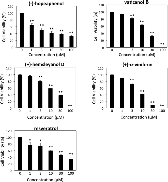
SK-MEL-28 cells were incubated with various concentrations of (−)-hopeaphenol, vaticanol B, (+)-hemsleyanol D, (+)-α-viniferin, and resveratrol for 48 h. Cell viability was assessed using the WST-8 assay. Data are represented as the mean±S.E.M. of three independent experiments. * p<0.05 and ** p<0.01 compared to control (0 µM).
| Cell viability (% of control) | |||||
|---|---|---|---|---|---|
| 1 µM | 3 µM | 10 µM | 30 µM | 100 µM | |
| 1′S-Dihydrophayomphenol A | 116.9±6.9 | 100.1±9.0 | 121.4±1.7 | 106.1±6.8 | 114.1±2.6 |
| Phayomphenol A1 | 105.1±2.2 | 95.1±6.1 | 79.7±7.6 | 109.1±4.3 | 121.5±4.0 |
| Phayomphenol A2 | 114.2±9.2 | 113.4±4.9 | 106.5±3.7 | 106.7±5.8 | 98.2±1.4 |
| (−)-Hopeaphenol | 61.6±4.0 | 50.9±6.3 | 40.0±5.5 | 33.3±1.9 | 26.4±1.5 |
| (+)-Isohopeaphenol | 82.1±6.5 | 93.3±5.6 | 74.3±8.8 | 67.4±4.3 | 20.3±0.9 |
| Hemsleyanol D | 96.0±1.4 | 80.0±3.0 | 58.9±1.6 | 38.4±0.8 | 0 |
| (−)-Ampelopsin H | 101.0±0.8 | 109.0±0.9 | 118.1±4.8 | 121.7±1.4 | 97.3±2.7 |
| Vaticanol B | 93.9±3.0 | 81.8±4.6 | 64.1±4.2 | 32.1±1.4 | 0 |
| Vaticanol A | 91.0±0.2 | 78.8±14.3 | 66.4±2.2 | 79.2±9.0 | 49.6±3.0 |
| Vaticanol E | 112.7±7.7 | 108.4±6.0 | 95.3±4.6 | 85.9±8.8 | 29.7±8.2 |
| Vaticanol G | 113.9±6.5 | 108.8±6.7 | 106.7±3.7 | 89.7±8.0 | 62.4±2.3 |
| (+)-α-Viniferin | 91.0±9.6 | 71.9±3.7 | 41.6±1.4 | 8.2±0.6 | 0 |
| Pauciflorol A | 99.3±0.3 | 83.9±7.5 | 68.5±0.3 | 79.6±7.3 | 71.0±7.3 |
| Hopeafuran | 120.8±4.9 | 103.0±6.0 | 112.2±1.5 | 85.8±4.8 | 35.4±0.3 |
| (−)-Balanocarpol | 119.5±1.6 | 117.0±3.5 | 143.0±2.8 | 137.5±2.6 | 120.1±1.5 |
| Malibatol A | 94.8±9.9 | 91.7±7.2 | 108.7±6.0 | 77.1±4.2 | 30.0±3.1 |
| Malibatol B | 81.3±3.9 | 88.1±6.9 | 85.8±9.3 | 82.0±3.5 | 49.2±0.5 |
| (+)-Parviflorol | 88.4±5.6 | 86.7±2.3 | 92.6±0.9 | 78.0±3.8 | 52.7±1.6 |
| trans-Resveratrol 10-C-Glc | 98.3±4.3 | 91.3±3.3 | 90.9±4.2 | 79.5±7.9 | 55.5±7.0 |
| cis-Resveratrol 10-C-Glc | 110.2±5.0 | 99.5±7.4 | 80.7±0.8 | 75.7±5.9 | 64.2±10.6 |
| trans-Piceid | 97.5±6.4 | 79.3±2.8 | 97.1±5.3 | 72.1±5.7 | 63.9±6.6 |
| Resveratrol | 77.6±7.5 | 74.4±8.8 | 60.0±1.6 | 46.7±1.9 | 34.8±5.1 |
Each value represents the mean±S.E.M. of three independent experiments.
In order to elucidate whether the decrease in cell viability induced by the oligostilbenoids was caused by their antiproliferative action, we next performed an EdU incorporation assay combined with cell cycle analysis based on DNA content. After treatment with 100 µM (+)-α-viniferin and resveratrol, the percentage of cells in the S phase were dramatically reduced, suggesting that these compounds inhibited SK-MEL-28 melanoma cell proliferation at the G1/S phase (Fig. 3, Table 2). Treatment with 100 µM (−)-hopeaphenol and hemsleyanol D also resulted in the accumulation of the cells in the G1 phase and in the decrease of cell population in S phase. However, the effect was moderate compared with (+)-α-viniferin and resveratrol (Fig. 3, Table 2). In contrast, the treatment of 100 µM vaticanol B had little effect on the cell cycle (Fig. 3, Table 2).

SK-MEL-28 cells were incubated with 100 µM of (−)-hopeaphenol, vaticanol B, (+)-hemsleyanol D, (+)-α-viniferin, and resveratrol for 24 h. Incorporation of EdU and DNA content were measured by flow cytometry. The data shown are representative of three independent experiments.
| Sample | Cell-cycle phase | |||
|---|---|---|---|---|
| Sub-G1 (%) | G1 (%) | S (%) | G2/M (%) | |
| Control | 0.51±0.01 | 73.8±1.54 | 21.1±0.78 | 4.64±0.04 |
| (−)-Hopeaphenol | 1.01±0.06 | 84.8±0.82 | 11.0±0.42 | 3.26±0.02 |
| Vaticanol B | 1.30±0.03 | 77.3±1.61 | 17.8±1.28 | 3.58±0.03 |
| Hemsleyanol D | 1.35±0.03 | 82.5±1.95 | 10.8±0.55 | 5.34±0.02 |
| (+)-α-Viniferin | 19.0±1.46 | 71.4±2.89 | 0.03±0.00 | 9.58±0.42 |
| Resveratrol | 2.23±0.07 | 88.1±0.45 | 0±0.00 | 9.69±0.16 |
Each value represents the mean±S.E.M. of three independent experiments.
The cell cycle analysis as shown in Fig. 3 and Table 2 also revealed that the cell populations in the sub-G1 area were increased in oligostilbenoid-treated melanoma cells, suggesting that these oligostilbenoids could induce apoptotic cell death in melanoma cells. This effect was especially pronounced upon (+)-α-viniferin treatment. In order to confirm whether the cell populations in the sub-G1 area were in fact apoptotic cells, we performed Western blot analysis with an antibody against cleaved caspase-3. As expected, increased expression level of cleaved caspase-3 was observed in cells treated with (+)-α-viniferin (Fig. 4). Furthermore, the expression level of p21 cyclin-dependent kinase inhibitor was also analyzed by Western blot. As shown in Fig. 4, p21 expression was significantly upregulated in cells treated with vaticanol B, hemsleyanol D, and resveratrol. On the other hand, the level of phosphorylated ERK1/2 decreased when cells were treated with all the compounds we tested (Fig. 4), demonstrating that these oligostilbenoids induced the inactivation of the ERK/MAPK cascade in malignant melanoma cells. To further investigate the cell cycle status in SK-MEL-28 cells, the expression level of cyclin D1 and cyclin B1 was examined by Western blot analysis. Treatment with all the compounds used in this study significantly diminished the expression of cyclin D1 (Fig. 4), suggesting that these compounds led to accumulation of cells in the G1 phase. Treatment with hemsleyanol D, (+)-α-viniferin, and resveratrol significantly diminished the expression of cyclin B1 (Fig. 4), suggesting that these compounds led to accumulation of cells in the G2 phase. Furthermore, treatment with hemsleyanol D and resveratrol resulted in DNA damage in SK-MEL-28 cells since phosphorylated form of Chk1 was observed in these cells (Fig. 4). Intriguingly, γ-H2AX, a sensitive indicator of DNA damage, was significantly induced by (+)-α-viniferin, and to a lesser extent by resveratrol (Fig. 4). These data indicated that hemsleyanol D, (+)-α-viniferin, and resveratrol might be DNA-damaging agents, and the cells treated with (+)-α-viniferin failed to induce G2/M DNA damage checkpoint.

SK-MEL-28 cells were incubated with 100 µM of (−)-hopeaphenol, vaticanol B, (+)-hemsleyanol D, (+)-α-viniferin, and resveratrol for 20 h. The extracted proteins were immunoblotted with the indicated antibodies. Graphs indicate the relative band intensities as determined by ImageJ software and plotted as the mean ±S.E.M. of three independent experiments. * p<0.05 and ** p<0.01 compared to control.
Based upon these findings, we proposed a model describing the mechanism of action of these oligostilbenoids, shown in Fig. 5.

SK-MEL-28 melanoma cells possess an inactivating mutation of p53 and an activating mutation of BRAF (BRAFV600E), both of which promote cell cycle progression through the activation of cyclin D/Cdk complex (left). Oligostilbenoids could upregulate p21 and suppress the ERK1/2 pathway, which in turn suppresses the cyclin D/Cdk complex and causes cell cycle arrest at the G1 phase. In addition, hemsleyanol D, (+)-α-viniferin, and resveratrol could also suppress cyclin B1 expression, resulting in cell cycle arrest at the G2/M phase (right). Hemsleyanol D, (+)-α-viniferin, and resveratrol might be DNA-damaging agents, and the cells treated with (+)-α-viniferin that fail to induce G2/M DNA damage checkpoint die by apoptosis.
In the present study, we have demonstrated that oligostilbenoids isolated from S. roxburghii possess potent chemotherapeutic properties against SK-MEL-28 malignant melanoma cells. Among the isolates, four resveratrol oligomers, i.e., (−)-hopeaphenol, vaticanol B, hemsleyanol D, and (+)-α-viniferin exhibited more efficient antiproliferative action than resveratrol on SK-MEL-28 cells (Fig. 2, Table 1). Although these compounds possess similar structures and strong growth inhibitory activities, the mechanisms underlying their respective effects were different from each other.
Significant cell cycle arrest at the G1 phase was observed in SK-MEL-28 cells treated with (+)-α-viniferin and resveratrol (Fig. 3, Table 2). A recent study has demonstrated that treatment of human melanoma cells with resveratrol resulted in inhibition of cell proliferation by inducing G1/S cell cycle arrest and apoptosis, possibly via the p53 pathway.19) Our present data indicated that resveratrol treatment triggered cell cycle arrest at the G1 phase possibly via the upregulation of p21 and the induction of apoptosis (Fig. 5). In contrast, increased expression of cleaved caspase 3, but not p21 was observed in cells treated with (+)-α-viniferin (Fig. 5). p21 is a potent inhibitor of cell cycle progression that inhibits cyclin D-Cdk4/6 complexes; however, its roles in apoptosis are controversial. It has been reported that p21 exhibits pro-apoptotic functions under certain conditions in specific systems.20,21) On the other hand, p21 can also protect cells and inhibit apoptosis via induction of cell cycle arrest, as the downregulation of p21 usually increases the sensitivity of tumor cells, including melanoma, to apoptosis induced by different chemotherapeutic agents.21,22) From this point of view, (+)-α-viniferin could thus be an excellent candidate for a chemotherapeutic agent against melanoma. Nevertheless, further analysis is required to clarify this issue.
Upregulation of p21 was also observed in SK-MEL-28 melanoma cells treated with vaticanol B and hemsleyanol D (Fig. 4). Treatment by these two compounds resulted in cell cycle arrest at G1 phase; however, their effect on cell cycle arrest is much milder than that of resveratrol (Fig. 3, Table 2). Of note, the induction of p21 by resveratrol, vaticanol B, and hemsleyanol D is p53-independent, because the SK-MEL-28 melanoma cells used in this study carried an inactivating mutation of p5323) (Fig. 5). In addition to the up-modulation of p21 expression, inactivation of the ERK/MAPK pathway was also observed upon treatment with all the compounds used in this study (Fig. 4). SK-MEL-28 melanoma cells possess an activating mutation of BRAF (BRAFV600E), resulting in the constitutive activation of the ERK/MAPK pathway.23) It is well known that ERK/MAPK promotes cell cycle progression through upregulation of cyclin D protein level.7) Indeed, downregulation of cyclin D1 was observed upon treatment with all the compounds used in this study (Fig. 4). Therefore, combination of ERK/MAPK inactivation and p21 induction by these oligostilbenoids could induce cell cycle arrest in melanoma cells. Furthermore, inactivation of the ERK/MAPK pathway by these oligostilbenoids may affect cytotoxicity against melanoma cells, as the Ras-Raf-MEK-ERK pathway plays a critical role in melanoma cell survival.24,25)
Hemsleyanol D, (+)-α-viniferin, and resveratrol treatments induced the downregulation of cyclin B1 (Fig. 4). Cyclin B is necessary for the G2/M phase transition during cell cycle progression. Overexpression of cyclin B1 has been reported in a variety of tumors, including breast, colon, prostate, oral, non-small cell lung cancers, and melanoma,26–29) suggesting that dysregulation of cyclin B1 may contribute to the loss of normal cell cycle control during tumorigenesis. In particular, disruption in cyclin B1 expression resulted in the induction of cell cycle arrest followed by apoptotic cell death.30,31) Increase in cell population at G2/M phase and induction of apoptosis (Fig. 3, Table 2) observed in cells treated with hemsleyanol D, (+)-α-viniferin, and resveratrol, as demonstrated by our data, were thus consistent with previous reports. It is likely that the down-modulation of cyclin B1 by these compounds might have contributed to these effects.
A large subset of cancer cells contains large quantities of mutant p53 protein, leading to the incapability to establish the G1 checkpoint. Recently, checkpoint kinase 1 (Chk1) inhibitors have thus emerged as promising novel therapeutic agents in order to increase DNA-damaging chemotherapeutic drug, e.g., cisplatin or gemcitabine, induced tumor cell death in p53 pathway defective cancers by abrogating the remaining intact checkpoint.32,33) In this study, we found the potential of hemsleyanol D and resveratrol as DNA-damaging agents since phosphorylated form of Chk1 was observed in cells treated with these compounds (Fig. 4). Therefore, combination treatment of these compounds with Chk1 inhibitors is expected to be efficacious against tumor. However, significant expression of γ-H2AX was only observed in cells treated with (+)-α-viniferin, and these cells failed to induce G2/M DNA damage checkpoint (Fig. 4). These data suggest that (+)-α-viniferin may be used alone as a combination therapy with a DNA-damaging agent/Chk1 inhibitor, although precise points of action and molecular mechanisms remain to be elucidated. From this point of view, (+)-α-viniferin could thus be an excellent candidate for a chemotherapeutic agent against melanoma.
In summary, our current findings uncovered the potent chemotherapeutic properties of oligostilbenoids isolated from S. roxburghii against malignant melanoma cells. Although further studies are required to verify this conclusion, our findings may aid the development of new strategies to prevent cell cycle progression and survival of malignant melanoma cells.
We thank K. Sawaragi, T. Michiyama, K. Inoue, A. Tahara, R. Morita, A. Goto, and T. Morita for their technical assistance. This work was supported in part by Grants from the Japan Agency for Medical Research and Development (AMED) and by the MEXT-Supported Program for the Strategic Research Foundation at Private Universities, 2014–2018. The authors also acknowledge the financial support by Kobayashi International Scholarship Foundation.
The authors declare no conflict of interest.

