Abstract
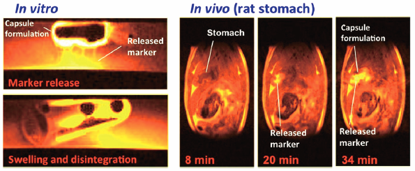
Although magnetic resonance imaging (MRI) has potential in assessments of formulations, few studies have been conducted because of the size and expense of the instrument. In the present study, the processes of in vitro and in vivo release in a gelatin capsule formulation model were visualized using a compact MRI system with 1.5 T permanent magnets, which is more convenient than the superconducting MRI systems typically used for clinical and experimental purposes. A Gd-chelate of diethylenetriamine-N,N,N′,N″,N″-pentaacetic acid, a contrast agent that markedly enhances proton signals via close contact with water, was incorporated into capsule formulations as a marker compound. In vitro experiments could clearly demonstrate the preparation-dependent differences in the release/disintegration of the formulations. In some preparations, the penetration of water into the formulation and generation of bubbles in the capsule were also observed prior to the disintegration of the formulation. When capsule formulations were orally administered to rats, the release of the marker into the stomach and its transit to the duodenum were visualized. These results strongly indicate that the compact MRI system is a powerful tool for pharmaceutical studies.