Abstract
We investigate the inhibitory effect of marketed drugs for treatment of inflammatory bowel disease (IBD) such as ulcerative colitis (UC) and Crohn’s disease (CD) on the uptake transporters of peptide transporter 1 (PEPT1), which are up-regulated under the inflamed condition. The uptake transport of glycylsarcosine, a typical substrate for PEPT1, was reduced to 60% only by 5-aminosalicylate at the clinically relevant concentration among tested marketed drugs in PEPT1 transfected HEK293 cell lines. These findings suggest that the inhibition of PEPT1, which were up-regulated in inflamed or non-inflamed site on UC and CD patients, contribute to the clinical effect of commercially available drugs for IBD patients through the inhibition of uptake of antigenic proinflammatory oligopeptides such as formyl-methionine (Met)-leucine (Leu)-phenylalanine (Phe) via PEPT1.
Inflammatory bowel disease (IBD) such as ulcerative colitis (UC) and Crohn’s disease (CD) are well-known as a refractory intestinal disease and as the typical lower gastrointestinal diseases. These diseases are chronic relapsing disorders characters by an aberrant inflammatory response with mucosal inflammation and organ damage. IBD is well associated with diarrhea, malnutrition, and weight loss through inhibited absorption of nutrition, water and so on,1) suggesting an important role of the active intestinal transporters for nutrition in disease. Among the solute carriers expression level of Peptide transporter 1 (PEPT1) was reported to be the highest in human duodenum and ileum, but significantly lower levels in all colon segments compared with duodenum and ileum.2,3) The mRNA expression levels of PEPT1 in the inflamed site on CD and UC in colon was significantly higher than those of non-inflamed site.4) Indeed, the expression and activity of PEPT1 has been shown to be increased by pro-inflammatory cytokines such as tumor necrosis factor-alpha and interferon-gamma.5) Intestinal inflammation, which occurs both in human IBD and in some types of mouse models of colitis, has been shown to increase colonic PEPT1 protein expression,6) On the other hand, there has been one report showing decreased expression of PEPT1 in descending colon of patients with IBD.7) However, in both in vitro and in vivo animal model, it was also suggested that increased uptake of bacterial peptide such as formyl-methionine (Met)-leucine (Leu)-phenylalanine (Phe) (fMLP) via PEPT1 stimulated expression of MHC Class 1 Molecules6) and PepT1-mediated uptake of bacterial peptides was involved in the colonic inflammation process in dextran sodium sulfate-induced colitis mouse.8) Collectively, increased uptake of bacterial peptides via increased expression of PEPT1 is suggested to be involved in the disease state. Marketed drugs for IBD have been considered to exert the pharmacological action through different mode of action. However, precise mode of action of 5-aminosalicylate (5-ASA) has not been clarified.9) Recently, 5-ASA was reported to decrease bacterial polyphosphate accumulation, leading to diminishing the capacity of bacteria to persist within chronically inflamed environments.10) Thus, considering the important role of bacteria in IBD, interaction of marketed drugs for IBD with PEPT1 can be a mechanism to exert their pharmacological actions. Then, in this study we determined the inhibitory effect of commercial drugs for IBD on the uptake transport of using glycylsarcosine (GlySar) using HEK293-PEPT1 cells.
MATERIALS AND METHODS
Materials3H-Glycylsarcosine (GlySar) was purchased from Moravek Biochemicals (Brea, CA, U.S.A.). Unless otherwise stated, all culture media and supplements were purchased from Invitrogen (Carlsbad, CA, U.S.A.). Twenty four-well plate was purchased from Corning (Tokyo, Japan). Azathioprine, 5-aminosalicylate (5-ASA), tacrolimus, prednisolone and cyclosporine A were purchased from Sigma-Aldrich Inc. (St. Louis, MO, U.S.A.). Cephalexin was purchased from Wako Pure Chemical Industries, Ltd. (Osaka, Japan). Rebamipide was obtained from Otsuka Pharmaceutical Co., Ltd. (Tokyo, Japan). All other regents used were of regent grade.
Cell CultureHuman PEPT1-expressing HEK293 cells, HEK293 cells transfected with a vector containing human PEPT1 cDNA, and control cells, HEK293 cells transfected with the empty vector, developed at Drug Development Solutions Division, Sekisui Medical Co., Ltd. (Ibaraki, Japan) were used. The cells were seeded in 24-well plates (Corning Japan Co., Ltd., Tokyo, Japan) at a density from 2.1 to 2.3×10⁵ cells/well and incubated in 5% CO2 incubator at 37°C for 2 d. The medium was composed of Dulbecco’s modified Eagle’s medium (DMEM) containing fetal bovine serum (FBS), antibiotic–antimycotic, and L-glutamine.
3H-GlySar UptakeCells were cultured for 2 d, after that, the medium from the plate seeded with cells was removed and replaced with 1 mL of The Hank’s Balanced Salt Solution (HBSS) in pH 6.0 for PEPT1. HBSS in the plate was then removed and replaced by 300 µL of the test solutions, HBSS containing test compounds, maintained at 37°C and the plate was preincubated at 37°C for 15 min. After preincubation, the test solutions were removed and replaced with 300 µL of 3H-GlySar, 0.1 µM, the typical substrate for PEPT1, containing test compounds, followed by incubation at 37°C for 2 min. Then, the solution was removed and the cells were rinsed once with 1 mL of ice-cold phosphate buffered saline (PBS) containing 0.2% bovine serum albumin (BSA) and twice with 1 mL of ice-cold PBS. After PBS removal, the cells were dissolved in 500 µL of 0.1 M NaOH. Subsequent to pipetting the cell lysate, 300 µL portion of the solution was collected into a glass vial. 10-mL Scintillator (Hionic-Fluor, PerkinElmer, Inc., Japan, Kanagawa, Japan) was added to each vial and the radioactivity was measured using a liquid scintillation counter (1900CA, PerkinElmer, Inc., Japan). Cephalexin was used a typical inhibitor to guarantee PEPT1 activities (data not shown).
Protein AssayThe protein content was determined by BCA protein assay kit (Thermo Fisher Scientific, Tokyo, Japan) using the bovine BSA as a standard according to a manufacturer’s instructions.
Drug SolubilityThe drugs and HBSS at pH 6.0 and pH 7.4 were incubated at 37°C for 30 min and the absorbance was measured at 620 nm with a plate reader.
Calculation of Uptake Clearance, % of Control, IC50 and Gut ConcentrationThe uptake clearance of each substrate was calculated by the uptake concentrations of the substrates divided by the protein volume and initial concentration and uptake clearance was calculated using the following Eq. 1.
 | (1) |
where d
Q/d
t,
A and
C0 mean the amount of the test substrates taken in the cells within a given time period, protein concentration and initial concentrations of the substrates, respectively.
The value of % of control was calculated using the following Eq. 2.
 | (2) |
where
A,
B,
C and
D mean uptake clearance without the addition of substrates and inhibitors in control cells, uptake clearance without the addition of substrates and inhibitors in transporter expressed cells, uptake clearance with the addition of substrates and inhibitors in control cells and uptake clearance with the addition of substrates without inhibitors in transporter expressed cells, respectively.
The theoretical concentration in the gastrointestinal tract was calculated by dividing the daily dose used in the clinical practice by the volume of water of 1650 mL in gastrointestinal tract reported by Davies and Morris.11)
Statistical AnalysisData are expressed as the mean±standard deviation (S.D.) of more than three experiments. ANOVA was used to test the statistical significance of differences among groups. Statistical significance in the differences of the means was determined by Student’s t-test.
RESULTS
Inhibitory Effect of Commercially Available Drugs for IBD on Uptake Transport of 3H-Glycylsarcosine (GlySar)The examined concentrations from the gut concentration in Table 1 were set, but maximum solubility in pH 6.0 of cyclosporine A and 5-ASA were lower than the gut concentration. Clearance and % of control value of GlySar uptake in HEK293-PEPT1 cells were shown in Figs. 1 and 2. Clearance and % of control value of GlySar uptake with 5-ASA was no more than 60% when compared to that without inhibitor (Fig. 1). However, the inhibitory effect wasn’t observed for the uptake transport of GlySar by the addition of azathioprine, tacrolimus, prednisolone, cyclosporine A and rebamipide in Fig. 2.
Table 1. Theoretical Drug Concentration in Human Intestine on Commercial Drugs for UC and CD Patients
| Compound | Clinical daily dose (mg) | Molecular weight | Gut concentration (µM) | Maximum solubility in pH 6.0 (µM) | Maximum solubility in pH 7.4 (µM) |
|---|
| Azathioprine | 50–100 | 277.26 | 30–61 | 250 | 250 |
| 5-ASA | 2400–3600 | 153.14 | 9498–14247 | 5000 | 10000 |
| Tacrolimus | 2.5–3 | 822.03 | 1.8–2.2 | 5 | 5 |
| Prednisolone | 5–60 | 360.44 | 8.4–101 | 500 | 500 |
| Cyclosporine A | 840–960 | 1202.61 | 423–484 | 5 | 5 |
| Rebamipide | 300 | 370.79 | 491 | 500 | 500 |
Gut concentrations on each drug were calculated utilizing the human intestinal volume data from the ref. 11.
DISCUSSION
PEPT1 plays a major part on the intestinal absorption for various substances. Under IBD state, the expression level of mRNA of PEPT1 on inflamed site in UC and CD patients were significantly higher than those in healthy volunteers.4) Thus, we hypothesized that PEPT1 would be involved in uptake of causative substances exacerbating the condition of IBD and investigated the inhibitory effect on PEPT1 by the commercial available drugs of IBD.
Anti-inflammatory drugs, immune-suppressing drugs, 5-ASA and so on are well utilized in clinical field in IBD.12–17) We focused on six commercial drugs, azathioprine, 5-ASA, tacrolimus, prednisolone, cyclosporine A and rebamipide. Rebamipide has been reported that it has a superior efficacy to the rat colitis model induced by dextran sulfate,18,19) although rebamipide did not have a clinical application to IBD yet.
Only 5-ASA among the tested drugs showed inhibition of Gly-Sar uptake in HEK293-PEPT1 cells (Fig. 1). The value of IC50 couldn’t be calculated because % of control value of 5-ASA was still remained more than 50% at the highest tested concentration of 5-ASA of 5000 µM. However, 5-ASA would inhibit the intestinal PEPT1 more efficiently, considering estimated clinically relevant gut concentration of 5-ASA was higher than tested maximum concentration (Table1).
Clinical efficacy of 5-ASA has not been observed when it is administrated by a conventional oral route so that it is absorbed in upper small intestine.20) Accordingly, the investigation of pH responsive and/or colon targeted formulations for 5-ASA was utilized in the clinical practice.20,21) Thus, in case utilizing the modified release 5-ASA formulation in clinical practice, colonic concentration of 5-ASA must be higher, suggesting the inhibitory effect of 5-ASA on PEPT1 would be more effective. Still, however, it hardly seems difficult that almost complete inhibition for PepT1-mediated transport by 5-ASA would occur. Then, we should clarify whether partial inhibition is clinically relevant effect in animal models.22)
It has been reported that serum amino acid concentration of IBD patients was significantly high when compared to health volunteers,23,24) which is consistent with the increased expression level of PEPT1 in IBD patients. Furthermore, serum amino acid concentrations were lower in IBD patients in remission status than in IBD active patients.24) Thus, the inhibition for PEPT1 could be useful to improve the clinical effect by reducing the absorption of causative substances such as bacterial peptide from the intestine.
Here in this study, we identified the inhibitory effect of 5-ASA on HEK293-PEPT1 cells. These results indicate that 5-ASA could obtain the pharmacological activity by the inhibitory effect on inflamed site which up-regulated PEPT1 in IBD patients. Further studies are required to assess in greater detail the contribution of the transporter inhibition to therapeutic effect of the numerous clinical drugs for IBD.
Conflict of Interest
The authors declare no conflict of interest.
REFERENCES
- 1) Sands BE. Inflammatory bowel disease: past, present, and future. J. Gastroenterol., 42, 16–25 (2007).
- 2) Englund G, Rorsman F, Rönnblom A, Karlbom U, Lazorova L, Gråsjö J, Kindmark A, Artursson P. Regional levels of drug transporters along the human intestinal tract: co-expression of ABC and SLC transporters and comparison with Caco-2 cells. Eur. J. Pharm. Sci., 29, 269–277 (2006).
- 3) Meier Y, Eloranta JJ, Darimont J, Ismair MG, Hiller C, Fried M, Kullak-Ublick GA, Vavricka SR. Regional distribution of solute carrier mRNA expression along the human intestinal tract. Drug Metab. Dispos., 35, 590–594 (2007).
- 4) Wojtal KA, Eloranta JJ, Hruz P, Gutmann H, Drewe J, Staumann A, Beglinger C, Fried M, Kullak-Ublick GA, Vavricka SR. Changes in mRNA expression levels of solute carrier transporters in inflammatory bowel disease patients. Drug Metab. Dispos., 37, 1871–1877 (2009).
- 5) Vavricka SR, Musch MW, Fujiya M, Kles K, Chang L, Eloranta JJ, Kullak-Ublick GA, Drabik K, Merlin D, Chang EB. Tumor necrosis factor-α and interferon-γ increase PepT1 expression and activity in the human colon carcinoma cell line Caco-2/bbe and in mouse intestine. Pflugers Arch., 452, 71–80 (2006).
- 6) Merlin D, Si-Tahar M, Sitaraman SV, Eastburn K, Williams I, Liu X, Hediger MA, Madara JL. Colonic epithelial PepT1 expression occurs in inflammatory bowel disease: transport of bacterial peptides influences expression of MHC class 1 molecules. Gastroenterology, 120, 1666–1679 (2001).
- 7) Wuensch T, Ullrich S, Schulz S, Chamaillard M, Schaltenberg N, Rath E, Goebel U, Sartor RB, Prager M, Büning C, Bugert P, Witt H, Haller D, Daniel H. Colonic expression of the peptide transporter PEPT1 is downregulated during intestinal inflammation and is not required for NOD2-dependent immune activation. Inflamm. Bowel Dis., 20, 671–684 (2014).
- 8) Dai X, Chen X, Chen Q, Shi L, Liang H, Zhou Z, Liu Q, Pang W, Hou D, Wang C, Zen K, Yuan Y, Zhang CY, Xia L. MicroRNA-193a-3p reduces intestinal inflammation in response to microbiota via down-regulation of colonic PepT1. J. Biol. Chem., 290, 16099–16115 (2015).
- 9) Hauso Ø, Martinsen TC, Waldum H. 5-Aminosalicylic acid, a specific drug for ulcerative colitis. Scand. J. Gastroenterol., 50, 933–941 (2015).
- 10) Dahl J-U, Gray MJ, Bazopoulou D, Beaufay F, Lempart J, Koenigsknecht MJ, Wang Y, Baker JR, Hasler WL, Young VB, Sun D, Jakob U. The anti-inflammatory drug mesalamine targets bacterial polyphosphate accumulation. Nat. Microbiol., 2, 16267 (2017).
- 11) Davies B, Morris T. Physiological parameters in laboratory animals and humans. Pharm. Res., 10, 1093–1095 (1993).
- 12) Present DH. 6-Mercaptopurine and other immunosuppressive agents in the treatment of Crohn’s disease and ulcerative colitis. Gastroenterol. Clin. North Am., 18, 57–71 (1989).
- 13) Pearson DC, May GR, Fick GH, Sutherland LR. Azathioprine and 6-mercaptopurine in Crohn disease. A meta-analysis. Ann. Intern. Med., 123, 132–142 (1995).
- 14) Lowry PW, Franklin CL, Weaver AL, Pike MG, Mays DC, Tremaine WJ, Lipsky JJ, Sandborn WJ. Measurement of thiopurine methyltransferase activity and azathioprine metabolites in patients with inflammatory bowel disease. Gut, 49, 665–670 (2001).
- 15) Xin HW, Schwab M, Klotz U. Transport studies with 5-aminosalicylate. Eur. J. Clin. Pharmacol., 62, 871–875 (2006).
- 16) Mendoza JL, Urcelay E, Lana R, Martín MC, López N, Guijarro LG, Mayol JA, Taxonera C, de la Concha EG, Peña AS, Díaz-Rubio M. MDR1 polymorphisms and response to azathioprine therapy in patients with Crohn’s disease. Inflamm. Bowel Dis., 13, 585–590 (2007).
- 17) Jahnel J, Fickert P, Hauer AC, Högenauer C, Avian A, Trauner M. Inflammatory bowel disease alters intestinal bile acid transporter expression. Drug Metab. Dispos., 42, 1423–1431 (2014).
- 18) Kishimoto S, Haruma K, Tari A, Sakurai K, Nakano M, Nakagawa Y. Rebamipide, an antiulcer drug, prevents DSS-induced colitis formation in rats. Dig. Dis. Sci., 45, 1608–1616 (2000).
- 19) Murai R, Kanbe T, Mukoyama T, Shimomura T, Hashiguchi K, Yoshida Y, Tsuchiya H, Hoshikawa Y, Kurimasa A, Shiota G. Effect of rectal administration of rebamipide on dextran sulfate sodium-induced colitis: role of hepatocyte growth factor. Inflamm. Res., 56, 240–245 (2007).
- 20) Criscuoli V, Modesto I, Orlando A, Cottone M. Mesalazine for the treatment of inflammatory bowel disease. Expert Opin. Pharmacother., 14, 1669–1678 (2013).
- 21) Böhm SK, Kruis W. Long-term efficacy and safety of once-daily mesalazine granules for the treatment of active ulcerative colitis. Clin. Exp. Gastroenterol., 7, 369–383 (2014).
- 22) Buyse M, Tsocas A, Walker F, Merlin D, Bado A. PepT1-mediated fMLP transport induces intestinal inflammation in vivo. Am. J. Physiol. Cell Physiol., 283, C1795–C1800 (2002).
- 23) Schicho R, Shaykhutdinov R, Ngo J, Nazyrova A, Schneider C, Panaccione R, Kaplan GG, Vogel HJ, Storr M. Quantitative metabolomic profiling of serum, plasma, and urine by (1)H NMR spectroscopy discriminates between patients with inflammatory bowel disease and healthy individuals. J. Proteome Res., 11, 3344–3357 (2012).
- 24) Dawiskiba T, Deja S, Mulak A, Ząbek A, Jawień E, Pawełka D, Banasik M, Mastalerz-Migas A, Balcerzak W, Kaliszewski K, Skóra J, Barć P, Korta K, Pormańczuk K, Szyber P, Litarski A, Młynarz P. Serum and urine metabolomic fingerprinting in diagnostics of inflammatory bowel diseases. World J. Gastroenterol., 20, 163–174 (2014).
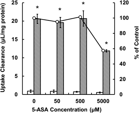
 , PEPT1; ○, % of control.
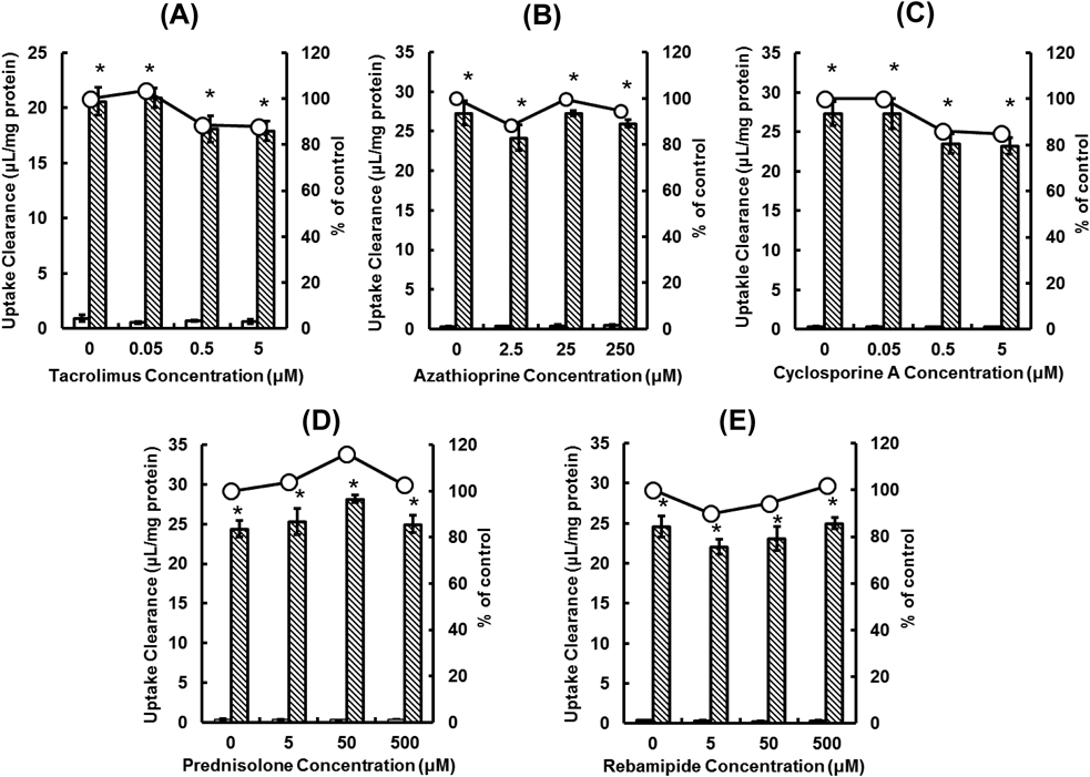
 , PEPT1, ○, % of control.