Abstract
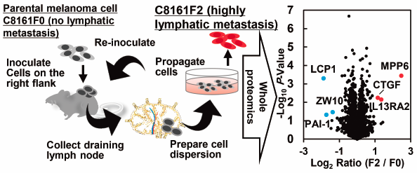
Metastasis of cancer cells to lymph nodes (LN) is a common modality of metastasis in clinical settings, but the mechanisms involved in lymphatic metastasis remain unclear compared to hematogenous metastasis to bones and the brain. To elucidate the molecular mechanisms responsible for melanoma LN metastasis, we first generated LN metastasis-prone melanoma cells (C8161F2) by the sequential in vivo transplantation of parental melanoma cells (C8161F0). Although the in vitro/in vivo proliferative potential of these melanoma cells were similar, the metastatic potential of the C8161F2 for LNs was significantly enhanced. We then conducted a proteomics analysis to identify the proteins and pathways that contribute to LN metastasis. We identified six proteins (three: up-regulated and three: down-regulated) whose expressions were statistically significantly different by more than 2-fold in the two cell groups. Some of these genes are responsible for the activation of the transforming growth factor-β (TGF-β)-related pathway, a well-known inducer of epithelial–mesenchymal transition (EMT). In addition, a gene ontology analysis revealed that the enhanced cell–cell adhesion appears to be involved in lymphatic metastasis. In conclusion, we established highly lymphatic metastatic melanoma cells, which would be valuable for studies of the molecular mechanisms responsible for lymphatic metastasis.