Notes
Activation of HSP90/HSF1 Signaling as an Adaptive Response to an Electrophilic Metabolite of Morphine

2023 年 46 巻 2 号 p. 334-337
詳細
2023 年 46 巻 2 号 p. 334-337
Morphinone (MO) is an electrophilic metabolite of morphine that covalently binds to protein thiols, resulting in toxicity in vitro and in vivo. We have previously identified a variety of redox signaling pathways that are activated during electrophilic stress. However, the role of MO in such activation remains unknown. In this study, we examined whether MO could activate heat shock protein (HSP) 90/heat shock factor (HSF) 1 signaling in HepG2 cells. MO exposure caused S-modification of HSP90 (determined using biotin-PEAC5-maleimide labeling) and nuclear translocation of transcription factor HSF1, thereby up-regulating its downstream genes encoding B-cell lymphoma 2-associated anthanogene 3 and heat shock 70 kDa protein 1. However, dihydromorphinone, a non-electrophilic metabolite of morphine, had little effect on HSF1 activation or upregulation of these genes, suggesting that covalent modification plays a role in this process and that the HSP90/HSF1 pathway is a redox-signaled adaptive response to morphine metabolism.
Morphinone (MO; Fig. 1A) is a metabolite of morphine, which is used as a narcotic analgesic throughout the world, and is produced by enzymatic reactions with nicotinamide adenine dinucleotide (NAD)(P)-dependent enzymes such as morphine-6 dehydrogenase, a member of the aldo-keto reductase 1C subfamily of isozymes.1,2) MO can covalently bind to thiols of glutathione and proteins, yielding MO–glutathione and MO–protein adducts, respectively, through the electrophilic carbon of its α,β-unsaturated carbonyl group.3) Modification by electrophiles changes protein functions, resulting in activation of redox signaling pathways.4) We reported recently that MO activates the Kelch-like ECH-associated protein 1 (Keap1)/nuclear factor (erythroid-derived 2)-like 2 (Nrf2) redox signaling pathway in HepG2 cells.5) However, the contribution of MO to other redox signaling pathways is unknown. Because morphine is reported to be endogenously produced in vivo,6) understanding of cellular responses against the reactive metabolite of morphine is required. In addition, high dose of morphine is used to reduce persistent pain because of analgesic tolerance of the opioid,7) suggesting that its metabolite, MO, potentially causes electrophilic stress in such condition.

(A) Structure of morphinone (MO) and dihydromorphinone (DHMO). (B) HepG2 cells were exposed to MO for 1 h, then protein thiols were labeled with biotin-PEAC5-maleimide (BPM). The precipitated HSP90 and total HSP90 in the lysate were detected using Western blotting. The bands were quantified using ImageJ software. Each value is the mean ± standard error (S.E.) of three determinations. * p < 0.05 vs. untreated cells. The cells were exposed to (C) MO (0–100 µM) or (D) DHMO (0–100 µM) for 1 h. Proteins in the cytosol and nuclear fraction were analyzed using Western blotting with antibodies against HSF1, HSF90, histone deacetylase 1 (HDAC1), and glyceraldehyde 3-phosphate dehydrogenase (GAPDH). Representative blots are shown.
Heat shock protein (HSP) 90, a negative regulator of heat shock factor 1 (HSF1), responds to electrophilic and oxidative stresses.4,8) Once HSP90 is modified by electrophiles and/or oxidants, HSF1 disassociates from HSP90 and then translocates into the nucleus, leading to upregulation of HSP expression.9,10) Carbone et al. showed that the endogenous electrophile 4-hydroxy-2-nonenal modifies HSP90 in a rat model of alcohol-induced oxidative stress.11) We reported previously that 1,4-naphthoquinone and cadmium, an exogenous electrophilic quinone and heavy metal, respectively, activate the HSP90/HSF1 pathway through S-modification of the cysteine (Cys) residues of HSP90 and induction of its downstream signaling proteins.12–14) These findings suggest that MO also activates the HSP90/HSF1 pathway. In this study, we therefore aimed to identify MO-mediated activation of the HSP90/HSF1 pathway in HepG2 cells.
MO and dihydromorphinone (DHMO; Fig. 1A) were synthesized using the methods of Rapoport et al.15,16) Biotin-PEAC5-maleimide (BPM), anti-glyceraldehyde-3-phosphate dehydrogenase antibody, and protease inhibitor cocktail were purchased from Dojindo (Kumamoto, Japan), Santa Cruz Biotechnology (Dallas, TX, U.S.A.), and Sigma-Aldrich (St. Louis, MO, U.S.A.), respectively. Anti-HSF1 antibody, anti-histone deacetylase 1 antibody, horseradish peroxidase (HRP)-conjugated anti-rabbit immunoglobulin G (IgG), and HRP-conjugated anti-mouse IgG were obtained from Cell Signaling Technology (Beverly, MA, U.S.A.). All other reagents were of the highest grade available.
Cell CultureHepG2 cells (RIKEN Cell Bank, Ibaraki, Japan) were cultured in Dulbecco’s modified Eagle’s medium (DMEM, #11995065, Thermo Fisher Scientific, Waltham, MA, U.S.A.) containing 10% (v/v) fetal bovine serum (FBS; Biowest, Nuaillé, France) and 1% (v/v) penicillin–streptomycin (Thermo Fisher Scientific) at 37 °C in 5% CO2. The cells were seeded on 12-well plates or 35 mm dishes (5 × 10⁵ cells/well or 8 × 10⁵ cells/dish) for 24 h, then incubated in serum-free DMEM for a further 24 h prior to treatment with MO or DHMO. Following treatment, cells were washed three times with ice-cold phosphate-buffered saline (PBS).
Western BlottingThe cells were collected by scraping in a lysis buffer (50 mM Tris–HCl (pH 7.5), 6 M urea, 1% (v/v) Triton X-100, 5 mM ethylenediaminetetraacetic acid (EDTA), and 1% (v/v) protease inhibitor cocktail). The protein concentration was determined using the bicinchoninic acid assay (Thermo Fisher Scientific), following the manufacturer’s instructions. Each sample was adjusted to the same protein concentration, mixed with half the volume of loading buffer (10% (v/v) sodium dodecyl sulfate (SDS), 62.5 mM Tris–HCl (pH 6.8), 20% (v/v) glycerol, 5 mM 2-mercaptoethanol, and 0.015% (v/v) bromophenol blue), then heat-denatured. The proteins were separated using SDS-polyacrylamide gel electrophoresis and transferred onto polyvinylidene difluoride membranes (Millipore-Sigma, St. Louis, MO, U.S.A.). After blocking with 5% (v/v) skim milk, the membranes were incubated with primary antibodies and then with HRP-conjugated secondary antibodies. Amersham ECL Western blotting Detection Reagent (GE Healthcare, Buckinghamshire, U.K.) was used to detect the immunoreactive proteins. The band intensities were measured using ImageJ software.17)
BPM Labeling AssayS-Modified HSP90 was detected using BPM labeling as previously described.18) Briefly, cells were collected in a lysis buffer (50 mM Tris–HCl (pH 6.8), 0.5% (w/v) deoxycholate, 1% (w/v) NP-40, 150 mM NaCl, and 1% (v/v) protease inhibitor cocktail) containing 100 µM BPM, reacted on ice for 1 h, then centrifuged at 15000 × g for 10 min at 4 °C. BPM-labeled proteins were precipitated using avidin-agarose, washed, and then detected using Western blotting.
Nuclear and Cytoplasmic ExtractionAfter being washed by PBS, the cells were incubated with trypsin and collected in DMEM containing 10% (v/v) FBS. After centrifugation at 1000 rpm for 10 min, the cell pellet was re-suspended in PBS and then centrifuged again at 4 °C. The pellet was gently re-suspended and incubated in 300 µL of suspension buffer (10 mM N-(2-hydroxyethyl)piperazine-N′-2-ethanesulfonic acid (HEPES)–KOH (pH 7.9), 1.5 mM MgCl2, 10 mM KCl, 0.5 mM dithiothreitol, 100 µM digitonin, and 1% (v/v) protease inhibitor cocktail) at 4 °C for 10 min, then centrifuged at 15000 rpm for 10 min at 4 °C. The cytoplasmic and nuclear fractions were collected from the supernatant and pellet, respectively.
Quantitative PCR (qPCR)Total RNA was extracted using TRIzol (Thermo Fisher Scientific), cDNA was generated from 1 µg total RNA using an Affinity Script qPCR cDNA Synthesis kit (Agilent Technologies, Santa Clara, CA, U.S.A.), and then qPCR was performed following the manufacturer’s instructions. The following primers were used: HSF1, forward 5′-TGAAAAGTGCCTCAGCGTAGCC-3′ and reverse 5′-TGCTCAGCATGGTCTGCAGGTT-3′; heat shock 70 kDa protein 1A (HSPA1A/HSP70), forward 5′-ACCTTCGACGTGTCCATCCTGA-3′ and reverse 5′-TCCTCCACGAAGTGGTTCACCA-3′; B-cell lymphoma 2-associated anthanogene 3 (BAG3), forward 5′-TGCCAGAAACCACTCAGCCAGA-3′ and reverse 5′-TGAGGATGAGCAGTCAGAGGCA-3′; acidic ribosomal phosphoprotein P0 (36B4), forward 5′-TCTACAACCCTGAAGTGCTTGAT-3′ and reverse 5′-CAATCTGCAGACAGACACTGG-3′. The expression of mRNAs was normalized to that of 36B4.
Small Interfering RNA (siRNA) TransfectionsiRNAs were transfected using Lipofectamine RNAiMAX (Thermo Fisher), following the manufacturer’s protocol. Briefly, HepG2 cells were cultured in 12-well plates until they reached 70–80% confluency and then incubated in fresh DMEM with 10% (v/v) FBS in the presence of the siRNA/Lipofectamine RNAiMAX mixture. After 48 h, the medium was removed and the cells were treated with MO or DHMO. The siRNA sense and antisense sequences for HSF1 (siHSF1) were: 5′-GUGACCACUUGGAUGCUAUdTdT-3′ (sense) and 5′-AUAGCAUCCAAGUGGUCACdTdT-3′ (antisense). The control siRNA sense and antisense sequences for β-galactosidase were: 5′-UGGCGAUUACCGUUGAUGUTTdTdT-3′ (sense), and 5′-ACAUCAACGGUAAUCGCCAdTdT-3′ (antisense).
Data AnalysisStatistical analysis was performed using one-way ANOVA followed by Sidak’s or Dunnett’s multiple-comparison test using GraphPad Prism version 8.4.3 (GraphPad Software, San Diego, CA, U.S.A.), and p < 0.05 was considered to indicate a significant difference.
First, we assessed modification of HSP90 during exposure to MO using BPM labeling. BPM does not modify electrophile-bound protein thiols and thus the band intensity in the resulting Western blot is inversely proportional to the degree of S-modification. Exposure of HepG2 cells to MO (100 µM) for 1 h significantly modified HSP90 in the cells (Fig. 1B), and tendency of such an S-modification at 50 µM MO was observed. This electrophilic metabolite of morphine (10–100 µM) enhanced translocation of HSF1 into the nucleus (Fig. 1C). While sensitivity of BPM assay was low, it is expected that even at concentrations lower than 100 µM MO, a small amount of binding of HSP90 to MO occurs, resulting in nuclear translocation of HSF1. While In contrast, DHMO (Fig. 1A), which is non-electrophilic because it lacks the C7–C8 double bond found in MO, did not enhance the nuclear localization of HSF1 (Fig. 1D). These results suggest that HSF1 dissociates from its complex with HSP90, presumably in response to S-modification of the latter by MO. HSP90 has two isoforms, stress-induced HSP90α and constitutively expressed HSP90β, that have six Cys residues.19) We have previously shown that the environmental electrophiles cadmium and 1,4-naphthoquinone modify the cysteine residues at Cys412 and Cy564 of HSP90β.14,18) Cys564 is also reported to modify by an endogenous electrophile 4-hydroxynonenal.11) Shibata et al. have reported that 6-(methylsulfinyl)hexyl isothiocyanate binds to Cys521 of HSP90β.20) We therefore speculate MO with electrophilic property also binds to multiple cysteines such as Cys412, Cy564, and/or Cys521 of HSP90.
To examine the effects on downstream genes of HSF1, we determined the levels of HSPA1A (HSP70) and BAG3, which are known to be regulated by HSF1.21) As shown in Figs. 2A and B, MO exposure of HepG2 cells transiently enhanced expression levels of HSPA1A and BAG3, whereas DHMO exposure did not. To determine whether the MO-mediated expression of these genes depended on HSF1, HSF1 in HepG2 cells was knocked down using siRNA. Transfection of siHSF1 markedly reduced expression of HSF1 to less than 20% of that in control siRNA-treated cells (Fig. 2C). Compared with control siRNA, siHSF1 also diminished MO-dependent upregulation of HSPA1A and BAG3 (Figs. 2D, E), indicating that MO activated HSF1 to induce its downstream genes. It has been reported that HSF1 protects cells from both endogenous and exogenous electrophiles, such as 4-hydroxy-2-nonenal, 1,4-naphthoquinone, and cadmium.13,14,22) HSP70 is a chaperone that plays an important role in protein maintenance and cell survival,23,24) and a co-chaperon BAG3 interacts with HSP70 to promote cell survival.25) These observations suggest that activation of HSF1 by MO may help cells to suppress cell death caused by the quinone. In summary, this study suggests that MO activates HSF1 through covalent modification of HSP90, leading to upregulation of its downstream genes, such as HSPA1A and BAG3. Since it was recently found that MO activates the Keap1/Nrf2 pathway,5) we speculate that activation of Keap1/Nrf2 and HSP90/HSF1 signaling is an adaptive response to electrophilic stress caused by morphine metabolism.
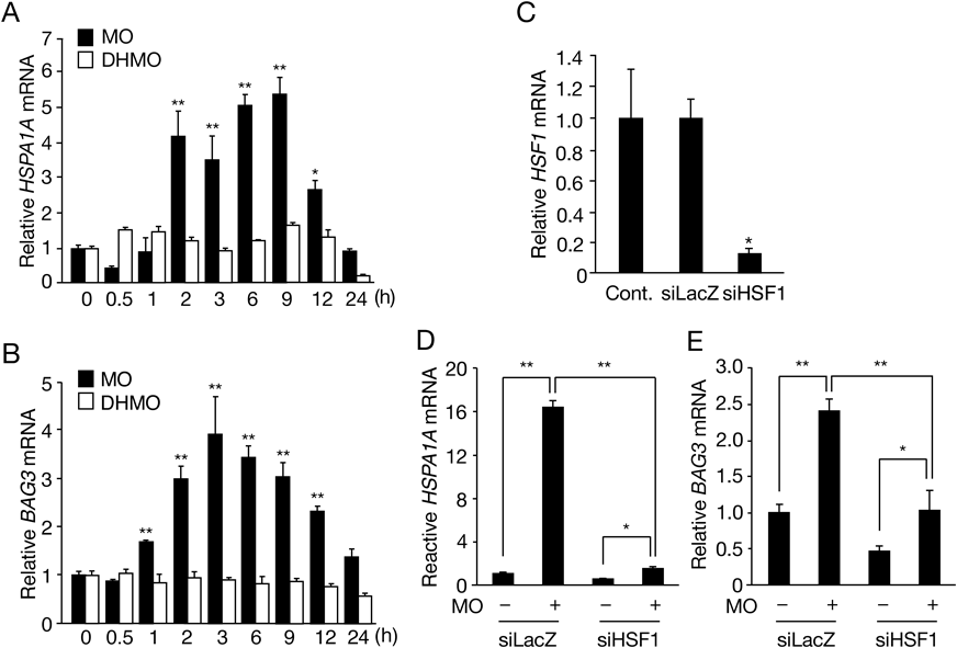
(A, B) HepG2 cells were exposed to 10 µM MO or 10 µM DHMO for 0 to 24 h. The total RNA was converted to cDNA for quantitative PCR (qPCR) analysis of (A) HSPA1A and (B) BAG3. Each value is the mean ± standard deviation (S.D.) of three or six determinations. * p < 0.01, ** p < 0.001 vs. 0 h. (C) Cells were transfected with either siRNA for β-galactosidase (siLacZ) or HSF1 (siHSF1) for 48 h or not transfected (Cont.), then qPCR analysis of HSF1 was performed. Each value is the mean ± S.D. of three determinations. * p < 0.05 vs. Cont. (D, E) HepG2 cells were transfected with siLacZ and siHSF1 for 48 h, then treated with or without 30 µM MO for 6 h. Expression levels of (D) HSPA1A and (E) BAG3 were analyzed using qPCR. Each value is the mean ± S.D. of three determinations. * p < 0.05, ** p < 0.001.
This work was supported in part by Grants-in-Aid (#22K15657 to K.M., #20K12180 to Y.A., and#18H05293 to Y.K.) for Scientific Research from the Ministry of Education, Culture, Sports, Science and Technology of Japan.
The authors declare no conflict of interest.

