1. INTRODUCTION: HEALTH FOOD IN JAPAN AND CURRENT PREVENTION OF CARDIAC DISEASE THROUGH NATURAL FOOD INGREDIENTS
In Japan, health consciousness has been steadily increasing, with the market for health foods reaching approximately 900 billion yen in fiscal year 2023.1) This trend will continue as companies intensify their product development, leading to further market growth. A key area of expansion is the food with functional claims category, which was introduced in 2015 to replace food for specified health uses and food with nutrient function claims. Functional claims regulations aim to address consumer health needs by providing comprehensive information to support health-conscious purchasing decisions.
As the demand for innovative health claims has increased, the variety of ingredients used in health foods has continuously increased, with food and pharmaceutical companies actively exploring and developing food-derived ingredients. Many ingredients with antioxidant properties, such as vitamins and flavonoids, have been extensively studied. Flavonoids are a class of naturally occurring compounds that have minimal adverse effects on humans when administered at moderate doses.2,3) Representative flavonoids include quercetin,4) a flavonol found in onions and apples, catechin, epicatechin, and epigallocatechin gallate,5) a flavanol present in green tea and cacao; anthocyanidins6) found in blueberries and grapes; daidzein,7) an isoflavone derived from soybeans; nobiletin and sudachitin,8) flavones found in citrus fruits such as shikuwasa and sudachi.
Antioxidants support immune function by neutralizing reactive oxygen species, which impair immune cell activity. Antioxidants can enhance immunity by activating immune cells, potentially preventing various lifestyle-related diseases. Additionally, they are associated with benefits, such as fatigue recovery, skin health, and antiaging effects. As societies age, lifestyle-related diseases increase in developed countries.
Heart failure, known as the “heart failure pandemic,” is a global issue affecting countries such as Japan.9,10) The increasing number of older patients with heart failure requiring hospitalization puts pressure on healthcare facilities. Therefore, the prevention of heart failure in daily life is especially important. Dietary recommendations in combination with pharmacotherapy have recently become crucial components in the treatment and management of heart failure.11) Chocolate is a popular food that has gained global recognition. Dark chocolate is composed of catechins and epicatechins, which are classified as flavanols and are found in cacao. Dark chocolate consumption significantly reduces N-terminal pro-B-type (brain) natriuretic peptide, a biomarker of heart failure pathology, with a possible preventive effect on heart failure in humans.12) Endothelium-dependent peripheral vasodilation via nitric oxide mediated by catechin and epicatechin vascular endothelial cells has been considered a possible mechanism of action.
In an animal model of phenylephrine-induced cardiac hypertrophy, nobiletin, a polymethoxyflavone (PMF), prevented both impaired myocardial contractile function and left ventricular hypertrophy.13) Although its mechanism of action has been investigated using proteomic analysis to identify the nobiletin-binding protein and molecular biological methods to examine its function, the ion channels, receptors, and enzymes involved in basic myocardial function remain unclear.
As described above, evidence supporting the efficacy of antioxidants and their related benefits has been growing with findings from both in vivo animal models and human clinical trials. However, the pharmacological and toxicological effects of dietary supplements cannot be fully explained by their antioxidant properties alone. For a more detailed analysis of the cardiovascular mechanisms of action of natural products, their direct effects on the cardiovascular system need to be ascertained. One promising approach is the use of isolated myocardium. It enables the measurement of chronotropic, inotropic and lusitropic effects, which are directly related to myocardial function. Electrophysiological measurements and ion imaging would provide insight into their mechanism of action. In this review, Chapter 2 delineates the basic mechanical response of the myocardium, which encompasses the chronotropic, inotropic, and lusitropic effects. Subsequent chapters delve into PMFs, such as sudachitin, demethoxysudachitin, and nobiletin (Chapter 3); quercetin, a flavonol skeleton (Chapter 4); and hesperidin, a flavanone skeleton (Chapter 5). Each chapter comprehensively examines the direct effects of these components on the myocardium. Chapter 6 provides a comprehensive conclusion and future prospects.
2. MEASUREMENT OF CHRONOTROPIC, INOTROPIC, AND LUSITROPIC EFFECTS WITH ISOLATED MYOCARDIAL PREPARATIONS
Application of agents to the isolated myocardium can be used to evaluate the acute effect on cellular targets such as ion channels, transporters and enzymes. The right atria contain the sinus node, the orthotopic pacemaker. The chronotropic effect of agents can be evaluated through measurement of the spontaneous beating rate, which reflects the firing rate of action potentials in the sinus node region.14–20) The sinus node action potential contains a rapid depolarization phase formed by the L-type Ca2+ current and a repolarization phase formed by the delayed rectifier potassium current (IK). This is preceded by a diastolic depolarization phase, triggers the rapid upstroke and is thus the determinant of the firing rate. The diastolic depolarization is formed by multiple ionic mechanisms including the deactivation of the delayed rectifier IK, the L type and T type Ca2+ currents (ICaL and ICaT), the hyperpolarization activated cyclic nucleotide activated cation current (If), the Ca2+ activated chloride channel current (IClCa), the Na+–Ca2+ exchanger (NCX) current. Thus, agents that affect these ionic mechanisms may show chronotropy. Agents that accelerate or decelerate the diastolic depolarization shows positive or negative chronotropy, respectively. Noradrenaline, the sympathetic neurotransmitter, induces positive chronotropy through an increase in the current density of multiple membrane currents including ICaL, If, and IK. This is mimicked by β-adrenergic agonists and phosphodiesterase inhibitors, agents which increase intracellular cAMP. A diastolic depolarization may arise at sites other than the sinus node and result in emergence of ectopic pacemakers. For example, the pulmonary vein myocardium is the origin of extra stimulus causing atrial fibrillation.21–23) The diastolic depolarization causing such ectopic activity may be formed by ionic different from that of the sinus node. This means that the ectopic focus may be selectively inhibited with pharmacological agents.24–29)
The inotropic effect of agents of on the working myocardium can be evaluated by contractile force measurements in atrial and ventricular myocardial tissue preparations driven at a constant frequency by external electrical stimulation. The Ca2+ that enters the cell during the action potential plateau stimulates Ca2+ release from the sarcoplasmic reticulum through the ryanodine receptor/Ca2+ release channel; a process known as the Ca2+-induced Ca2+ release (CICR) mechanism. The simultaneous rise in throughout the cytoplasm, which is called the Ca2+ transient, causes myocardial contraction through activation of the contractile proteins. The balance between transsarcolemmal Ca2+ entry and CICR as a source of Ca2+ varies among animal species, developmental stage of the animal.30–35) In the adult mammalian ventricular myocardium, CICR accounts for the most part of the Ca2+ forming the Ca2+ transient. Agents which affect the Ca2+ transient cause inotropy. Noradrenaline increases Ca2+ influx through Ca2+ channels, increases CICR and causes positive inotropy. Such effect is mimicked by β-adrenoceptor agonists and phosphodiesterase inhibitors, which increase cAMP. Cardiac glycosides, such as digoxin and ouabain, cause positive inotropy through a different mechanism. Cardiac glycosides inhibit the sarcolemmal Na+–K+ ATPase causing an increase in subsarcolemmal Na+ concentration. This in turn causes a decrease in Ca2+ efflux through the Na+–Ca2+ exchanger leading to an increase in the amount of Ca2+ handled by the sarcoplasmic reticulum (SR) and positive inotropy. The positive inotropy caused by cardiac glycosides differs from that caused by agents acting through an increased in intracellular cAMP, the latter but not former, is accompanied by positive chronotropy.
Contractile force measurements also enable evaluation of the lusitropic effects of agents, which reflects changes in the ability of the myocardium to relax; the shortening of the time required from peak tension to basal tension is considered to reflect relaxing activity.35–38) The two major mechanisms for decrease in cytoplasmic Ca2+ concentration on a Ca2+ transient is sequestration of Ca2+ into the SR through the sarcoendoplasmic Ca2+ ATPase (SERCA) and transsarcolemmal Ca2+ efflux through the NCX; the relative contribution of these two mechanisms differs among various myocardia. In the case of the mammalian ventricular muscle, the time course of myocardial relaxation is determined by SERCA. The relaxation is accelerated (positive lusitropy) by activators of SERCA, and is decelerated by inhibitors of SERCA (negative lusitropy). NCX, despite its limited role in the removal of cytoplasmic Ca2+ after each Ca2+ transient, affects the amount of Ca2+ handled by the SR in the long term and thus may be involved in inotropy rather than lusitropy. The activity of SERCA is regulated by phospholamban, a small protein present on the SR membrane which, in its unphosphorylated form, inhibits SERCA activity; the phosphorylated form of phospholamban lacks such inhibitory effect. Noradrenaline, which causes phosphorylation of phospholamban through activation of A kinase causes an increase in SERCA activity and, thus, positive lusitropy. Such an effect is mimicked by β-agonists and phosphodiesterase inhibitors, but not by cardiac glycosides, which shows inotropy that is not mediated by an increase in cAMP (Fig. 1). Another mechanism for positive lusitropy is the decrease in the Ca2+ sensitivity of the contractile proteins which can be induced by cAMP-dependent phosphorylation of troponin I.39,40) The contributions of SERCA activation and phosphorylation of troponin I appears to vary depending on the experimental conditions.
Thus, studies of the chronotropic, inotropic, and lusitropic effects of agents with isolated myocardial preparations will describe their mode of action on the myocardium and provide information concerning their mechanism of action.
3. PMFs
3.1. Positive Chronotropic and Inotropic Effects of Sudachitin, Demethoxysudachitin, and NobiletinCitrus peels are rich in PMFs,41) which have various pharmacological properties including anti-dementia,42) anti-inflammatory,43) anticancer,44) hepatoprotective,45) and lipolysis-promoting effects.46) Consequently, the interest in citrus fruits with high PMF content for the development of health food products has increased.
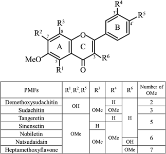
Sudachi (Citrus sudachi Hort. ex Shirai), a traditional Japanese citrus fruit widely consumed in Tokushima Prefecture, contains PMFs in its peels, the most abundant being sudachitin (Fig. 2), followed by demethoxysudachitin47) (Fig. 2). Both PMFs are unique to C. sudachi, as they are not found in other citrus species. A clinical study has indicated that sudachitin-containing sudachi peel extract powder can help reduce visceral fat in individuals at risk of developing diabetes, highlighting its potential to prevent lifestyle-related diseases.8)
Shikuwasa (Citrus depressa Hayata), a citrus species native to Japan and Taiwan, is particularly popular in the Okinawa Prefecture, Japan. Its peel is rich in PMFs, with nobiletin being the most abundant, followed by tangeretin48) (Fig. 2). Nobiletin exhibits various pharmacological effects, including promoting mitochondrial function through upregulation of the SIRT1/PGC-1α signaling pathway, a mechanism associated with longevity,49) making it a functional ingredient in health food.
However, limited research has been conducted on the direct cardiac effects of PMFs such as sudachitin and nobiletin. Hence, we investigated the chronotropic and inotropic effects of sudachitin, demethoxysudachitin, and nobiletin in rat atrial preparation.50) Our findings showed that sudachitin at 0.3–30 µM concentrations induced a concentration-dependent positive chronotropic effect in spontaneously beating right atrial preparations, with a maximum efficacy of approximately 40%, lower than the maximum efficacy (100%) elicited by 1 µM isoproterenol, a β-adrenoceptor full agonist. Similarly, sudachitin at 0.3–30 µM concentrations induced a concentration-dependent positive inotropic effect in electrically stimulated left atrial preparations, with a maximum efficacy of approximately 20%, which is also lower than the maximum efficacy elicited by isoproterenol. Propranolol (0.1 µM), a β-receptor antagonist did not affect the positive chronotropic and inotropic effects of sudachitin. The concentration–response curves for the chronotropic and inotropic effects of dibutyryl-cAMP (1–100 µM) were shifted to the left upon pretreatment with sudachitin (3–10 µM). The investigation revealed that phosphodiesterase (PDE) inhibitors (3-isobutyl-1-methylxanthine (IBMX) at 1 µM or milrinone at 10 µM) exhibited positive chronotropic and inotropic effects when administered alone. Similarly, sudachitin (10–30 µM) demonstrated positive chronotropic and inotropic effects when administered alone. However, a notable finding was the absence of any interaction between the PDE inhibitors and sudachitin, suggesting an additive effect between these two substances.
The inhibition of PDE subtypes by sudachitin has been reported using various human recombinant proteins.51) Sudachitin demonstrated the potency to inhibit PDE3A, PDE5A, PDE8A1, and PDE10A2, with IC50 values of 5.19, 6.54, 26.3, and 9.99 µM, respectively. The IC50 value of sudachitin for PDE3A, which is expressed in the myocardium and involved in the regulation of its contraction, is approximately 5 µM, which is within the concentration range (0.3–30 µM) at which the sudachitin-induced positive chronotropic effects.50)
The binding of sudachitin to PDE has been reported through computer simulations, as follows.51) An analysis of PDE3A and PDE3B revealed that sudachitin shares binding sites with selective PDE3 inhibitors, such as milrinone and amrinone, as well as non-selective PDE inhibitors, including IBMX. Notably, sudachitin demonstrated lower binding energies than milrinone and amrinone, suggesting its potential for stronger interactions with PDE3 in the simulations.
Thus, the mechanism of action of sudachitin for chronotropy and inotropy has been linked to the enhancement of cAMP-dependent pathways via inhibition of PDE3, without β-adrenoceptor involvement.50) Moreover, the mechanism of action of sudachitin in the myocardium is similar to that in glucose metabolism, i.e., an increase in cAMP-dependent pathways via PDE inhibition by sudachitin regulates insulin secretory capacity.51,52)
Similarly, both demethoxysudachitin and nobiletin at 0.3–30 µM concentrations induced concentration-dependent positive chronotropic effects, with a maximum efficacy of approximately 30%, which is also lower than that induced by isoproterenol. Additionally, both demethoxysudachitin and nobiletin produced concentration-dependent positive inotropic effects in left atrial preparations, with maximum efficacies of approximately 10 and 40%, respectively. Although demethoxysudachitin and nobiletin had similar positive chronotropic and inotropic effects to those of sudachitin, their maximum efficacy was lower than that of isoproterenol. The similar effects observed with demethoxysudachitin, nobiletin, and sudachitin indicate that these cardiac effects are not specific to the three PMFs but are common to PMFs.
The mechanism of action of demethoxysudachitin, which has a chemical structure similar to sudachitin (Fig. 2), in the myocardium is unknown. Thus, determining whether demethoxysudachitin and sudachitin affect cAMP-dependent pathways via PDE inhibition would be interesting.
Although numerous studies have reported on the bioactivity of nobiletin, no reports on the mechanisms underlying its chronotropic and inotropic effects exist. Among the PDE subtypes expressed in the myocardium, PDE3 and PDE4 are predominant in mediating the cardiac pacemaker and excitation–contractile coupling functions, and their activation leads to the degradation of cAMP.53,54) Therefore, if nobiletin inhibits PDE3 and PDE4, the amount of cAMP in the myocardium would increase. Nobiletin inhibits PDE3A, PDE4A1A, and PDE4B1, and the IC50 values of nobiletin for these subtypes were 16.0, 5.23, and 6.08 µM, respectively.55) The IC50 value of nobiletin for PDE3 and 4 were within the concentration range (0.3–30 µM) in which nobiletin induced positive chronotropic effects.50) Based on the above reports, the relationship between the direct mechanical action of nobiletin on the myocardium and PDE inhibition should be elucidated in future studies.
3.2. Positive Chronotropic and Inotropic Effects of Other PMFs and PolyphenolsReports on the direct myocardial effects of PMFs, other than ours, are scarce. One report is from Itoigawa et al. who investigated the inotropic effects of 3,5,6,7,8,3′,4′-heptamethoxyflavone (HMF) and natsudaidain using guinea-pig papillary muscle56,57) (Fig. 2). Both compounds (1–300 µM) produced concentration-dependent positive inotropic effects, with pD2 values of 4.98 and 4.33, respectively, indicating that natsudaidain was more potent than HMF. However, the maximum positive inotropic effect of HMF was 2.5 times greater than that of natsudaidain. The mechanism underlying HMF’s positive inotropic effect involves the endogenous release of catecholamines from cardiac tissue. Meanwhile, the mechanism of action of natsudaidain is unclear.
Although we focused on the myocardial effects of PMFs among polyphenols, positive inotropic effects have also been reported for other polyphenol compounds, such as naringin (a flavanone)58) and resveratrol (a stilbene),59) which have gained global attention as health food ingredients. In addition, the positive inotropic effects of these ingredients involve enhancement of the β-adrenoceptor/protein kinase A-dependent mechanism via inhibition of PDE activity.58,59) Thus, PMFs are not the only polyphenol-derived food ingredient with direct cardiac effects.
3.3. Structure–Activity Relationships of PMFsAlthough many researchers are interested not only in the pharmacological activity of PMFs, but also in the structure–activity relationships that arise depending on the number and position of the functional groups, reports are limited to their anticancer effects.60,61)
In a study investigating the effects of many flavonoids, including five citrus PMFs, on the proliferative potential of various cancer cells, natsudaidain had the strongest antiproliferative effect, followed by tangeretin, nobiletin, and HMF.60) By contrast, sinensetin, without a methoxy group at the 8-position of the A ring (R3, Fig. 2), showed no effect. Thus, PMFs with a methoxy group at the 8-position of the A ring have strong physiological activity. In a report on the anti-proliferative potential of the promyelocytic leukemia HL60 cell line, natsudaidain with six methoxy groups and one hydroxyl group showed the strongest effect, followed by tangeretin (containing five methoxy groups), nobiletin (six methoxy groups), and HMF (seven methoxy groups).61) These results suggest that the anticancer effect of PMF is not only dependent on the number of methoxy groups, but also that the position and combination of arrangements is an important factor for its activity.
Methoxy groups are functional groups that increase hydrophobicity. In general, increased hydrophobicity facilitates passage through the lipid bilayers of cell membranes and improves the cell permeability of compounds. This may increase the overall binding to target molecules, resulting in increased activity. Natsume et al. have reported that structural correlation analysis was performed on the absorbability of 73 types of PMFs using Caco-2 cells.62) PMFs with methoxy groups at positions 5 and 7 of the A-ring (R1 and R2, Fig. 2), at positions 3′ and 4′ of the B-ring (R4 and R5, Fig. 2), and at positions 3 of the C-ring (R6, Fig. 2) improved absorption in vitro.62)
We have previously reported that sudachitin (containing three methoxy groups) exhibits higher relaxant potency (EC50: 16.9 µM) than demethoxysudachitin (two methoxy groups, EC50: 29.9 µM) on phenylephrine-induced contractions of rat aorta with endothelium.63) Kaneda et al. have reported that nobiletin, with six methoxy groups, exhibited significant vasorelaxation on phenylephrine-induced contractions of rat aorta with endothelium, with an EC50 of 3.6 µM.64) These results indicate that the potency of PMF to induce relaxation of vascular smooth muscle may depend on the number of methoxy groups. However, the relationship between the vasorelaxant effect and the number of methoxy groups, as well as the combination of their arrangement, needs to be compared not only with the results of the PMFs described above but also with the effects of PMFs with different numbers of methoxy groups, such as tangeretin,65) sinensetin,66) and HMF, under the same experimental conditions.
Regarding the modulation of cardiac function by PMFs, we reported sudachitin, demethoxysudachitin, and nobiletin in rat left atria, and Itoigawa et al. have reported natsudaidain and HMF on the right ventricular papillary muscle of guinea pigs.57) It may not be appropriate to compare the results across different systems because the electrophysiological, Ca2+ handling and pharmacological properties of the myocardium varies among different chambers, animal species and the developmental stage.31,32,67–70) In the future, the structure–activity relationships of chronotropic and inotropic effects should be considered by comparing the potency of many PMF in the same test system. Such research on the myocardium, combined with previous findings, may help determine which PMFs are safer or more effective in improving the disease status.
3.4. Balance between Cardiac Stimulation and Vasodilation by PMFsUsing sudachitin as an example, we propose that its primary cardiocirculatory targets are vascular smooth muscles, with secondary effects on the heart50) (Fig. 3). Our previous research has demonstrated that sudachitin exhibits endothelium-independent vasorelaxation, with an EC50 value of 15.0 µM for relaxation of phenylephrine-induced contraction in the rat aorta (without endothelium), with the maximum relaxation reaching approximately 100%, indicating complete relaxation.63) When applied at 10 µM, a concentration close to its EC50 value for vasorelaxation, sudachitin demonstrated a positive chronotropic effect of approximately 20% and a positive inotropic effect of less than 10%. Thus, although sudachitin has some stimulatory effects on the heart, such effects are relatively mild at vasodilatory concentrations, unlike β-adrenoceptor full agonists, such as isoproterenol, which strongly stimulates cardiac function. This mild cardiostimulatory effect of sudachitin may help increase cardiac output through a moderate increase in heart rate, enhanced blood flow, and improved peripheral circulation. Therefore, C. sudachi intake may help prevent lifestyle-related diseases by promoting overall cardiovascular health.
PMFs in general have positive chronotropic and inotropic effects on the heart through multiple mechanisms including PDE inhibition50,51,55,58,59) and release of endogenous catecholamines.56) In the blood vessels, PMFs show endothelium-independent dilatory action through both cAMP- and cGMP-mediated mechanisms.63,64) This overall profile of PMFs appear to resemble that of PDE inhibitors. Positive inotropes with PDE inhibitory activity, such as milrinone71) and pimobendan,72,73) improved the hemodynamic profile but did not improve the survival rate; thus, these drugs are not recommended for chronic heart failure in Western countries. The positive inotropic response caused by PDE inhibition is milder than that by β-adrenoceptor agonists and is effective even in the failing heart in which the β-adrenoceptor-mediated inotropy is weakened. There are cases in which PDE inhibitors were applied under β-adrenoceptor blockade.74,75) It is recently proposed that, in the evaluation of positive inotropes for the treatment of heart failure, the improvement in functional status and the QOL, rather than survival rate alone, should be emphasized.10,76) PMFs, with their mild cardiostimulatory and vasodilatory effects mentioned above, together with their well-known antioxidant effects, may be of potential as medical treatments and/or dietary supplements, and further research progress is anticipated.
4. QUERCETIN
Quercetin is a flavonoid abundantly contained in onions and apples known for its anti-oxidant, anti-virus and anti-cancer activities.77–79) Long-term application of quercetin was reported to exert beneficial effects on the cardiovascular system including antihypertension and cardioprotection. On the other hand, information concerning the acute effects of quercetin on the myocardium is limited. We examined the acute effects of quercetin on isolated myocardial tissue from the mouse, which is known to be highly dependent on sarcoplasmic reticulum function and has been used for the studies of myocardial contraction and relaxation.31,67,80)
The beating rate of the right atria was slightly increased by quercetin; the increase after treatment with 10 and 30 µM quercetin was about 5 and 15%, respectively (unpublished data); this positive chronotropic effect was affected neither by prazosin nor by propranolol, which rules out the involvement of adrenoceptor stimulation. A positive chronotropic effect of quercetin was also observed in the Langendorff perfused mouse heart81) and in the supine mouse under pentobarbital anesthesia.82) Quercetin was reported to inhibit the type 3 phosphodiesterase activity with IC50 values from 5 to 30 µM,83–85) and constitutive phosphodiesterase activity has been detected in the mouse atrium.86) Thus, the mild positive chronotropic effect of quercetin may be caused by its phosphodiesterase-inhibiting activity.
In the mouse ventricular myocardium, quercetin, at 10 and 30 µM, decreased the contractile force by about 10 and 25%, respectively36) (Fig. 1). On the contrary, quercetin increased the shortening and Ca2+ transient amplitude in isolated mouse cardiomyocytes.87) Stimulation of α-adrenoceptors in the mouse ventricular myocardium is known to result in negative inotropy,88) but the negative inotropy by quercetin was inhibited neither by prazosin nor by propranolol, ruling out the involvement of adrenoceptors. Both augmentation and inhibition of myocardial L-type Ca2+ channel current density has been reported for quercetin; at 30 µM, the current amplitude was increased by 25%81) or decreased by less than 10%.82) Quercetin glycoside was reported to decrease the sensitivity of contractile proteins to Ca2+.89) These factors, as well as inhibitory effects on the phosphodiesterase, probably affects the overall inotropic effect of quercetin.
Quercetin showed a positive lusitropic effect in the mouse ventricular myocardium; the time for relaxation was decreased by about 3.5 ms and 5.5 ms, by 10 and 30 µM quercetin, respectively36) (Fig. 1). This positive lusitropy was blocked neither by prazosin nor by propranolol. The SERCA inhibitor cyclopiazonic acid markedly prolonged the time required for relaxation. Under this condition, the lusitropic effect of quercetin was completely blocked.36) On the other hand, the NCX inhibitor SEA0400 affected neither the time for relaxation nor the lusitropic effect of quercetin.36) These results indicated that quercetin produces a positive lusitropic effect by activation of SERCA through mechanisms unrelated to β-adrenoceptors. In cardiac sarcoplasmic reticulum membrane preparations, quercetin, as well as structurally related compounds, stimulated the Ca2+-dependent ATPase activity90); the mechanism of action appeared to be release of inhibition by phospholamban. Thus, it is probable that quercetin acts at the interface between phospholamban and SERCA. However, as quercetin has phosphodiesterase inhibitory activity, the possibility that quercetin releases the inhibition by phospholamban through enhancement of the cAMP-dependent protein kinase activity cannot be ruled out at present.
A major risk factor for the diastolic dysfunction of the heart is diabetes mellitus.91) In the diabetic mouse myocardium, diastolic dysfunction is observed regardless of the presence or absence of contractile dysfunction and is accompanied with defects in the intracellular Ca2+ handling mechanisms.37,40,92) In the ventricular myocardium of the streptozotoin-induced diabetic mouse, the contractile force was decreased, while the time for relaxation was prolonged, compared with that from the normal mouse.36,37) In the isolated ventricular cardiomyocytes from the diabetic mouse, the Ca2+ transient amplitude was decreased, while the time for the decline in Ca2+ concentration was prolonged, compared with those from normal mouse, indicating abnormalities in Ca2+ handling. The prolongation of the time required for relaxation by cyclopiazonic acid was larger in the normal mouse compared with the diabetic mouse, which implies that the diabetic mouse myocardium is in a state of diastolic dysfunction due to a decrease in SERCA activity. Quercetin, at 10 and 30 µM accelerated relaxation both in the normal and diabetic myocardium; the effect was larger in the diabetic myocardium, which implies that quercetin may be of therapeutic benefit in the treatment of myocardial diastolic dysfunction.
Quercetin had a positive lusitropic effect with mild positive chronotropic and negative inotropic effects in the normal and diabetic myocardium. Although its mechanism of action is not clarified yet, the pharmacological profile of quercetin was different from that of β-agonists and phosphodiesterase inhibitors which have both strong positive chronotropic and inotropic effects. It rather resembled ellagic acid and gingerol, the two compounds of natural origin which showed positive lusitropy both in normal and diabetic myocardium.37) The lack of strong positive chronotropy and inotropy implies that quercetin would not increase myocardial oxygen demand, which may be an advantage in the pharmacotherapy of heart failure.
In isolated vascular tissue preparations, quercetin showed vasodilative effects. It caused vasodilation when applied alone and also enhanced the action of nitrovasodilators. The endothelium-dependent action of quercetin in the mouse aorta involved the promotion of endothelial nitric oxide synthase (eNOS)-mediated NO production.93) The vasorelaxant effect of quercetin on the porcine coronary artery resembled that of UK-114,542, a selective inhibitor of the cGMP selective phosphodiesterase 5, although the precise mechanism of action of quercetin was not specified.94) Recent progress in the function of phosphodiesterase isoforms have demonstrated the broad therapeutic potential of phosphodiesterase inhibitors.95,96) The phosphodiesterase-5A inhibitor vardenafil was reported to prevent the development of HFpEF in rats with type 2 diabetes.97) Further investigation of quercetin’s intracellular site of action including phosphodiesterase isoforms would reveal its therapeutic potential for various cardiovascular disorders.
Plant-derived agents such as digitalis and forskolin have been used in the treatment of heart failure; their positive inotropic potency has been considered useful for the treatment of systolic dysfunction with decreased left ventricular ejection fraction (LVEF). On the other hand, the number of patients suffering from diastolic dysfunction is recently increasing, and is attracting attention as heart failure with preserved ejection fraction (HFpEF).92,98) SGLT2 inhibitors and the mineralocorticoid receptor antagonist finerenone are recommended and used in the pharmacological treatment for diastolic dysfunction10); these agents are expected to provide protective effects by inhibiting fibrosis in the heart and kidneys.99,100) Drugs which directly improve diastolic dysfunction through enhancement of myocardial relaxation are not yet developed and are highly anticipated.101,102) SERCA activity is the major mechanism for myocardial relaxation30) and is reported to be decreased in various types of heart failure.103,104) Thus, agents like quercetin, which is characterized by its lusitropic effects, may provide an interesting compound for future drug development.
5. HESPERETIN
Flavonoids such as hesperidin and its aglycone hesperetin, found in citrus fruits like oranges and grapefruits, have been reported to possess various pharmacological effects, including antioxidant,105,106) anti-inflammatory,106) antimicrobial,106) and antitumor107) activities. Epidemiological studies have shown that long-term intake of hesperidin/hesperetin has a protective effect against cardiovascular diseases such as coronary artery disease and ischemic stroke.108) It is generally thought that the antioxidant effects of hesperidin/hesperetin, as well as related flavonoids, cause various beneficial effects in the long term. Regarding acute effects, hesperetin has been reported to block multiple ion channels involved in myocardial excitation including the Na+ channel current and the delayed rectifier K+ current.109,110) However, information on the direct effects of hesperidin/hesperetin on myocardial excitation and contraction is scarce.
We examined the acute effects of hesperetin in isolated myocardial tissue preparations from the guinea pig111) (Fig. 4). Hesperetin did not affect the spontaneous beating rate of the right atria at concentrations up to 30 µM and showed only a slight decrease in beating rate at 100 µM. In the ventricular myocardium, hesperetin had no effect on the contractile force at concentrations up to 30 µM and showed a slight increase at 100 µM. Hesperetin had no effect on the time course of relaxation at concentrations up to 100 µM. Thus, hesperetin had no chronotropic, inotropic or lusitropic effects at concentrations up to 30 µM. Moreover, hesperetin at 30 µM was reported to have no effect on the action potential duration in canine ventricular myocytes.110)
The pulmonary vein myocardium is recognized as a major origin of ectopic activity responsible for the majority of atrial fibrillation.23) The pulmonary vein myocardium contains various ion channels involved in the generation of the diastolic depolarization and can spontaneously generate action potentials that are accelerated by several neurohumoral factors.24,25,112–114) Recent studies have demonstrated that both the initiation and maintenance of atrial fibrillation are induced by persistent triggers, including those from the pulmonary vein myocardium.115) We performed microelectrode measurement of action potentials to examine whether hesperetin exerts any influence on this potential ectopic activity111) (Fig. 4). About a half of the isolated pulmonary vein preparations from the guinea pig showed spontaneous firing of action potentials. Hesperetin at concentrations of 10–100 µM significantly decreased the slope of the diastolic depolarization and reduced the frequency of spontaneous action potentials. This effect was dependent on the concentration of hesperetin, EC50 values being 41.6 µM for slope and 56.2 µM for firing frequency. Hesperetin suppressed spontaneous activity by reducing the slope of the diastolic depolarization while minimally altering the overall waveform of the action potential. Therefore, the primary effect of hesperetin in pulmonary vein myocardium was the inhibition of ion channels involved in the diastolic depolarization that functions specifically in pulmonary vein myocardium. An ionic mechanism that meets these conditions include the persistent component of the voltage dependent Na+ channel current.
The rapidly activating and rapidly inactivating component of the voltage dependent Na+ channel current (Peak INa) is responsible for the rapid upstroke of the action potential and the conduction of excitation through the working myocardium. Class I antiarrhythmics block peak INa and thereby exerts antiarrhythmic effects through inhibition of the propagation of arrhythmogenic excitation and prevention of re-entrant excitation.116) Following the peak INa, a persistent component of the voltage dependent Na+ channel current flows, which is referred to as the persistent Na+ current, the sustained Na+ current, or the late Na+ current (late INa). The late INa can be induced either by a sustained depolarization or by a slow depolarization in the voltage range overlapping with that of the diastolic depolarization of the pulmonary vein myocardium.117) Some of the conventional class I antiarrhythmic drugs have inhibitory effects on late INa, but to what extent it contributes to the overall effect of the drugs is not clear.118) On the other hand, some of the recently developed blockers of the Na+ channel such as GS-458967 and NCC-3902 have a high selectivity towards late INa. We applied these blockers to isolated pulmonary vein preparation and found that they reduce the slope of the diastolic depolarization and inhibit the spontaneous firing of action potentials.29,119) At concentrations to inhibit the automaticity of the pulmonary vein myocardium, these blockers had minimum effects on the spontaneous beating of the right atria and the contractile force of the ventricular myocardium. Thus, the effects of hesperetin and the late INa blockers were very similar, suggesting that they share common mechanisms of action. In fact, hesperetin blocked the slowly inactivating component of the human NaV1.5 Na channel current expressed in HEK293 cells.109) This provides direct evidence that hesperetin inhibits the automaticity of the pulmonary vein myocardium through blockade of late INa.
Class I antiarrhythmic drugs such as pilsicainide are commonly used for the treatment of atrial fibrillation, but its effectiveness is not always satisfactory.116) This may be because they are not designed to affect the pulmonary vein myocardium, the origin of ectopic electrical activity triggering atrial fibrillation. Further, class I antiarrhythmic drugs have cardio-suppression as a side effect which limit their use in patients with a risk of decreased cardiac output.19,120,121) Blockers with selectivity for late INa appears to have ideal properties as an antiarrhythmic agent for atrial fibrillation; it has selective inhibitory effects on pulmonary vein automaticity and has minimum effects on myocardial conduction and contraction. NCC-3902, a highly selective blocker of late INa, was reported to terminate experimental atrial fibrillation in a canine rapid pacing atrial model.122) Hesperetin, which inhibits the automaticity of the pulmonary vein myocardium through blockade of late INa may be promising as an antiarrhythmic drug. It also teaches us that some naturally derived compounds might have such an excellent pharmacological profile as antiarrhythmic drugs.