Experimental
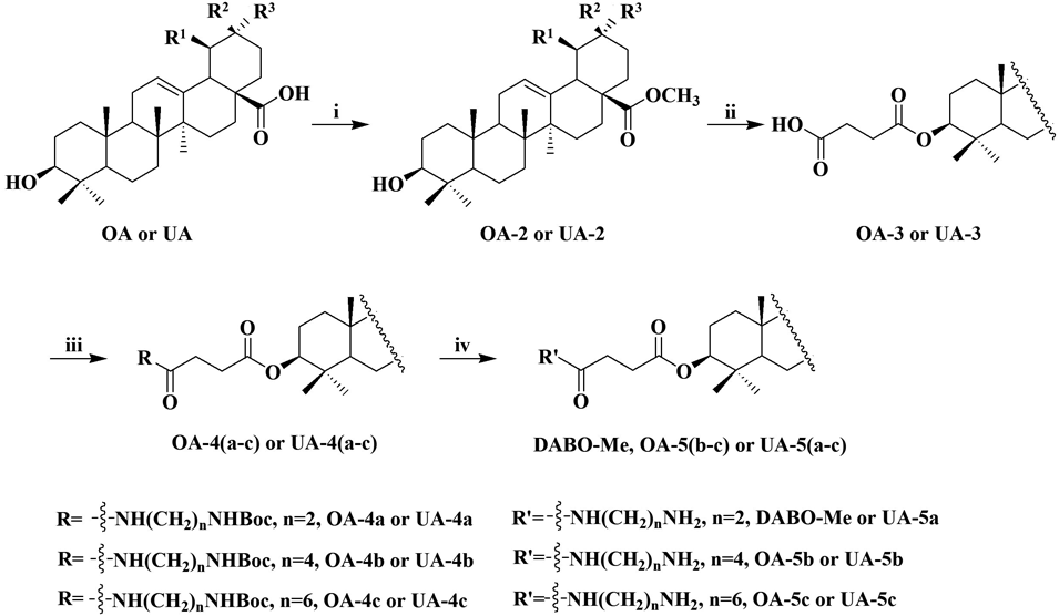
GeneralTarget compounds were purified on a silica gel column chromatography (200–300 mesh, Qingdao Marine Chemical Factory, China) using petroleum ether/ethyl acetate or methylene chloride/anhydrous methanol as eluent. Their structures were confirmed using NMR on a AVANCE 500 MHz spectrometer (BRUKER, Switzerland) in CDCl3. Chemical shifts are expressed in ppm and the coupling constants (J) in Hz. IR were recorded in KBr pellets on a WGH-30 (Shanghai Yonggui analysis instrument Co., Ltd.). Melting points were determined on an X-5 fiber melting point detector (temperature-controlled, Beijing Tektronix Instrument Co., Ltd.). Mass spectra were recorded on a GC-TOF high-resolution mass spectrometer (HR-MS) and an Agilent 6540 RRLC/Q-TOF MS (Agilent, Santa Clara, CA, U.S.A.). Most chemicals and solvents were purchased from commercial sources. Further purification and drying by standard methods were employed when necessary. All the reagents and chemicals were of analytical grade or chemically pure. The synthesis routes are presented in Chart 1.
ChemistrySynthesis of OA and UA Derivatives OA and UA derivatives (compounds OA-2, UA-2, OA-3 and UA-3, structures shown in Chart 1) were synthesized as previously described.6)
Synthesis and Characterization of OA DerivativesMethyl 3β-O-[4-(2-N-tert-Butoxycarbonyl (Boc)-aminoethylamino)-4-oxo-butyryl]olean-12-ene-28-oate (OA-4a)To a stirred solution of OA-3 (300.0 mg, 0.53 mmol), N-Boc-ethylenediamine (95.1 mg, 0.63 mmol) and dimethylaminopyridine (DMAP) (72.6 mg, 0.63 mmol) in anhydrous methylene chloride (10 mL) was added 1-ethyl-(3-(3-dimethylamino)propyl)-carbodiimide hydrochloride (EDCI) (379.5 mg, 2.10 mmol) which dissolved in anhydrous methylene chloride (10 mL) at 0°C. The reaction mixture was stirred for 5 min, and then at room temperature for 3 h. The reaction mixture was extracted with methylene chloride and water, washing to neutral with saturated NaCl solution. The organic layer was dried over anhydrous magnesium sulfate. Filtration and evaporation of solvent at reduced pressure gave light yellow solid, which was purified by silica gel chromatography with a gradient elution of methylene chloride–methanol (20 : 1, v/v) to yield a white solid (365.8 mg, 97.5%), mp 111.9–112.5°C; IR (KBr) νmax 3393, 2949, 2861, 1721, 1375, 1248, 1173 cm−1; 1H-NMR (500 MHz, CDCl3) δ: 6.39 (s, 1H, –CONH–), 5.29 (s, 1H, H-12), 5.05 (s, 1H, –NH-Boc), 4.50–4.46 (m, 1H, H-3), 3.60 (s, 3H, –COOCH3), 3.31–3.22 (m, 4H, –NHCH2CH2NH–), 2.83 (d, J = 13.3 Hz, 1H, H-18), 2.63–2.62 & 2.45–2.44 (m, 4H, –COCH2CH2CO–), 1.41 (s, 9H, –C(CH3)3), 0.90, 0.87, 0.82, 0.78, 0.74, 0.70 (each s, 3H); 13C-NMR (125 MHz, CDCl3) δ: 178.26, 172.68, 172.14, 156.71, 143.79, 122.25, 81.37, 79.63, 55.33, 51.48, 47.55, 46.71, 45.85, 41.64, 41.30, 40.58, 40.46, 40.37, 39.29, 38.10, 37.74, 36.92, 33.85, 33.07, 32.59, 32.37, 31.15, 30.66, 29.99, 28.37, 28.04, 27.68, 25.88, 23.62, 23.50, 23.39, 23.21, 18.19, 16.82, 16.69, 15.32. HR-MS Calcd for C42H68N2O7 [M + H]+ 713.5105, Found 713.5100.
Methyl 3β-O-[4-(4-N-Boc-aminobutylamino)-4-oxo-butyryl]olean-12-ene-28-oate (OA-4b)The OA-3 (462.7 mg, 0.75 mmol) was connected with N-Boc-1,4-diaminobutane hydrochloride (201.6 mg, 0.90 mmol) according to the same procedure used in the preparation of OA-4a, to give OA-4b (576.8 mg, 96.0%) as a white solid: mp 101.2–101.9°C; IR (KBr) νmax 3388, 2945, 2864, 1730, 1376, 1249, 1174 cm−1; 1H-NMR (500 MHz, CDCl3) δ: 6.01 (s, 1H, –CONH–), 5.26 (s, 1H, H-12), 4.64 (s, 1H, –NH-Boc), 4.49–4.46 (m, 1H, H-3), 3.60 (s, 3H, –COOCH3), 3.24–3.10 (m, 4H, –NHCH2CH2CH2CH2NH–), 2.83 (d, J = 12.4 Hz, 1H, H-18), 2.64–2.63 & 2.44–2.43 (m, 4H, –COCH2CH2CO–), 1.42 (s, 9H, –C(CH3)3), 1.11, 0.91, 0.88, 0.83, 0.70 (each s, 3H); 13C-NMR (125 MHz, CDCl3) δ: 178.25, 172.82, 171.56, 156.12, 143.79, 122.25, 81.37, 79.24, 55.33, 51.48, 47.55, 46.72, 45.86, 41.64, 41.30, 39.29, 39.20, 38.09, 37.75, 36.93, 33.85, 33.07, 32.59, 32.37, 31.23, 30.66, 30.07, 28.41, 28.06, 27.68, 27.53, 26.66, 25.88, 23.62, 23.52, 23.39, 23.06, 18.20, 16.82, 16.70, 15.32. HR-MS Calcd for C44H72N2O7 [M + H]+ 741.5418, Found 741.5418.
Methyl 3β-O-[4-(6-N-Boc-aminohexylamino)-4-oxo-butyryl]olean-12-ene-28-oate (OA-4c)The OA-3 (500 mg, 0.88 mmol) was connected with N-Boc-hexanediamine hydrochloride (227.2 mg, 1.05 mmol) according to the same procedure used in the preparation of OA-4a, to give OA-4c (579.8 mg, 86.1%) as a white solid: mp 89.8–90.5°C; IR (KBr) νmax 3413, 2946, 2869, 1731, 1377, 1251, 1175 cm−1; 1H-NMR (500 MHz, CDCl3) δ: 5.79 (s, 1H, –CONH–), 5.27 (t, J = 3.5 Hz, 1H, H-12), 4.51–4.48 (m, 2H, H-3 × 1 & –NH-Boc × 1), 3.62 (s, 3H, –COOCH3), 3.23–3.21 & 3.20–3.09 (s, 4H, –NHCH2CH2CH2CH2CH2CH2NH–), 2.84 (d, J = 13.9 Hz, 1H, H-18), 2.67–2.64 & 2.47–2.44 (m, 4H, –COCH2CH2CO–), 1.44 (s, 9H, –C(CH3)3), 1.12, 0.92, 0.88, 0.85, 0.84, 0.72 (each s, 3H); 13C-NMR (125 MHz, CDCl3) δ: 178.25, 172.82, 171.43, 156.09, 143.79, 122.25, 81.35, 55.34, 51.48, 47.55, 46.71, 45.86, 41.64, 41.30, 39.29, 38.10, 37.74, 36.92, 33.85, 33.07, 32.60, 32.37, 31.28, 30.66, 30.30, 30.12, 29.94, 29.41, 28.42, 28.06, 27.68, 26.49, 26.24, 26.12, 25.88, 23.82, 23.62, 23.51, 23.39, 23.06, 18.19, 16.82, 16.70, 15.32. HR-MS Calcd for C46H76N2O7 [M + H]+ 769.5731, Found 769.5727.
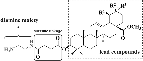
Methyl 3β-O-[4-(2-Aminoethylamino)-4-oxo-butyryl]olean-12-ene-28-oate (DABO-Me)To a stirred solution of OA-4a (200 mg, 0.28 mmol) in anhydrous methylene chloride (5 mL) was added trifluoroacetic acid (TFA) (0.62 mL, 8.42 mmol) at 0°C. The reaction mixture was stirred for 2 h at room temperature. After the reaction, with 0.1 mol L−1 NH3⋅H2O adjust pH to 9 under the ice water bath. The reaction mixture was extracted with methylene chloride and water. The organic layer was dried over anhydrous magnesium sulfate. Filtration and evaporation of solvent at reduced pressure gave light yellow solid, which was purified by silica gel chromatography with a gradient elution of methylene chloride–methanol (10 : 1 v/v) to yield a white solid (158.6 mg, 92.2%), mp 110.5–111.2°C; IR (KBr) νmax 3431, 2925, 2862, 1727, 1385, 1145 cm−1; 1H-NMR (500 MHz, CDCl3) δ: 6.87 (s, 1H, –CONH–), 5.29 (s, 1H, H-12), 4.50 (s, 1H, H-3), 3.64 (s, 3H, –COOCH3), 3.39–2.94 (m, 4H, –NHCH2CH2NH2), 2.73–2.40 (m, 6H, –COCH2CH2CO– ×4 & –NH2 × 2), 2.16 (d, J = 11.3 Hz, 1H, H-18), 1.14, 0.97, 0.89, 0.84, 0.83, 0.75, 0.72 (each s, 3H); 13C-NMR (125 MHz, CDCl3) δ: 177.33, 138.23, 138.09, 127.97, 127.87, 81.52, 79.00, 55.36, 51.51, 50,76, 48.51, 44.35, 41.41, 41.14, 38.87, 38.52, 37.83, 37.24, 36.87, 35.72, 34.94, 33.09, 32.63, 32.10, 29.92, 28.08, 27.37, 27.10, 25.18, 24.15, 23.63, 21.64, 21.07, 18.33, 17.34, 16.65, 16.35, 15.49. HR-MS Calcd for C37H60N2O5 [M + H]+ 613.4580, Found 613.4580.
Methyl 3β-O-[4-(4-Aminobutylamino)-4-oxo-butyryl]olean-12-ene-28-oate (OA-5b)The OA-4b (401.32 mg, 0.55 mmol) was reacted with TFA (1.23 mL, 16.61 mmol) according to the same procedure used in the preparation of DABO-Me, to give OA-5b (260.6 mg, 75.1%) as a white solid: mp 89.8–90.5°C; IR (KBr) νmax 3419, 2951, 2862, 1736, 1388, 1186 cm−1; 1H-NMR (500 MHz, CDCl3) δ: 6.92 (s, 1H, –CONH–), 5.23–5.03 (m, 3H, H-12 × 1 & –NH2 × 2), 4.44 (d, J = 4.9 Hz, 1H, H-3), 3.61 (s, 3H, –COOCH3), 3.17–2.66 (m, 4H, –NHCH2CH2CH2CH2NH2), 2.57–2.36 (m, 4H, –COCH2CH2CO–), 2.13 (d, J = 12.3 Hz, 1H, H-18), 1.11, 1.08, 0.88, 0.83, 0.80, 0.68 (each s, 3H); 13C-NMR (125 MHz, CDCl3) δ: 178.21, 172.88, 172.11, 143.76, 127.90, 122.19, 81.34, 55.29, 51.46, 48.46, 47.51, 46.67, 45.82, 44.30, 41.59, 41.26, 39.25, 38.78, 38.06, 37.70, 36.89, 33.82, 33.07, 32.56, 32.34, 30.63, 29.99, 28.02, 27.65, 26.34, 25.88, 24.13, 23.60, 23.37, 23.03, 18.17, 16.79, 16.68, 15.30. HR-MS Calcd for C39H64N2O5 [M + H]+ 641.4893, Found 641.4899.
Methyl 3β-O-[4-(6-Aminohexylamino)-4-oxo-butyryl]olean-12-ene-28-oate (OA-5c)The OA-4c (369.0 mg, 0.55 mmol) was reacted with TFA (1.64 mL, 22.1 mmol) according to the same procedure used in the preparation of DABO-Me, to give OA-5c (279.2 mg, 87.0%) as a white solid: mp 141.2–141.6°C; IR (KBr) νmax 3422, 2936, 2860, 1722, 1376, 1247, 1183 cm−1; 1H-NMR (500 MHz, CDCl3) δ: 6.42 (s, 1H, –CONH–), 5.27 (s, 1H, H-12), 4.49–4.46 (m, 1H, H-3), 3.61 (s, 3H, –COOCH3), 3.21 (d, J = 5.8 Hz, 2H, –NH2), 3.01–2.84 (m, 5H, –NHCH2 CH2CH2CH2CH2CH2NH2 × 4 & H–18 × 1), 2.65–2.62 & 2.50–2.47 (m, 4H, -COCH2CH2CO–), 1.25, 1.06, 0.92, 0.87, 0.83, 0.72 (each s, 3H); 13C-NMR (125 MHz, CDCl3) δ: 178.24, 172.86, 171.57, 143.79, 138.10, 127.93, 122.23, 81.37, 55.33, 51.47, 50.65, 48.49, 47.55, 46.70, 45.85, 44.33, 41.63, 41.39, 39.32, 37.73, 37.23, 36.91, 33.07, 32.61, 31.15, 30.65, 30.08, 29.65, 29.26, 28.05, 27.67, 26.32, 25.88, 24.13, 23.61, 23.05, 18.19, 17.32, 16.81, 16.38, 15.3. HR-MS Calcd for C41H68N2O5 [M + H]+ 669.5206, Found 669.5205.
Synthesis and Characterization of UA DerivativesMethyl 3β-O-[4-(2-N-Boc-aminoethylamino)-4-oxo-butyryl]urs-12-ene-28-oate (UA-4a)The UA-3 (582 mg, 1.02 mmol) was connected with N-Boc-ethylenediamine (196 mg, 1.22 mmol) according to the same procedure used in the preparation of OA-4a, to give UA-4a (659.8 mg, 82.5%) as a white solid: mp 87.3–87.9°C; IR (KBr) νmax 3374, 2931, 2868, 1729, 1375, 1239, 1176 cm−1; 1H-NMR (500 MHz, CDCl3) δ: 6.24 (s, 1H, –CONH–), 5.23 (t, J = 3.6 Hz1H, H-12), 4.94 (s, 1H, –NH-Boc), 4.52–4.49 (m, 1H, H-3), 3.60 (s, 3H, –COOCH3), 3.35–3.24 (m, 4H, –NHCH2CH2NH–), 2.69–2.64 & 2.47–2.44 (m, 4H, –COCH2CH2CO–), 2.22 (d, J = 11.3 Hz, 1H, H-18), 1.44 (s, 9H, –C(CH3)3), 1.07, 0.94, 0.93, 0.86, 0.85, 0.84, 0.74 (each s, 3H); 13C-NMR (125 MHz, CDCl3) δ: 178.03, 172.69, 172.16, 156.72, 138.19, 125.45, 81.40, 79.68, 55.33, 52.66, 51.41, 48.09, 47.49, 41.59, 40.63, 40.35, 39.51, 39.04, 38.87, 38.28, 37.75, 36.87, 36.63, 32.89, 31.17, 30.64, 30.00, 28.37, 28.08, 28.01, 24.22, 23.56, 23.29, 21.15, 18.19, 17.03, 16.90, 16.74, 15.46. HR-MS Calcd for C42H68N2O7 [M + H]+ 713.5105, Found 713.5101.
Methyl 3β-O-[4-(4-N-Boc-aminobutylamino)-4-oxo-butyryl]-urs-12-ene-28-oate (UA-4b)The UA-3 (600 mg, 1.05 mmol) was connected with N-Boc-1,4-diaminobutane hydrochloride (283.5 mg, 1.26 mmol) according to the same procedure used in the preparation of OA-4a, to give UA-4b (699.8 mg, 89.8%) as a white solid: mp 82.1–82.9°C; IR (KBr) νmax 3403, 2935, 2872, 1711, 1376, 1239, 1175 cm−1; 1H-NMR (500 MHz, CDCl3) δ: 6.23–6.18 (m, 1H, –CONH–), 5.17 (s, 1H, H-12), 4.77–4.70 (m, 1H, –NH-Boc), 4.43–4.42 (m, 1H, H-3), 3.54 (s, 3H, –COOCH3), 3.18–3.05 (s, 4H, –NHCH2CH2 CH2CH2NH–), 2.82 (d, J = 13.3 Hz, 1H, H-18), 2.68–2.42 (m, 4H, –COCH2CH2CO–), 1.36 (s, 9H, –C(CH3)3), 1.11, 1.10, 0.88, 0.79, 0.78, 0.68, 0.67 (each s, 3H); 13C-NMR (125 MHz, CDCl3) δ: 177.21, 172.72, 171.53, 155.95, 138.12, 125.37, 81.21, 78.96, 55.26, 53.41, 52.82, 51.34, 48.01, 47.41, 41.92, 39.44, 38.96, 38.81, 38.21, 37.67, 36.80, 36.56, 32.82, 31.06, 29.99, 29.59, 28.92, 28.38, 28.10, 27.94, 27.41, 27.37, 24.93, 24.15, 23.52, 23.48, 23.23, 22.98, 21.10, 18.13, 17.13, 16.99, 16.70, 15.40. HR-MS Calcd for C44H72N2O7 [M + H]+ 741.5418, Found 741.5420.
Methyl 3β-O-[4-(6-N-Boc-aminohexylamino)-4-oxo-butyryl]urs-12-ene-28-oate (UA-4c)The UA-3 (600 mg, 1.05 mmol) was connected with N-Boc-hexanediamine hydrochloride (318.6 mg, 1.26 mmol) according to the same procedure used in the preparation of OA-4a, to give UA-4c (774.9 mg, 95.9%) as a white solid: mp 71.3–71.9°C; IR (KBr) νmax 3387, 2933, 2865, 1720, 1373, 1244, 1168 cm−1; 1H-NMR (500 MHz, CDCl3) δ: 5.90(s, 1H, –CONH–), 5.22 (s, 1H, H-12), 4.58 (s, 1H, –NH-Boc), 4.48 (t, J = 7.5 Hz, 1H, H-3), 3.58 (s, 3H, –COOCH3), 3.20–3.07 (m, 4H, –NHCH2CH2CH2CH2CH2CH2NH–), 2.65–2.62 & 2.45–2.43 (m, 4H, –COCH2CH2CO–), 2.21 (d, J = 11.3 Hz, 1H, H-18), 1.41 (s, 9H, –C(CH3)3), 1.05, 0.92, 0.86, 0.83, 0.82, 0.71 (each s, 3H); 13C-NMR (125 MHz, CDCl3) δ: 178.00, 172.78, 171.42, 156.08, 138.17, 125.43, 81.30, 79.01, 55.31, 53.39, 52.86, 51.38, 48.06, 47.47, 41.97, 39.49, 39.27, 39.01, 38.85, 38.26, 37.72, 36.84, 36.61, 32.87, 31.22, 30.62, 30.08, 29.91, 29.40, 28.41, 28.08, 27.99, 26.11, 24.20, 23.55, 23.52, 23.28, 21.14, 18.17, 17.03, 16.88, 16.74, 15.44. HR-MS Calcd for C46H76N2O7 [M + H]+ 769.5731, Found 769.5724.
Methyl 3β-O-[4-(2-Aminoethylamino)-4-oxo-butyryl]urs-12-ene-28-oate (UA-5a)The UA-4a (320 mg, 0.45 mmol) was reacted with TFA (1.33 mL, 17.95 mmol) according to the same procedure used in the preparation of DABO-Me, to give UA-5b (248.6 mg, 90.4%) as a white solid: mp 98.7–99.3°C; IR (KBr) νmax 3416, 2936, 2872, 1727, 1388, 1234, 1186 cm−1; 1H-NMR (500 MHz, CDCl3) δ: 6.41 (s, 1H, –CONH–), 5.25–5.23 (m, 1H, H-12), 4.51–4.48 (m, 1H, H-3), 3.60 (s, 3H, –COOCH3), 3.35–3.30 & 2.88–2.86 (m, 4H, –NHCH2CH2NH2), 2.67–2.47 (m, 4H, –COCH2CH2CO–), 2.34 (s, 2H, –NH2), 2.22 (d, J = 11.3 Hz, 1H, H-18), 1.07, 0.94, 0.93, 0.86, 0.85, 0.74 (each s, 3H); 13C-NMR (125 MHz, CDCl3) δ: 178.64, 173.14, 172.31, 156.79, 138.16, 125.58, 80.93, 79.05, 55.24, 52.90, 51.41, 48.10, 47.58, 42.01, 39.51, 39.06, 38.88, 38.75, 38.63, 36.98, 36.64, 32.99, 30.66, 29.67, 28.13, 28.04, 27.24, 24.24, 23.60, 23.30, 21.16, 18.32, 17.01, 16.91, 15.60, 15.42. HR-MS Calcd for C37H60N2O5 [M + H]+ 613.4580, Found 613.4576.
Methyl 3β-O-[4-(4-Aminobutylamino)-4-oxo-butyryl]urs-12-ene-28-oate (UA-5b)The UA-4b (500 mg, 0.68 mmol) was reacted with TFA (1.5 mL, 20.24 mmol) according to the same procedure used in the preparation of DABO-Me, to give UA-5b (341.6 mg, 79.0%) as a white solid: mp 115.3–115.9°C; IR (KBr) νmax 3433, 2931, 2867, 1386, 1210 cm−1; 1H-NMR (500 MHz, CDCl3) δ: 7.70 (s, 2H, –NH2), 7.16 (s, 1H, –CONH–), 5.23 (s, 1H, H-12), 4.46 (s, 1H, H-3), 3.60 (s, 3H, –COOCH3), 3.17–2.04 (m, 4H, –NHCH2CH2CH2CH2NH2), 2.60–2.47 (m, 4H, –COCH2CH2CO–), 2.22 (d, J = 13.1 Hz, 1H, H-18), 1.07, 0.94, 0.93, 0.86, 0.84, 0.81, 0.74 (each s, 3H); 13C-NMR (125 MHz, CDCl3) δ: 178.05, 173.57, 155.33, 138.20, 125.44, 81.91, 55.23, 52.88, 51.42, 48.08, 47.44, 41.97, 39.50, 39.04, 38.87, 38.20, 37.65, 36.82, 36.63, 32.87, 31.90, 30.64, 30.01, 29.67, 29.33, 28.03, 27.92, 25.74, 24.21, 23.56, 23.29, 22.66, 21.17, 18.18, 17.06, 16.87, 16.61, 15.42. HR-MS Calcd for C39H64N2O5 [M + H]+ 641.4893, Found 641.4896.
Methyl 3β-O-[4-(6-Aminohexylamino)-4-oxo-butyryl]urs-12-ene-28-oate (UA-5c)The UA-4c (300 mg, 0.39 mmol) was reacted with TFA (1.2 mL, 15.61 mmol) according to the same procedure used in the preparation of DABO-Me, to give UA-5c (232.1 mg, 89.0%) as a white solid: mp 99.1–99.6°C; IR (KBr) νmax 3439, 2925, 2862, 1719, 1208 cm−1; 1H-NMR (500 MHz, CDCl3) δ: 7.81 (s, 2H, –NH2), 6.63 (s, 1H, –CONH–), 5.28 (s, 1H, H-12), 4.48–4.45 (m, 1H, H-3), 3.59 (s, 3H, –COOCH3), 3.16–2.92 (m, 4H, –NHCH2CH2CH2CH2CH2CH2 NH2), 2.61–2.46 (m, 4H, –COCH2CH2CO–), 2.22 (d, J = 11.1 Hz, 1H, H-18), 1.06, 0.93, 0.92, 0.85,0.84, 0.82, 0.73 (each s, 3H); 13C-NMR (125 MHz, CDCl3) δ: 178.03, 173.11, 158.74, 138.20, 125.42, 81.58, 65.80, 55.30, 55.38, 52.88, 51.40, 48.07, 47.47, 41.97, 39.50, 38.86, 38.24, 37.69, 36.84, 36.62, 32.87, 30.63, 30.06, 29.66, 28.68, 28.02, 25.66, 24.21, 23.56, 23.28, 21.15, 18.18, 17.04, 16.88, 16.68, 15.43. HR-MS Calcd for C41H68N2O5 [M + H]+ 669.5206, Found 669.5205.
Biological ActivityReagentsThiazolyl blue tetrazolium bromide (MTT) was purchased from Sigma-Aldrich (St. Louis, MO, U.S.A.). The Annexin V-propidium iodide (PI) staining kit and cell cycle detection kit were purchased from Nanjing KeyGen Biotech. Co., Ltd., China.
Cell CultureHuman breast cancer MCF-7 cells, human cervical carcinoma HeLa cells, human NSCLC A549 cells, and a non-malignant cell line (Chang liver) were purchased from the Cell Bank of the Chinese Academy of Sciences (Shanghai, China). The cells were cultured and maintained in RPMI 1640 medium (Hyclone) supplemented with 10% fetal bovine serum (FBS), penicillin (100 U/mL) and streptomycin (0.1 mg/mL) at 37°C in a humidified atmosphere with 5% CO2.
Cell Viability AssayThe proliferative activities of A549, MCF-7, Hela, and Chang liver cells exposed to different treatments were assessed using the MTT assay. Cells were seeded in 96-well plates at a density of 2 × 10³ cells per well in complete medium and cultured for 24 h, after which media were replaced with RPMI-1640 containing 10% FBS with or without pentacyclic triterpenoid derivatives. Gefitinib and test compounds were dissolved in dimethyl sulfoxide (DMSO) at a final concentration of <0.1% before addition to cell culture assays. Following incubation at 37°C for 48 h, 20 µL MTT (5 mg/mL) was added to each well, and the plates were incubated for 4 h. The reaction was terminated by adding 150 µL DMSO, and the optical density at 492 nm was measured on a microplate reader after 10 min of low-speed shaking. The cell viability after exposure to each test compound was calculated as follows: (OD570 treated cells − OD570 blank control)/(OD570 control − OD570 blank control) × 100%. Dose response curves were plotted for the samples, and the IC50 values were calculated as the concentrations of the test compounds resulting in 50% reduction of absorption compared with the control cells. More than three independent experiments were performed.
Analysis of Apoptosis and Cell Cycle Progression by Flow CytometryApoptosis and cell cycle progression in MCF-7 cells were analyzed after incubating cells with DABO-Me (15 µM) for 48 h on a CytoFLEX flow cytometer (Beckman Coulter, Brea, CA, U.S.A.). For cell cycle analysis, MCF-7 cells were harvested using trypsin in ethylenediaminetetraacetic acid (EDTA), washed twice with phosphate buffered saline (PBS), resuspended with 70% cold ethanol, and incubated at 4°C overnight in the dark. Cells were then treated with RNaseA for 30 min at 37°C and stained with PI at 4°C in the dark for 30 min. Samples were then put on ice for analysis by flow cytometry. For apoptosis analysis, the cells were collected with EDTA-free trypsin and washed twice with cold PBS. Cells were then resuspended in binding buffer and stained with annexin V-fluorescein isothiocyanate (FITC) and propidium iodide for 15 min. The number of annexin V-FITC-positive apoptotic cells was expressed as the percentage of the total number of cells counted.
Western Blotting AnalysisTotal protein was extracted from cells using RIPA buffer (Sigma). After treatment with 15 µM DABO-Me for 48 h, MCF-7 cells were washed with ice-cold PBS solution and lysed in lysis buffer at 0°C for 20 min. Cell lysates were scraped and centrifuged at 14000 rpm for 30 min at 4°C, and the supernatant was collected. Equivalent amounts of protein were subjected to sodium dodecyl sulfate-polyacrylamide gel electrophoresis and transferred to polyvinylidene difluoride membranes. After blocking for 1 h in 5% skim milk in Tris-buffered saline, the membrane was incubated with the indicated primary antibodies (1 : 2000 dilution) overnight at 4°C, followed by treatment with horseradish peroxidase-coupled secondary antibody (1 : 5000 dilution) for 1 h at room temperature. Immunoreactive proteins were detected using an enhanced chemiluminescent substrate kit (Thermo Scientific, MA, U.S.A) and visualized using the ChemiDoc MP imaging system (Bio-Rad, Hercules, CA, U.S.A.). Antibodies against the following proteins were used for Western blotting: caspase-3, caspase-9, cytochrome-c, Bcl-2, Bax, cyclin D1, CDK4, TIMP-1, matrix metalloproteinase (MMP)-2, MMP-9, and β-actin (all from Proteintech Group, SanYing Biotechnology, Wuhan, China). β-Actin was used as an internal control.
Cell Migration and Invasion AssaysMCF-7 cells were plated at a density of 2 × 10⁵ cells per well in 6-well plates and incubated overnight in complete medium. After 24 h of treatment with 15 µM DABO-Me, cells were digested by trypsinization, and 2 × 10⁴ cells were seeded in 200 µL medium with 1% FBS in the upper chambers of a 24-well Transwell plate containing polycarbonate filters with 8 µm pores (Corning Inc., Corning, NY, U.S.A.). In the lower chambers, 500 µL medium containing 10% FBS was added as the chemoattractant. The cells were incubated for 24 h, the chamber was washed with PBS, and the cells on the upper surface of the insert were wiped away gently with a cotton swab. Cells adhering to the lower surface were fixed with methanol, stained with Giemsa solution, and counted under an inverted microscope in nine random fields of view (40× objective).
The cell invasion assay procedure was similar to that of the migration assay, except that the Transwell membranes were coated with Matrigel (BD Biosciences, San Jose, CA, U.S.A.) at a dilution of 1 : 4 and plates were incubated for 1 h at 37°C. Cells adhering to the lower surface were counted as described for the cell migration assay.
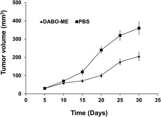
Animal ExperimentsAll animal experiments were performed according to the rules approved by the Animal Experimental Ethics Committee of Dalian Medical University. Female nude mice (BALB/c, 4–6 weeks old, 15–20g) were provided by Dalian Medical University Experimental Animal Center. Mice were bred and housed in laminar-flow cabinets under specific pathogen-free conditions at room temperature. MCF-7 cells (5 × 10⁶ cells in 0.1 mL PBS) at log growth-phase were injected subcutaneously into the athymic nude mice on day 0 to establish a tumor-bearing mouse model. On day 5 after implantation, DABO-Me were injected into the tumor daily with sterile PBS as a negative control. Tumor size was measured every 5 d. On day 30, all tumors of significant size were harvested and weighed to determine the tumor burden. Tumor volume was calculated with the following formula: (length × width2)/2.
Statistical AnalysisData were analyzed using statistical methods and expressed as the mean ± standard deviation (S.D.). Statistical comparisons were made using the Student’s t-test. Differences were considered statistically significant at p ≤ 0.05 (*), p ≤ 0.01 (**) and p ≤ 0.0001 (****). Three independent experiments were performed to confirm data and results.