Chemical and Pharmaceutical Bulletin
Online ISSN : 1347-5223
Print ISSN : 0009-2363
ISSN-L : 0009-2363
Regular Articles
A New Inhibitor of ADAM17 Composed of a Zinc-Binding Dithiol Moiety and a Specificity Pocket-Binding Appendage
Hiroshi TateishiMika TateishiMohamed O RadwanTakuya MasunagaKosuke KawatashiroYasunori ObaMisato OyamaNatsuki Inoue-KitahashiMikako FujitaYoshinari OkamotoMasami Otsuka
著者情報
ジャーナル フリー HTML
電子付録

2021 年 69 巻 11 号 p. 1123-1130

詳細
Abstract

A disintegrin and metalloproteinase 17 (ADAM17) is a zinc-dependent enzyme that catalyzes the cleavage of the extracellular domains of various transmembrane proteins. ADAM17 is regarded as a promising drug target for the suppression of various diseases, including cancer metastasis. We synthesized a new ADAM17 inhibitor, SN-4, composed of a zinc-binding dithiol moiety and an appendage that specifically binds to a pocket of ADAM17. We show that SN-4 inhibits the ability of ADAM17 to cleave tumor necrosis factor α (TNF-α) in vitro. This activity was reduced by the addition of zinc, indicating the importance of the zinc chelating dithiol moiety. Inhibition of TNF-α cleavage by SN-4 in cells was also observed, and with an IC50 of 3.22 µM, SN-4 showed slightly higher activity than the well-studied ADAM17 inhibitor marimastat. Furthermore, SN-4 was shown to inhibit cleavage of CD44 by ADAM17, but not by ADAM10, and to suppress cell invasion. Molecular docking showed good fitting of the specificity pocket-binding group and one SH of SN-4 and hinted at possible means of structural optimization. This study provides clues for the development of potent and selective ADAM17 inhibitors.

Introduction

Despite recent progress in the development of new therapies, cancer remains one of the leading causes of death worldwide.1) Cancer is notoriously difficult to treat once it has metastasized.24) Therefore, the molecular mechanisms behind metastasis have been extensively studied, and the development of anti-metastasis drugs24) has been attempted, albeit with limited success.

A well-studied protein related to metastasis is a disintegrin and metalloproteinase 17 (ADAM17), a member of the ADAM family of metalloproteases, which has been identified as a tumor necrosis factor-α (TNF-α)-converting enzyme.5,6) ADAM17 is a membrane protein and contains a catalytic center with zinc7) in its extracellular domain. It functions by catalyzing the cleavage of the extracellular domains of other transmembrane proteins and is triggered by activation of signaling proteins such as protein kinase C, Rac, and mitogen-activated protein kinase.8,9) Some of its substrates are important for cell adhesion; for example, CD44, which interacts with hyaluronic acid, is cleaved by ADAM17 to induce metastatic spread and invasion of cancer cells.10) Thus, ADAM17 is regarded as a promising drug target for the suppression metastasis. Many inhibitors of ADAM17 have been synthesized,1114) most of which comprise hydroxamic acid, which is a general zinc-binding group, and a moiety that interacts with a pocket in the vicinity of the zinc ion. However, the hydroxamic acid moiety has been shown to cause side effects in vivo,15) and some ADAM17 inhibitors with hydroxamic acid have failed clinical testing because of this toxicity.11,14) We herein synthesized a new inhibitor of ADAM17, and its activity was evaluated.

Results and Discussion

Design and Synthesis

We previously reported a zinc-binding molecule SN-1 (Fig. 1) containing two SH groups16) and demonstrated that SN-1 and its derivatives bind and regulate the function of various zinc proteins, including human immunodeficiency virus-1 (HIV-1) enhancer binding protein,16) apolipoprotein B mRNA-editing enzyme, catalytic polypeptide-like 3G (APOBEC3G),17,18) farnesyltransferase,19,20) and TNF receptor-associated factor 6 (TRAF6).21,22) Molecular docking indicated that one SH binds to the zinc ion and the other interacts with neighboring amino acids.18,2022) We reasoned that SN-1 would attain ADAM17 selectivity by the introduction of a 4-(but-2-yn-1-yloxy)benzenesulfonyl amide group, which selectively binds to the S1′ pocket of ADAM17,2325) and designed such a derivative, SN-4 (Fig. 1).

Fig. 1. Design of the New ADAM17 Inhibitor. Structures of SN-1, SN-4 and SN-4(Nps)2 Were Shown

Dithiol is easily air-oxidized to disulfide. We previously reported that an oxidized form of SN-1, produced by the introduction of two Nps groups, works as a prodrug and releases SN-1 under reducing conditions inside cells more efficiently than intra- or inter-molecular oxidized forms of SN-1.22) Thus, SN-4(Nps)2 (Fig. 1) was synthesized as a prodrug of SN-4.

The synthesis was carried out as shown in Chart 1. Reductive amination of aldehyde 126) using 2-(tert-butylthio)ethan-1-amine afforded 2 (84%). Secondary amine 2 was treated with 4-(but-2-yn-1-yloxy)benzenesulfonyl chloride27) and Et3N to give acetal 3 (65%), which was converted to aldehyde 4 (80%) under acidic conditions. 2-(tert-Butylthio)ethan-1-amine was condensed with aldehyde 4 followed by reduction with NaCNBH3 to yield 5 (64%). Treatment of 5 with 2-nitrobenzenesulfenyl chloride (Nps-Cl) provided 6, and the Nps group on the amino nitrogen of 6 was selectively removed by 3-methylindole16) to afford the final SN-4(Nps)2 compound (54%).

Chart 1. Synthesis of SN-4(Nps)2

Reagents and conditions: (i) 2-(tert-butylthio)ethylamine hydrochloride, NaCNBH3, MeOH, MS3A, room temperature (r.t.), 2 d, (ii) 4-(2-butynyloxy)benzenesulfonyl chloride, Et3N, THF, 0 °C-r.t., 2 h, (iii) 2 M HCl, THF, 45 °C, 4.5 h, (iv) 2-(tert-butylthio)ethylamine hydrochloride, NaCNBH3, MeOH, MS3A, 50 °C, 2 d, (v) Nps-Cl, AcOH, DMF, 0 °C-r.t., overnight, (vi) 3-methylindole, CH2Cl2 : MeOH = 2 : 3, 0.5 M HCl, r.t., 6 h.

Biological Evaluation

The ADAM17 inhibitory activity of SN-4 was examined. TNF-α is a well-known substrate of ADAM17, and soluble TNF-α (sTNF-α) is released by ADAM17-mediated extracellular domain cleavage. To examine ADAM17 inhibition by SN-4 in vitro, ADAM17, full-length TNF-α (pro-TNF-α), and SN-4 generated from SN-4(Nps)2 and three equivalents16) of dithiothreitol (DTT) were used. Immunoblot analysis showed that pro-TNF-α was cleaved by ADAM17, and this activity was suppressed by the addition of SN-4 (Fig. 2A, lane 1–3). The same experiment in the presence of zinc ions resulted in a decrease in SN-4 activity (Fig. 2A, lane 4), demonstrating the importance of the zinc chelating function of the dithiol moiety. Next, cellular experiments were conducted. Stimulation of differentiated THP-1 cells by lipopolysaccharide (LPS) activates ADAM17 to release sTNF-α.28) Here, THP-1 cells were incubated with SN-4(Nps)2 or marimastat29) (control, Fig. 2B) before the addition of LPS. After incubation, the amount of sTNF-α in the supernatant was evaluated by immunoblotting. As shown in Figs. 2C and D, the increase of sTNF-α by LPS stimulation was inhibited by SN-4 and marimastat in a dose-dependent manner. Using enzyme-linked immunosorbent assay (ELISA), we then determined the IC50 values of SN-4 and marimastat, which were 3.22 and 4.75 µM, respectively (Figs. 2E, F). The IC50 value of marimastat was almost identical to that reported,30) and SN-4 showed slightly higher activity than marimastat. Furthermore, like marimastat, SN-4 showed no toxicity at 0.1–10 µM (Fig. 2G). Together, these results demonstrate the ability of SN-4 to inhibit ADAM17.

Fig. 2. Analysis of the Ability of SN-4 to Reduce TNF-α Cleavage

(A) In vitro inhibition of ADAM17 cleavage of Pro-TNF-α in the presence or absence of zinc ions. Mixtures of recombinant ADAM17 (100 µg/mL), recombinant Pro-TNF-α (100 µg/mL), SN-4(Nps)2 (10 µM), and DTT (30 µM) were incubated in the absence or presence of zinc chloride (20 µM) in Tris–HCl buffer (50 mM, pH 8.0) at 37 °C overnight and analyzed by immunoblotting. (B) Structure of marimastat. (C), (D) Ability to reduce TNF-α release from cells. THP-1 cells differentiated with PMA were incubated in the presence of SN-4(Nps)2 or marimastat (10, 1, or 0.1 µM) for 1 h before the addition of LPS (200 ng/mL). The cells were further incubated for 6 h, and the harvested supernatant was analyzed by immunoblotting (C). The band intensity was quantitated, and the value of TNF-α was normalized to β-actin. The relative values are shown in graph (D). (E), (F) IC50 of SN-4 (E) and marimastat (F) to reduce TNF-α release. The experimental procedure was the same as that shown in (C) except that various concentrations of the compounds were used and TNF-α was quantified by ELISA. (G) Cellular toxicity. Differentiated THP-1 cells with PMA were incubated in the presence of SN-4(Nps)2 or marimastat (10, 1, or 0.1 µM) for 1 d, and cellular viability was determined by MTT assay.

The selectivity of SN-4 was then examined using two highly similar metalloproteinases, ADAM17 and ADAM10.31) Cleavage of the extracellular domain of CD44 is known to be regulated by ADAM10 and ADAM17 in response to distinct stimulation; ionomycin and PMA activate ADAM10 and ADAM17, respectively.32) U251 MG cells were incubated with SN-4(Nps)2 or marimastat followed by addition of ionomycin or PMA. After incubation, CD44EXT, generated by cleavage together with soluble CD44, was evaluated by immunoblotting. In the case of ionomycin treatment, CD44 cleavage was inhibited by marimastat but not by SN-4 (Figs. 3A–C). On the other hand, both compounds inhibited CD44 cleavage by PMA stimulation (Figs. 3D–F). This is consistent with a previous report that showed that marimastat inhibits both ADAM10 and ADAM17.33) In contrast, SN-4 selectively inhibited ADAM17. Furthermore, fluorescent immunostaining confirmed that CD44 was maintained on the membranes of PMA-treated cells after addition of SN-4 or marimastat (Fig. 3G).

Fig. 3. Analysis of the Ability of SN-4 to Reduce CD44 Cleavage and to Suppress Cell Invasion

(A)–(F) Immunoblotting assessment of the reduction of CD44 (CD44 full) cleavage. The U251 MG cells were incubated in the presence of various concentrations of SN-4(Nps)2 or marimastat for 1 h before the addition of ionomycin (5 µg/mL) (A–C) or PMA (500 ng/mL) (D–F). The cells were further incubated for 30 min, and the cell lysate was analyzed by immunoblotting. The band intensity was quantified, and the value of CD44EXT was normalized to β-actin. The relative values are shown in graphs (B, C, E, F). (G) Immunostaining assessment of the reduction of CD44 (CD44 full) cleavage. The same experimental procedure was used as shown in (D) except with 50 µM of the compounds and with immunostaining (antibody BU52) and microscopic observation used to detect the CD44 extracellular domain. To stain nuclei, Hoechst 33342 was used. (H), (I) Ability to suppress cell invasion. U251 MG cells in cell culture inserts with pores were incubated in the presence of 10 µM of the compounds for 24 h. The non-invaded cells (upper surface of membrane) were removed, and invaded cells (lower surface of membrane) were stained with crystal violet (H). The cell numbers were evaluated by MTT assay. Relative cell invasion is shown in the graph (I). (Color figure can be accessed in the online version.)

Since CD44 is important for the adhesion of cells,10) we assessed whether SN-4 inhibits cellular invasion. U252 MG cells were cultured on a Transwell plate in the presence of SN-4 for 24 h, and invaded cells were stained with crystal violet (Fig. 3H) or evaluated with the 3-(4,5-dimethylthial-2-yl)-2,5-diphenyltetrazalium bromide (MTT) assay (Fig. 3I). The results showed that SN-4 clearly suppresses the invasion of cells to the same magnitude as that of marimastat.

Molecular Docking Study

Selective ADAM17 inhibition of SN-4 could be attributed to binding of its 4-(but-2-yn-1-yloxy)benzenesulfonyl amide group to the S1′ pocket of ADAM17.2325) To test this, a molecular docking study of ADAM17-SN-4 was performed. As the template, we employed the X-ray crystal structure (PDB code: 2OI0)24) of ADAM17 co-crystalized with an aryl sulfonamide 4b (Fig. 4A), which has the same ADAM17-binding 4-(but-2-yn-1-yloxy)benzenesulfonyl amide group. In this model, shown in Fig. 4A, the ADAM17 binding moiety of SN-4 can almost be overlaid on the co-crystalized compound 4b, occupying the S1′ specificity pocket, extending through a narrow channel, and reaching the S3′ subsite. This imparts a significant selectivity towards ADAM-17 over other metalloproteinases in which this channel is blocked by a conserved tyrosine.24) As shown in Fig. 4B, the sulfone linker contributes to overall complex stability via hydrogen bonding with Leu348 and Gly349. Furthermore, the SH group of SN-4 strongly coordinates with the active site zinc ion, affording a stable tetrahedron with amino acid residues His405, His409, and His415. Concomitantly, the pyridinium nitrogen is implicated in hydrogen bonding with water molecule HOH578. Of note, the other SH group lies in proximity to hydrophobic amino acid residues (Val314, Lys315, Leu350, and Ala351) in the neighboring subsite, but its interaction with the amino acids could not been observed.

Fig. 4. Molecular Docking Analysis of the Binding Mode of SN-4 to the Catalytic Site of ADAM17

(A) Superimposed SN-4 (green) and the co-crystalized native ligand 4b shown on the right (magenta) placed into the ADAM17 active site (PDB code: 2OI0). Molecular Operating Environment was used for the calculations as previously described.36) (B) 3D binding mode of SN-4 in the ADAM17 active site and neighboring subsite for the analysis conducted in (A). Amino acid residues and water molecule are shown in off-white, and the zinc ion is the cyan sphere. Hetero-atoms are colored by element and intermolecular bonds are represented by the black dashed lines. Amino acids, water molecule, and zinc ion, which directly interact with SN-4 are black-colored, and the others are red-colored. (Color figure can be accessed in the online version.)

Conclusion

In this study, an ADAM17 specificity pocket-binding moiety was introduced into zinc-binding dithiol molecule SN-1 to construct a new compound, SN-4. SN-4 was shown to selectively inhibit ADAM17, cleave the extracellular domains of TNF-α and CD44, and suppress cell invasion. The molecular docking study showed accommodation of the specificity pocket-binding group leading to specific ADAM17 inhibitory activity and interaction of one SH with the zinc ion, while the other SH group was shown to be free. Modification of the dithiol group to optimize fitting to neighboring amino acids could increase the inhibitory activity of SN-4. Recently, we reported compounds that suppress the expression of CD44 and migration of cells.34) Together with these compounds, SN-4 is considered to be a lead compound for the development of anti-metastasis drugs in the near future. Furthermore, since ADAM17 was reported to play an important role in symptoms of coronavirus disease 2019 (COVID-19),14,35) this study may also be useful in the fight against the ongoing pandemic.

Experimental

General Procedure Pertaining to Synthesis

All reagents were commercially available with high purity grade. TLC was performed on precoated plates, TLC sheets silica 60 F254 (Merck, Darmstadt, Germany) or TLC sheets Chromatorex NH silica (Fuji Silysia Chemical, Kasugai, Japan). Chromatography was carried out on Silica Gel 60N (40–100 mesh) (Kanto Chemical, Tokyo, Japan) and NH silica gel Chromatorex (NH, 100–200 mesh) (Fuji Silysia Chemical). NMR spectra were recorded on a JEOL (Tokyo, Japan) JNM-AL300 (300 MHz) and Bruker (Billerica, MA, U.S.A.) Avance 600 (600 MHz). Chemical shifts were referenced to tetramethylsilane (TMS). Mass spectrum (FAB) and high-resolution mass spectra (HRMS) were recorded by a JEOL JMS-DX303. HRMS were recorded by using positive fast atom bombardment (FAB) with 3-nitrobenzyl alcohol (NBA) as the matrix. IR-red spectra were recorded on a JASCO (Tokyo, Japan) FT/IR-410. The samples were prepared as KBr discs or thin films between sodium chloride discs. Melting points were determined on a Yanaco (Kyoto, Japan) melting point apparatus and were uncorrected.

Synthesis of SN-4(Nps)2

2-({[2-(tert-Butylthio)ethyl]amino}methyl)-6-(diethoxymethyl)-N,N-dimethylpyridin-4-amine (2):

Aldehyde 126) (1.6 g, 6.4 mmol) was dissolved in 45 mL of dist. MeOH, molecular sieves 4A and 2-(tert-butylthio)ethan-1-amine (2.7 g, 16 mmol) were added, and the mixture was stirred under an argon atmosphere for 25 h. Then, NaCNBH3 (1.2 g, 19 mmol) was added, and the mixture was stirred for 20 h. The mixture was filtered to remove molecular sieves, 10 mL of purified water was added, and the mixture was stirred for 30 min, and then the solvent was evaporated under reduced pressure. After adding 10 mL of purified water to the residue and extracting with CH2Cl2 (30 mL × 3), the organic layer was dried with MgSO4. The organic layer was concentrated under reduced pressure and purified by column chromatography (CH2Cl2 : MeOH = 20 : 1). 2 (2.0 g, 99%) was obtained as a brown oil. 1H-NMR (CDCl3) δ: 1.24 (6H, t, J = 7.03 Hz, CH3 ×2), 1.32 (9H, s, CH3 ×3), 2.73 (2H, t, J = 6.57, CH2), 2.84 (2H, t, J = 6.03 Hz, CH2), 3.01 (6H, s, CH3 ×2), 3.55–3.75 (4H, m, CH2 ×2), 3.87 (2H, s, CH2), 5.32 (1H, s, CH), 6.53 (1H, s, C5H2N (CH)), 6.71 (1H, s, C5H2N (CH)). 13C-NMR (CDCl3) δ: 15.1, 27.6, 30.9, 40.1, 42.6, 48.7, 63.1, 98.9, 103.7, 104.6, 151.4, 154.4, 157.4. FAB-MS (m/z): 370 (M + H)+.

4-(But-2-yn-1-yloxy)-N-[2-(tert-butylthio)ethyl]-N-{[6-(diethoxymethyl)-4-(dimethylamino)pyridin-2-yl]methyl}benzenesulfonamide (3):

Amine 2 (2.0 g, 5.4 mmol) was dissolved in 18 mL of tetrahydrofuran (THF) and 0.98 mL of Et3N, 4-(but-2-yn-1-yloxy)benzenesulfonyl chloride27) (2.0 g, 8.1 mmol) dissolved in 7 mL of THF was added at 0 °C, and the mixture was stirred at room temperature for 11 h. Then, water was added, the mixture was stirred for 10 min. The solution was concentrated under reduced pressure and extracted with CH2Cl2. The extract was dried over MgSO4 and concentrated under reduced pressure. The residue was purified by color chromatography (hexane : AcOEt = 1 : 1) to afford 3 (2.0 g, 65%) as a yellow liquid. 1H-NMR (CDCl3) δ: 1.13 (15H, m, CH3 ×5), 1.81 (3H, s, CH3), 2.49 (2H, t, J = 7.9 Hz, CH2), 2.93 (6H, s, CH3 ×2), 3.20 (2H, t, J = 7.8 Hz, CH2), 3.54 (4H, q, J = 7.3 Hz, CH2 ×2), 4.30 (2H, s, CH2), 4.64 (2H, s, CH2), 5.17 (1H, s, CH), 6.56 (1H, s, C5H2N (CH)), 6.65 (1H, s, C5H2N (CH)), 6.98 (2H, d, J = 8.6 Hz, C6H4 (CH ×2)), 7.72 (2H, d, J = 8.6 Hz, C6H4 (CH ×2)). 13C-NMR (CDCl3) δ: 3.65, 15.2, 27.4, 31.0, 39.2, 42.5, 49.8, 54.8, 56.6, 62.3, 73.0, 84.7, 102.6, 103.2, 104.8, 115.1, 129.2, 131.5, 155.7, 156.0, 157.9, 161.0. IR (neat) 2973, 2314, 1604, 1500, 1442, 1338, 1230, 1153, 1110, 732, 1002, 917, 836 cm−1. High resolution (HR)MS (FAB) m/z Calcd for C29H44N3O5S2 (M + H)+ 578.2722. Found: 578.2725.

4-(But-2-yn-1-yloxy)-N-[2-(tert-butylthio)ethyl]-N-{[4-(dimethylamino)-6-formylpyridin-2-yl]methyl}benzenesulfonamide (4):

Acetal 3 (0.30 g, 0.52 mmol) was dissolved in 3 mL of THF, 2M HCl was added at 0 °C, and the mixture was stirred at 50 °C for 3 h. The solution was neutralized with Et3N, concentrated under reduced pressure, extracted with CH2Cl2, dried with MgSO4 and concentrated under reduced pressure. The residue was purified by column chromatography (Hexane : AcOEt = 1 : 2) to afford 4 (0.18 g, 67%) as a yellow liquid. 1H-NMR (CDCl3) δ: 1.21 (9H, s, CH3 ×3), 1.88 (3H, s, CH3), 2.57 (2H, t, J = 8.1 Hz, CH2), 3.07 (6H, s, CH3 ×2), 3.33 (2H, t, J = 8.1 Hz, CH2), 4.45 (2H, s, CH2), 4.71 (2H, s, CH2), 6.89 (1H, s, C5H2N (CH)), 7.06 (2H, d, J = 8.8 Hz, C6H4 (CH ×2)), 7.08 (1H, s, C5H2N (CH)), 7.79 (2H, d, J = 8.8 Hz, C6H4 (CH ×2)), 9.85 (1H, s, CHO). 13C-NMR (CDCl3) δ: 3.70, 27.4, 30.98, 39.4, 42.6, 50.1, 54.5, 56.7, 84.8, 104.0, 108.5, 115.2, 129.2, 154.0, 155.5, 161.1, 194.2. IR (KBr) 2962, 2815, 2333, 1712, 1600, 1504, 1438, 1338, 1249, 1153, 998, 921, 840, 721 cm−1. HRMS (FAB) m/z Calcd for C25H33N3O4S2Na (M + Na)+ 526.1810. Found: 526.1830.

4-(But-2-yn-1-yloxy)-N-[2-(tert-butylthio)ethyl]-N-{[6-({[2-(tert-butylthio) ethyl]amino}methyl)-4-(dimethylamino)pyridin-2-yl]methyl}benzenesulfonamide (5):

Aldehyde 4 (1.4 g, 2.8 mmol) was dissolved in 30 mL of dist. MeOH and added molecular sieves 3A, and the mixture was stirred for 20 min. Then 2-(tert-butylthio)ethan-1-amine was added and the mixture was stirred at 50 °C. for 24 h. NaCNBH3 (0.55 g, 8.8 mmol) was added, and the mixture was stirred at room temperature for 24 h. Molecular sieves 3A were removed by filtration, 10 mL of water was added, and the mixture was stirred at room temperature for 30 min. The solution was concentrated under reduced pressure and extracted with CH2Cl2. The extract was dried over MgSO4 and concentrated under reduced pressure. The residue was purified by column chromatography (CH2Cl2 : MeOH = 20 : 1) to afford 5 (1.1 g, 64%) as a brown liquid. 1H-NMR (CDCl3) δ: 1.21 (9H, s, CH3 ×3), 1.32 (9H, s, CH3 ×3), 1.88 (3H, s, CH3), 2.23 (1H, s, NH), 2.57 (2H, t, J = 8.1 Hz, CH2), 2.72 (2H, t, J = 6.6 Hz, CH2), 2.83 (2H, t, J = 6.6 Hz, CH2), 2.99 (6H, s, CH3 ×2), 3.28 (2H, t, J = 8.1 Hz, CH2), 3.74 (2H, s, CH2), 4.35 (2H, s, CH2), 4.71 (2H, s, CH2), 6.46 (1H, s, C5H2N (CH)), 6.54 (1H, s, C5H2N (CH)), 7.05 (2H, d, J = 8.8 Hz, C6H4 (CH ×2)), 7.79 (2H, d, J = 8.8 Hz, C6H4 (CH ×2)). 13C-NMR (CDCl3) δ: 3.72, 27.5, 28.7, 31.0, 39.3, 42.1, 42.5, 49.2, 49.8, 54.7, 54.9, 56.7, 73.4, 84.8, 103.7, 103.9, 115.1,129.2, 131.6, 155.7, 156.2, 161.0. IR (neat) 2958, 2348, 1600, 1500, 1450, 1338, 1226, 1153, 998, 917, 833, 732 cm−1. HRMS (FAB) m/z Calcd for C31H49N4O3S3 (M + H)+ 621.2967. Found: 621.2963.

4-(But-2-yn-1-yloxy)-N-({4-(dimethylamino)-6-[({2-[(2-nitrophenyl)dithio]ethyl}[(2-nitrophenyl)thio]amino)methyl]pyridin-2-yl}methyl)-N-{2-[(2-nitrophenyl) dithio]ethyl}benzenesulfonamide (6):

Compound 5 (41 mg, 0.066 mmol) was dissolved in 0.7 mL of DMF and 1.5 mL of AcOH, 2-nitrobenzenesulfenyl chloride (Nps-Cl) (63 mg, 0.33 mmol) was added at 0 °C, and the mixture was stirred for 5 h. The resulting mixture was neutralized with sat. NaHCO3, and extracted with CH2Cl2. The extract was dried over MgSO4. and concentrated under reduced pressure. The residue was purified by column chromatography (Hexane : AcOEt = 1 : 1) to afforded 6 (45 mg, 70%) as a yellow solid. 1H-NMR (CDCl3) δ: 1.88 (3H, s, CH3), 2.81 (2H, t, J = 7.2 Hz, CH2), 2.92–2.97 (8H, m, CH3 ×2, CH2), 3.33–3.46 (4H, m, CH2 ×2), 4.19 (2H, s, CH2), 4.28 (2H, s, CH2), 4.71 (2H, s, CH2), 6.37 (1H, s, C5H2N (CH)), 6.46 (1H, s, C5H2N (CH)), 7.03 (2H, d, J = 8.9 Hz, C6H4 (CH ×2)), 7.27 (1H, m C6H4 (CH)), 7.30–7.33 (2H, m, C6H4 (CH ×2)), 7.61–7.64 (3H, m, C6H4 (CH)), 7.75 (2H, d, J = 8.9 Hz, C6H4 (CH)), 8.07 (1H, d, J = 12.0 Hz, C6H4 (CH)), 8.14 (1H, d, J = 8.2 Hz, C6H4 (CH)), 8.17 (1H, d, J = 8.2 Hz, C6H4 (CH)), 8.20–8.23 (2H, m, C6H4 (CH ×2)), 8.28 (1H, d, J = 8.2 Hz, C6H4 (CH)). 13C-NMR (CDCl3) δ: 3.7, 36.4, 36.8, 39.2, 47.9, 55.6, 56.8, 60.4, 64.4, 73.1, 76.8, 77.0,77.2, 104.6, 104.7, 114.9, 115.2, 124.9, 125.3, 125.9, 126.0, 126.1, 126.1, 126.2, 127.3, 129.4, 134.1, 137.4, 161.2. IR (KBr) 2341, 1600, 1511, 1446, 1338, 1153, 925, 848, 736 cm−1. HRMS(FAB) m/z Calcd for C41H42N7O9S6 (M + H)+ 968.1368. Found: 968.1352.

4-(But-2-yn-1-yloxy)-N-({4-(dimethylamino)-6-[({2-[(2-nitrophenyl)dithio]ethyl}amino)methyl]pyridin-2-yl}methyl)-N-{2-[(2-nitrophenyl)dithio]ethyl} benzenesulfonamide (SN-4(Nps)2):

Compound 6 (40 mg, 0.041 mmol) was dissolve in a mixture of CH2Cl2 and MeOH (4.5 mL, 2 : 3), 3-methylindole (22 mg, 0.16 mmol) and 0.5 M HCl (0.2 mL) were added at 0 °C, and the mixture was stirred at room temperature for 6 h. The solution was concentrated under reduced pressure, neutralized with sat. NaHCO3, and extracted with CH2Cl2. The extract was concentrated under reduced pressure. The residue was purified by amino silica gel column chromatography (CH2Cl2 : AcOEt = 15 : 1) to afford a yellow solid SN-4(Nps)2 (18 mg, 54%). 1H-NMR (CDCl3) δ: 1.88 (3H, s, CH3), 2.80 (2H, t, CH2), 2.86–2.93 (10H, m, CH3 ×2, CH2 ×2), 3.40 (2H, t, J = 7.7 Hz, CH2), 3.64 (2H, s, CH2), 4.29 (2H, s, CH2), 4.71 (2H, s, CH2), 6.34 (1H, s, C5H2N (CH)), 6.47 (1H, s, C5H2N (CH)), 7.02 (2H, d, J = 8.9 Hz, C6H4 (CH ×2)), 7.33–7.35 (2H, m, C6H4 (CH)), 7.64–7.66 (2H, m, C6H4 (CH ×2)), 7.74 (2H, d, J = 8.9 Hz, C6H4 (CH ×2)), 8.14 (1H, d, J = 9.4 Hz, C6H4 (CH)), 8.25 (2H, d, J = 9.6 Hz, C6H4 (CH ×2)), 8.30 (1H, d, J = 9.4 Hz, C6H4 (CH)). 13C-NMR(CDCl3) δ: 3.72, 39.2, 47.7, 48.0, 54.7, 55.0, 56.8, 73.0, 84.9, 104.0, 115.2, 126.1, 126.2, 127.2, 127.4, 129.4, 130.8, 134.1, 137.3, 137.7, 145.5, 145.7, 155.6, 155.6, 161.2. IR (KBr) 3085, 2337, 1600, 1511, 1446, 1334, 1160, 917, 848, 744 cm−1. HRMS(FAB) m/z Calcd for C35H39N6O7S5 (M + H)+ 815.1484. Found: 815.1501. mp: 105 °C. Purity: 97.1% (See supplementary materials).

Cell Culture, Differentiation, and Stimulation

The human monocytic cell line THP-1 was cultured in RPMI-1640 (FUJIFILM Wako Pure Chemical Corporation, Osaka, Japan) supplemented with 10% heat-inactivated fetal bovine serum (FBS) (Sigma-Aldrich, St. Louis, MO, U.S.A.) and 55 µM 2-mercaptoethanol. The human astrocytoma cell line U251 MG was cultured in Dulbecco’s modified Eagle’s medium (DMEM) (FUJIFILM Wako Pure Chemical Corporation) supplemented with 10% FBS. All media were supplemented with 89 µg/mL streptomycin (Meiji Seika Pharma, Tokyo, Japan), and cells were incubated at 37 °C in a humidified atmosphere of 95% air and 5% CO2. The THP-1 cells were differentiated by incubation for 1 d with phorbol 12-myristate 13-acetate (PMA) (Sigma-Aldrich) at 300 nM. The differentiated THP-1 cells were stimulated with lipopolysaccharide (LPS) (Sigma-Aldrich), and stimulation of the U251 MG cells was performed by ionomycin (LKT Laboratories, Saint Paul, MN, U.S.A.) or PMA (Sigma-Aldrich).
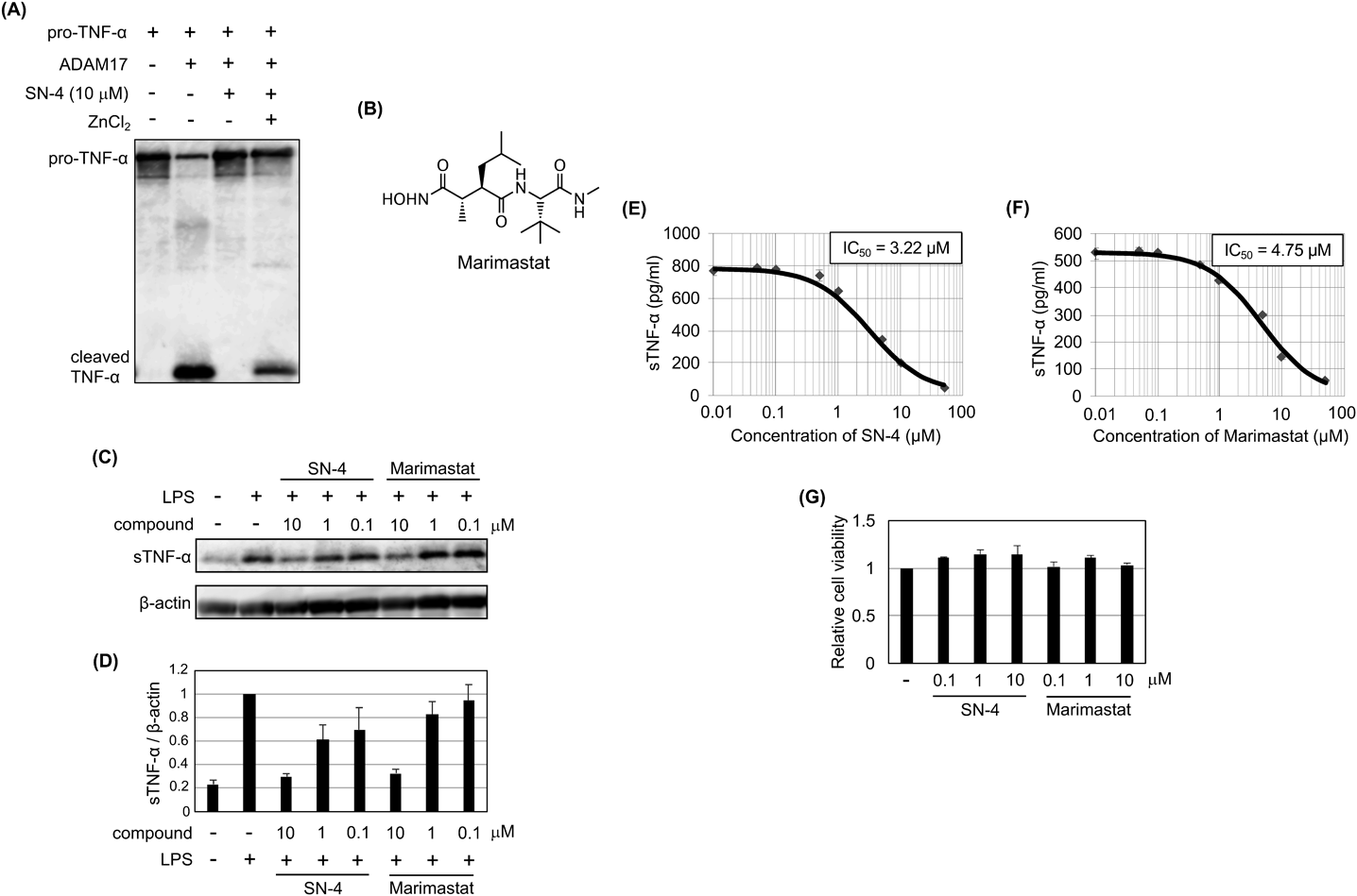
Chemicals

The compound SN-4(Nps)2 was synthesized as described above. A control inhibitor marimastat was purchased from AnaSpec (Fremont, CA, U.S.A.). The SN-4(Nps)2 or marimastat was dissolved in dimethyl sulfoxide (DMSO) (FUJIFILM Wako Pure Chemical Corporation), and the solution was added to culture medium or in vitro reaction solution in a 1/100 volume.

MTT Assay and Analysis of Cell Invasion

Amounts of viable cells were evaluated using MTT (Dojindo Molecular Technologies, Kumamoto, Japan) as previously described.37,38) Cell invasion was evaluated using Matrigel invasion chamber (BD Biosciences, Franklin Lakes, NJ, U.S.A.). U251 MG cells were seeded in upper transwell chamber (1 × 105 cells/well) and incubated for 1 d in presence of each compound. Noninvasive cells in upper surface of the membrane were removed by cotton tipped applicator, and invasive cells in lower surface of the membrane were stained with crystal violet (Nacalai Tesque, Kyoto, Japan) or their amounts were examined by MTT assay.

Immunostaining

U251 MG cells treated with compounds and PMA were fixed with 4% phosphate buffered saline (PBS) solution of paraformaldehyde (Tokyo Chemical, Tokyo, Japan) for 15 min at room temperature, and washed three times with PBS. The cells were incubated with PBS containing 5% normal goat serum (FUJIFILM Wako Pure Chemical Corporation) and 0.3% Triton X-100 (Nacalai Tesque) for 1 h. After removal of PBS, the cells were reacted with anti-CD44 monoclonal antibody BU52 (Ancell, Bayport, MN, U.S.A.) (1 : 400) in PBS containing 1% BSA (Nacalai Tesque) and 0.3% Triton X-100 overnight at 4 °C. The cells were then washed three times with PBS, reacted with goat anti-mouse immunoglobulin G (IgG) (H + L) Cross-Absorbed Secondary Antibody, Alexa Fluor 488 (Thermo Fisher Scientific, Waltham, MA, U.S.A.) at room temperature in the dark for 1 h. After washing three times with PBS, the cells were incubated with Hoechst 33342 (Dojindo Molecular Technologies) solution at RT for 15 min, followed by washing three times with PBS. Microscopic observation was performed using TCS SP5 confocal laser-scanning microscope (Leica, wetzlar, Germany).

Immunoblot Analysis and ELISA

Immunoblot analysis was performed using cells lysed in PBS/Laemmli sample buffer (1 : 1) or cell culture supernatant as described previously.39) As an antibody, Human TNF-alpha Antibody (R&D Systems, Minneapolis, MN, U.S.A.) (1 : 500), anti-CD44 ICD (intracellular domain) Polyclonal antibody (TransGenic, Kobe, Japan) (1 : 500), or anti-β-actin clone AC-15 (Sigma-Aldrich) (1 : 1000) was used. Immunoreactivity was detected by using ImmunoStar LD (FUJIFILM Wako Pure Chemical Corporation). Intensity of bands was quantified by ImageJ (National Institute of health, Bethesda, MD, U.S.A.). ELISA was conducted by using Human TNF-α ELISA kit (Thermo Fisher Scientific).

In Vitro TNF-α Cleavage Assay

Mixture of Recombinant Human TACE/ADAM17 Protein (R&D Systems), SN-4(Nps)2 and 1,4-dithiothreitol (DTT) (FUJIFILM Wako Pure Chemical Corporation) with or without zinc chloride (FUJIFILM Wako Pure Chemical Corporation) in Tris–HCl buffer (50 mM, pH 8.0) was incubated at 37 °C for 30 min. Then Recombinant Pro-TNF-alpha Fusion Protein (R&D Systems) was added. The solution was incubated at 37 °C overnight followed by immunoblot analysis. The final concentrations are as follows. ADAM17, 100 µg/mL; SN-4(Nps)2, 10 µM; DTT, 30 µM; zinc chloride; 20 µM, Pro-TNF-α, 100 µg/mL.
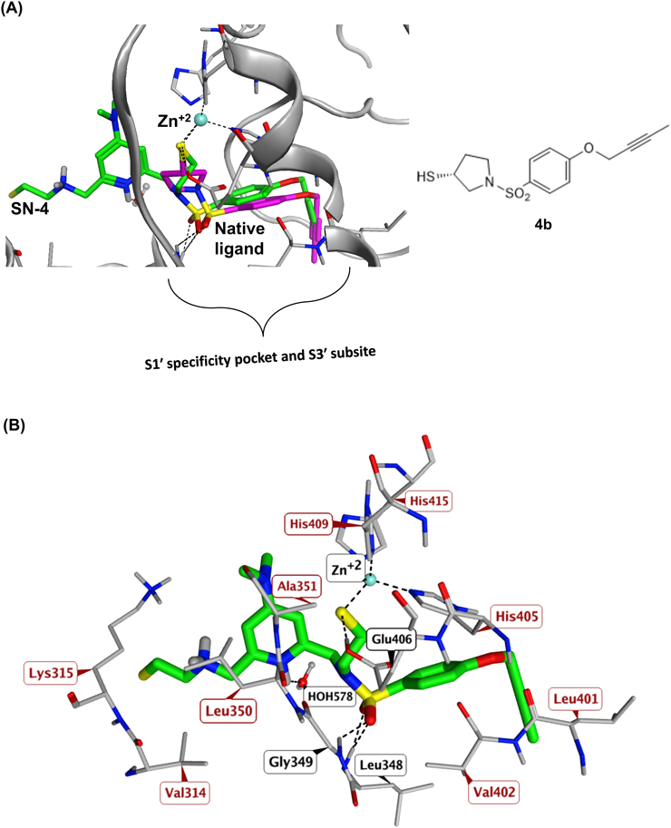
Molecular Docking Analysis

ADAM-17 X-ray crystal structure co-crystalized with an Aryl-sulfonamide ligand (PDB code 2OI0)24) was retrieved from Protein Data Bank to be utilized as a model in the present study. The protein structure was prepared using QuickPrep module of MOE (Version 2019.0) (Chemical Computing Group, Montreal, Canada). Pocket water molecules and zinc ion were conserved. The docking study was conducted using the rigid-receptor method.36,40) The co-crystallized ligand was defined as the center of the binding site. All other options were left at their default values. One hundred docking positions were generated for each ligand. The generated docking positions were visualized using MOE.41)

Acknowledgments

This work was supported by a Grant-in-Aid for Scientific Research (B) from the Japan Society for the Promotion of Science (17890030) (to M. O.) and by Grant-in-Aid for Scientific Research (C) from the Japan Society for the Promotion of Science (20590104, 23590131, and 26460148) (to Y. O.). This work was also supported by a Grant for Joint Research Project with Science Farm Ltd.

Conflict of Interest

The authors declare no conflict of interest.

Supplementary Materials

The online version of this article contains supplementary materials.

References
 
© 2021 The Pharmaceutical Society of Japan
feedback
Top