Results
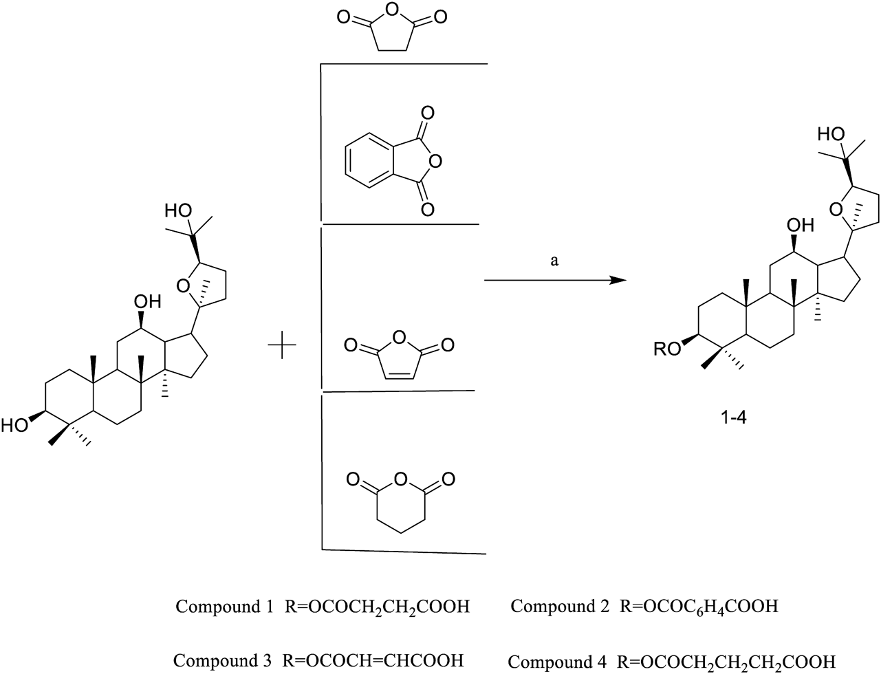
Synthesis of Compounds 1–4Compounds 1–4 were synthesized according to Chart 1. PDQ (Fig. 1) was dissolved in dry dichloromethane solution, and the reactions were initiated with butanedioic anhydride, phthalic anhydride, maleic anhydride and glutaric anhydride at room temperature, in the presence of trimethylamine to obtain the target compounds. The structural characterization of Compounds 1–4 was assessed by MS, 1H-NMR and 13C-NMR.
(20S, 24R)-Dammar-20, 24-Epoxy-12β, 25-Diol-3β-yl HemisuccinateCompound 1: A white crystal in ethyl acetate, Yield: 86%, mp 168.8–174.6 °C. 1H-NMR (500 MHz, Pyr) δ: 4.72 (1H, dd, J = 11.7, 4.8 Hz, H-24), 3.94 (1H, dd, J = 8.7, 6.7, Hz,H-12), 3.68 (1H, dd, J = 10.5, 4.7 Hz, H-3), 1.46 (s, 3H), 1.27 (s, 3H), 1.25 (s, 3H), 0.95 (s, 3H), 0.94 (s, 3H), 0.90 (s, 3H), 0.87 (s, 3H), 0.75 (s, 3H). 13C-NMR (126 MHz, Pyr) δ: 175.00(C-34), 172.65(C-31), 86.78(C-24), 85.72(C-20), 80.84(C-3), 71.10(C-12), 70.38(C-25), 56.22(C-5), 52.22(C-14), 50.63(C-9), 49.84(C-13), 48.44(C-17), 40.03(C-8), 38.71(C-4), 38.24(C-1), 37.21(C-10), 35.03(C-7), 32.87(C-15), 32.46(C-32), 31.76(C-33), 30.41(C-11), 30.07(C-22), 28.84(C-27), 28.13(C-23), 27.78(C-28), 27.33(C-2), 27.02(C-26), 25.58(C-21), 24.06(C-16), 18.48(C-6), 18.38(C-30), 16.81(C-19), 16.52(C-29), δ 15.57(C-18). MS, m/z: 577.41 [M + H]+.
(20S, 24R)-Dammar-20, 24-Epoxy-12β, 25-Diol-3β-yl HemiphthalateCompound 2: A white crystal in ethyl acetate, Yield: 90%, mp 226.1–227.6 °C. 1H-NMR (500 MHz, Pyr) δ: 5.02 (1H, dd, J = 11.9, 4.6 Hz, H-24), δ 3.96 (1H, td, J = 10.0, 4.5 Hz, H-12), δ 3.70 (1H, td, J = 10.4, 4.6 Hz, H-3), The hydrogen signals of the benzene ring [δ 7.54–8.13 (4H, m, C6 H4)], 8.13 (dd, J = 6.9, 2.0 Hz, 1H), 7.94 (dd, J = 6.7, 2.0 Hz, 1H), 7.54 (td, J = 6.3, 1.7 Hz, 2H), 1.49 (s, 3H), 1.29 (s, 3H), 1.27 (s, 3H), 1.09 (s, 3H), 0.96 (s, 3H), 0.94 (s, 3H), 0.90 (s, 3H), 0.73 (s, 3H). 13C-NMR (126 MHz, Pyr) δ: 170.25(C-38), 168.61(C-31), 134.55(C-34), 134.38(C-35), 131.07(C-32), 131.02(C-37), 129.67 (C-36), 129.05 (C-33), 86.78 (C-24), 85.72 (C-20), 82.39 (C-3), 71.10 (C-12), 70.38 (C-25), 56.35 (C-5), 52.21 (C-14), 50.64 (C-9), 49.83 (C-13), 48.44 (C-17), 40.04 (C-8), 38.78 (C-4), 38.42 (C-1), 37.23 (C-10), 35.03 (C-7), 32.87 (C-15), 32.46 (C-11), 31.74 (C-22), 28.84 (C-27), 28.27 (C-23), 27.78 (C-28), 27.33 (C-2), 27.02(C-26), 25.57(C-21), 23.68(C-16), 18.47(C-6), 18.38(C-30), 16.89 (C-19), 16.45 (C-29), 15.55 (C-18). MS, m/z: 625.41 [M + H]+.
(20S, 24R)-Dammar-20, 24-Epoxy-12β, 25-Diol-3β-yl HemimaleateCompound 3: A white crystal in ethyl acetate, Yield: 79%, mp 207.5–209.7 °C. 1H-NMR (500 MHz, Pyr) δ: 6.73 (1H, d, J = 12.1 Hz, –CH = CH–), 6.55 (1H, d, J = 12.1 Hz, –CH = CH–), 4.85 (1H, d, J = 7.3 Hz, H-24), 3.96 (1H, td, J = 10.0, 4.5 Hz, H-12), 3.69 (1H, dd, J = 10.4, 4.5 Hz, 3-H), 1.48 (s, 3H), 1.28 (s, 3H), 1.26 (s, 3H), 1.01 (s, 3H), 0.95 (s, 3H), 0.92 (s, 3H), 0.88 (s, 3H), 0.72 (s, 3H). 13C-NMR (126 MHz, Pyr) δ: 168.09(C-34), 165.79(C-31), 132.20(C-32), 128.66(C-33), 86.45(C-24), 85.40(C-20), 81.55(C-3), 70.77(C-12), 70.05(C-25), 55.96(C-5), 51.89(C-14), 50.30(C-9), 49.51(C-13), 48.12(C-17), 39.71(C-8), 38.41(C-4), 37.97(C-1), 36.87(C-10), 34.69(C-7), 32.54(C-15), 32.13(C-11), 31.44(C-22), 28.51(C-27), 27.76(C-23), 27.46(C-28), 27.01(C-2), 26.70(C-26), 25.26(C-21), 23.46(C-16), 18.14(C-6), 18.05(C-30), 16.49(C-19), 16.15(C-29), 15.23(C-18). MS, m/z: 575.39 [M + H]+.
(20S, 24R)-Dammar-20, 24-Epoxy-12β, 25-Diol-3β-yl HemiglutarateCompound 4: A white crystal in ethyl acetate, Yield: 81%, mp 176.7–182.3 °C. 1H-NMR (500 MHz, Pyr) δ: 4.70 (1H, dd, J = 11.6, 4.9 Hz, 24-H), 3.96 (1H, td, J = 10.0, 4.5 Hz, 12-H), 3.71 (1H, td, J = 10.4, 4.5 Hz, 3-H), 1.49 (s, 3H), 1.29 (s, 3H), 1.27 (s, 3H), 0.98 (s, 3H), 0.91 (s, 3H), 0.90 (s, 3H), 0.89 (s, 3H), 0.78 (s, 3H). 13C-NMR (126 MHz, Pyr) δ: 175.52(C-35), 173.01(C-31), 86.78(C-24), 85.72(C-20), 80.61(C-3), 71.11(C-12), 70.38(C-25), 56.17(C-5), 52.22(C-14), 50.65(C-9), 49.84(C-13), 48.45(C-17), 40.04(C-8), 38.72(C-4), 38.17(C-1), 37.22(C-10), 35.03(C-34), 34.24(C-32), 34.06(C-7), 32.87(C-15), 32.47(C-11), 31.76(C-22), 28.84(C-27), 28.15(C-23), 27.78(C-28), 27.32(C-2), 27.03(C-26), 25.58(C-21), 24.08(C-16), 21.33(C-33), 18.50(C-6), 18.39(C-30), 16.80(C-19), 16.54(C-29), 15.57(C-18). MS, m/z: 591.42 [M + H]+.
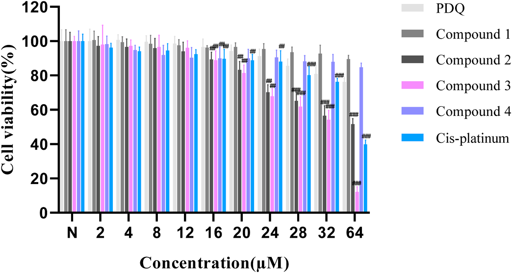
Anti-proliferative Activities of Compounds 1–4The effects of Compounds 1–4 on SKOV3 cell proliferation were analyzed by MTT assay. It was found that PDQ, Compounds 1 and 4 had no significant inhibitory activity against the proliferation of SKOV3 cells at 0–64 µM, while Compound 2, Compound 3 and cis-platinum exhibited significant anti-proliferative effects on SKOV-3 cells in concentration-dependent fashion (Fig. 2). The IC50 values of Compound 2, Compound 3 and cis-platinum against SKOV3 cells were 53.6 ± 4.35, 33.8 ± 2.21 and 70.1 ± 7.64 µM, respectively, at 48 h. These findings suggest that Compound 3 can suppress the proliferation of SKOV3 cells in vitro.
Effect of Compound 3 on SKOV3 Cell MorphologyAfter treatment with Compound 3 for 48 h, the growth rate of SKOV3 cells was reduced, intracellular vacuoles appeared, and a large number of cell fragments were floating on the surface of the culture medium (Fig. 3A). Compared with the normal group, the cell density was dose-dependently attenuated in Compound 3 treatment group, along with cell membrane rupture, nucleus dissociation and aggregation (Fig. 3B).
Compound 3 Promotes Cell Apoptosis in VitroApoptosis is considered as one of the major modes of cancer cell death. To examine whether Compound 3 can induce apoptosis, fluorescein isothiocyanate (FITC)-Annexin V/propidium iodide (PI) assays and flow cytometric analysis were performed. The results demonstrated that Compound 3 could induce SKOV3 cell apoptosis (Fig. 4), and the apoptotic cell populations were found to be 0.06, 6.63, 8.41, 34.27 and 43.74% after treatment with Compound 3 (0, 16, 20, 24 µM) and cis-platinum (32 µM) for 24 h, respectively. The findings indicated that treatment with Compound 3 decreased the apoptotic rate of SKOV3 cells in a concentration-dependent fashion. More importantly, the apoptotic rate of Compound 3 was relatively similar to that of cis-platinum.
Effect of Compound 3 on Reactive Oxygen Species (ROS) LevelsROS plays an essential role in regulating mitochondria-dependent cell death. Therefore, we assessed whether ROS level is increased in SKOV3 cells after treatment with Compound 3. 2,7-Dichlorodi-hydro fluorescein diacetate (DCFH-DA) and flow cytometric analysis were performed to detect the cellular levels of ROS. After exposure to 16, 20 and 24 µM Compound 3 for 24 h, the cellular levels of ROS in SKOV3 cells were 2.85 ± 0.21, 6.25 ± 0.33 and 17.63 ± 0.56%, respectively, when compared to the control group (0 µM). Meanwhile, the cellular level of ROS was 20.03 ± 0.71% after being exposed to 32 µM cis-platinum. Altogether, the results demonstrated that Compound 3 could induce SKOV3 cell apoptosis in a ROS-dependent fashion (Fig. 5).
Compound 3 Upregulates the Expression of Anti- and Pro-apoptotic Proteins in SKOV3 CellsGiven that Bcl-2 and Caspase family proteins are involved in the regulatory network of cell apoptosis, we further measured the expression levels of anti- and pro-apoptotic proteins in SKOV3 cells through Western blotting. From the results in Fig. 6, we found that Compound 3 increased the expression levels of cleaved-Caspase-3 (CC-3)/Caspase-3 (C-3), cleaved-Caspase-9 (CC-9)/Caspase-9 (C-9) and Cytochrome C (Cytc), while increased the ratio of Bax/Bcl-2 in a concentration-dependent fashion.
Molecular Docking Results of Compound 3The molecular docking results showed that Compound 3 could form hydrogen bond with poly(ADP-ribose)polymerase (PARP) receptor (PDB code: 5DSY). The 24-position OH of Compound 3 forms hydrogen bonds with the hydroxyl group of TYR-460, and the maleic acid structure of Compound 3 has two hydrogen bonds with the guanidine and amino sites of ARG-431 in the receptor. Thus, it is speculated that Compound 3 may induce SKOV3 cell apoptosis by targeting PARP.
Experimental
Reagents and InstrumentsPDQ was obtained by using (20S)-protopanaxadiol as a raw material through a chiral oxidation ring in our laboratory, and its purity was >95% by HPLC analysis. Other chemical reagents were procured from Macklin Biochemical Technology (Shanghai, China), and were used as supplied. To monitor the reactions, TLC analysis was conducted with pre-coated silica gel HSGF254 plates, and 10% sulfuric acid solution was used as a visualizing reagent. Column chromatography was conducted using a silica gel (200–300 mesh, Qingdao, China). The melting point was determined by YRT-3 melting point meter (Tianjin, China). 1H- and 13C-NMR spectral data were obtained using a Bruker Avance 500-MHz spectrometer (Bruker, Germany), with TMS in pyridine-d5 as an internal standard. High-resolution mass spectrometry detection was conducted on a Triple TOF 5600+ system coupled with an electrospray ionization (ESI) source (AB SCIEX, U.S.A.).
Preparation of Compounds 1–4The carboxylic acid (Compounds 1–4) was synthesized according to the route depicted in Chart 1. PDQ (0.5 g, 1.05 mmol, 1 equivalent (equiv.)) was dissolved in 10 mL dry 1,2-dichloromethane. Subsequently, 1 mL triethylamine and different anhydrides (Compound 1: 6 equiv. butanedioic anhydride, 0.6 g, 6.096 mmol; Compound 2: 6 equiv. phthalic anhydride, 0.9 g, 6.096 mmol; Compound 3: 6 equiv. maleic anhydride, 0.6 g, 6.096 mmol; and Compound 4: 6 equiv. glutaric anhydride, 0.72 g, 6.3 mmol) were mixed and stirred at room temperature for 50 h. After rinsing 3 times with distilled water, the organic phase was dried over Na2SO4 and concentrated in vacuo. The obtained crude products were analyzed by silica gel column chromatography (petroleum ether ethyl acetate solutions in different proportions). Finally, the pure Compounds 1–4 were obtained.
Cell CultureThe human ovarian cancer cell line SKOV3 was supplied by the American Type Culture Collection (VA, U.S.A.). After culturing in RPMI 1640 medium containing 10% fetal bovine serum (FBS), penicillin (100 U/mL) and streptomycin (100 mg/mL), the cells were maintained at 37 °C and 5% CO2.
Cytotoxicity AssayThe log-phase SKOV3 cells (5 × 10³ cells/mL) were subcultured in 96-well plates for 24 h, and then treated with 10 µL of Compound 3 (0–64 µM) for another 24 h. Cell viability was assessed using the MTT assay.44) The percentage of viable cells was calculated as follows: survival (%) = experimental absorbance/control absorbance × 100%.
Histological StainingThe SKOV3 cells (2 × 10⁵ cells/well) were grown in 6-well plates for 24 h, and then exposed to 16, 20 and 24 µM Compound 3 or 32 µM cis-platinum for another 24 h. Dimethyl sulfoxide (DMSO) (0.1%) was employed as a control group. The cells were fixed in paraformaldehyde (4%) for 4 h, dehydrated through a gradient concentration of alcohol, and then embedded in paraffin. After dewaxing in xylene and rehydrating through graded ethanol series, the sections were rinsed with phosphate-buffered saline (PBS) and subjected to hematoxylin and eosin (H&E) staining. Finally, cell morphology was examined using a phase-contrast microscope (Olympus, Tokyo, Japan).
Assessment of ROS LevelsThe levels of ROS we detected by DCFH-DA assay. Briefly, SKOV3 cells (1 × 10⁶) were exposed to either 16, 20 and 24 µM Compound 3 and cis-platinum (32 µM) or 1 mM H2O2 for 24 h. After harvesting and rinsing with ice-cold PBS, the cells were stained with DCFH-DA (100 µM) at 37 °C for 15 min in the dark. The stained cells were washed and resuspended in 1 mL PBS, and then examined using a FACS Caliber flow cytometer (BD Bioscience, NJ, U.S.A.) at 488/530 nm (excitation/emission). The fluorescent signals were analyzed by Cell Quest software. ROS levels are presented as mean fluorescence intensity.
Apoptosis AnalysisApoptotic cells were quantified by flow cytometry after staining with FITC-Annexin V/PI according to a previous method.45) Briefly, SKOV3 cells (6 × 10⁵) were grown in 25-cm2 flasks for 24 h, and then treated with Compound 3 for another 24 h. Afterwards, the floating and non-floating cells were harvested, rinsed twice with ice-cold PBS, resuspended in 200 mL of 1× binding buffer containing 40 ng/sample of PI and FITC-Annexin V (1 : 50), and incubated at 37 °C for 15 min in the dark. The FACS Caliber flow cytometer was used to quantify the total number of apoptotic cells at 610 nm for PI and 488/525 nm (excitation/emission) for FITC. Data analysis was performed with Cell Quest software (BD Bioscience).
Western Blot DetectionSKOV3 cells (1 × 10⁵ cells/well) were grown in 10-cm culture dishes for 24 h, followed by treatment with 0 (control), 16, 20 and 24 µM Compound 3 for 48 h. Cells lysed in radio immunoprecipitation assay (RIPA) buffer containing phenylmethylsulfonyl fluoride (PMSF) for Western blotting. Protein was separated through 12% sodium dodecyl sulfate-polyacrylamide gel electrophoresis (SDS-PAGE) and transferred onto polyvinylidene difluoride (PVDF) membranes. After blocking with 5% skimmed milk, the membranes were exposed to the following primary antibodies: Bax (1 : 1000), Bcl-2 (1 : 1000), C-3 (1 : 1000), CC-3 (1 : 1000), C-9 (1 : 1000), CC-9 (1 : 1000), Cytc (1 : 1000) and β-actin (1 : 1000) overnight, and then with secondary antibodies. The protein concentrations of Bax, Bcl-2, C-3, CC-3 C-9, CC-9, and Cytc in SKOV3 cells were determined.
Molecule Docking between Compound 3 and PARPThe binding of Compound 3 to PARP receptor was studied with Schrodinger molecular docking software. The three dimensional (3D) structures of PARP (PDB code: 5DSY46)) and Compound 3 were imported, and the interaction of the butt pose was analyzed by Sybyl 6.91 software package.
Statistical AnalysisThe GraphPad Prism v8.0.4 software was employed to perform statistical tests. Data are representative of 3 independent assays, and values are shown as mean ± standard deviation (S.D.). Statistically significant differences are represented by #, in which # p < 0.05, ## p < 0.01, ### p < 0.001.