ESSAY|TOWARD JES 100TH ANNIVERSARY
Celebration of 100th anniversary of the Japan Endocrine Society
2022 Volume 69 Issue 9 Pages 1027-1033
Details
2022 Volume 69 Issue 9 Pages 1027-1033
I heartily celebrate the 100th anniversary of the Japan Endocrine Society to express my respect about this milestone being reached. I celebrated the 80th anniversary of the Japan Endocrine Society as its 16th president at Hamarikyu Asahi Hall in Tokyo in 2007. Here, I convey a message to young doctors by looking back on my career as a physician scientist in the fields of endocrinology and metabolism.
Professor Ibayashi had a huge impact on my life (Fig. 1). He was appointed the 14th professor at the Department of Internal Medicine, Faculty of Medicine, Kyushu University, in 1971, after the end of campus disputes throughout Japan in the 1960s. After he passed on his knowledge of steroid hormones to me, I became hooked and engaged in medicine and research of steroid hormones for 50 years.

Prof. Ibayashi and collaborators of DHEAS study group of Ministry of Health, Labour and Welfare
The field of modern endocrinology came into being with the discovery of Addison’s disease in 1855. With the development of gene technology in the latter half of the 20th century, the genes encoding all adrenal steroidogenic enzymes and many related nuclear receptors and transcription factors were identified. I attended the annual meeting of the American Endocrine Society as an active member every year and absorbed the progress of the year greedily. As I was interested in the nuclear receptors essential for the action of steroid hormones, I became engaged in research on the estrogen receptor under the tutelage of Dr. Marc Lippman at the NIH in the USA for three years. From the latest research at the NIH, I realized that we were at the dawn of a new era, thanks to gene technology. Soon after returning to Japan and introducing gene techniques into clinical medicine to clarify the causes of disease as translational research, our research progressed dramatically. I was selected as 15th professor at the department after the retirement of Professor Ibayashi in 1988. The passing on of knowledge and building on previous findings are key to medical science. Professor Ibayashi taught me and my peers the importance of acquiring keen powers of observation and deep insights to precisely understand a patient’s condition. The describing and reporting of abnormal pathological conditions that have never previously been experienced are particularly important, and led to the discovery of new clinical entities such as Addison’s disease and Hashimoto’s disease. During my 17 years working as a professor, I obtained substantial funding in the form of Grants-in-Aid for Scientific Research (C, B, A, S), and from CREST, COE, Ministry of Health, Labour and Welfare, and METI to advance research via persistent efforts.
I examined a patient with complete testicular feminization (CTF) and wanted to clarify the cause of this disease, so I started researching the androgen receptor (AR). My colleagues and I identified AR gene mutations (SNP) in 10 families with AR abnormalities and clarified the relationship between these mutations and clinical disease type. In a case with no abnormality of the AR gene in CTF, we also found that a 90 kDa coactivator of AR was lacking. This new clinical entity of coactivator disease attracted attention from all over the world. In the process of this research, we succeeded in cloning a new 106 kDa coactivator gene of AR named ANT-1. We proposed compartment theory to explain how efficient transcription and splicing of AR proceed between the nuclear receptor compartment of AR and the splicing factor compartment of ANT-1 in the nucleus.
The risk of prostate cancer increases upon testosterone (T) supplementation in male patients with late-onset hypogonadism (LOH). We succeeded in identifying SARM (selective androgen receptor modulator) C12H28O with a steroid skeleton named S42, which exerts antagonistic activity on AR in the prostate and agonistic activity on it in the liver, skeletal muscle, bone, adipose tissue, and brain. S42 is expected to be useful for the treatment of LOH. SGRM (selective glucocorticoid receptor modulator), which dissociates anti-inflammatory transrepressing action and transactivating action, causing osteoporosis and diabetes by acting on glucocorticoid receptor (GR), has not been developed. We established guidelines on the management and treatment of glucocorticoid-induced osteoporosis (GIO) by following Japanese GIO cohorts to determine the factors conferring a risk for fracture.
Professor Ken-ichirou Morohashi (Kyushu University Medical School) succeeded in cloning the gene encoding the master transcription factor Ad4BP/SF-1, which is essential for ontogeny and differentiation of the adrenal gland and gonad. The adrenal gland differentiates via a cascade of the transcription factors Ad4BP/SF-1, and DAX-1 which is expressed in the adrenal gland and gonadotrophs in the pituitary gland. We reported three male cases of Addison’s disease with the complication of hypogonadotropic hypogonadism, in which the cause of the disease was unknown. We clarified gene mutations of DAX-1 in all cases for the first time in Japan. This shows the importance of describing and reporting abnormal pathological conditions that have never previously been experienced.
I performed a nationwide epidemiological study of adrenal disorders in Japan as principal investigator in research on disorders of adrenal hormone production via funding from the Ministry of Health, Labour and Welfare. A total of 109 cases of Addison’s disease were identified (estimated 660 cases in total in Japan), in which the rate of adrenal crisis was 37.4%, with induction by infection in 75% and discontinuation of cortisol in 7.5%. My colleagues and I succeeded in making adrenal steroid hormone-producing cells for the first time by expressing Ad4BP/SF-1cDNA in rat and human mesenchymal stem cells for the simultaneous supplementation of cortisol and DHEA to prevent sudden death by adrenal crisis and severe infection in Addison’s disease.
In her book Silent Spring, Rachel Carson described that environmental endocrine-disruptors (EEDs) might cause disorders of sex differentiation and threaten species with extinction. We engaged in research on EEDs through substantial funding from CREST. I hypothesized that EEDs interact with nuclear receptors including AR and DAX-1, according to our results on human sex differentiation disorders associated with mutations of AR, DAX-1, and WT-1 genes.
My colleagues and I developed a novel method for the three-dimensional (3D) reconstruction of confocal microscopic images to screen anti-androgenic EEDs. In this approach, transcriptionally active agonist DHT-bound AR produced fluorescence foci, but transcriptionally inactive antagonist OHF- or EED-bound AR showed the disruption of fluorescence foci in a homogeneous manner. Using this approach, we identified the anti-androgenic EEDs vincrozolin, pp'DDE, and nitrofen. We also established a new ovarian granulosa-like cell line named KGN with a high level of aromatase activity from a patient with invasive ovarian granulosa cell carcinoma. In addition, we developed a novel non-radioactive ELISA of aromatase activity using KGN cells, for which we obtained an international patent. It was subsequently decided that this assay system using KGN cells would be used to perform large-scale screening of EEDs affecting aromatase activity around the world. Moreover, we found that atrazine-induced aromatase expression was Ad4BP/SF-1-dependent, showing that atrazine bound directly to Ad4BP/SF-1 as a ligand for Ad4BP/SF-1. In this way, we contributed to identifying EEDs and clarifying the mechanisms behind their actions.
I also helped to host the 11th International Congress on Hormonal Steroids and the 17th International Congress on Hormones and Cancer as chairman of the local organizing committee at Fukuoka City for the first time in Japan on 21st October, 2002 (with General Secretary Toshihiko Yanase). About 1,000 researchers from 25 countries attended this event. The program included six plenary lectures by BM O’Malley, DJ Mangelsdorf, RJ Santen, JS Richards, S Kato, and K Morohashi, 30 symposia, and 231 general presentations. The event was widely appreciated for the high quality of its program. We were able to disseminate excellent Japanese results on steroidogenesis, nuclear receptors, and transcription factors to the whole world (Fig. 2).

11th International Congress on Hormone Steroids (ICHS)
17th International Congress on Hormone and Cancer (ICHC)
Professor Ibayashi was a pioneer of DHEAS research. He clarified that DHEAS was the most abundant circulating C19 steroid hormone, with about 90% of the hormone synthesized in the adrenal gland and referred to as adrenal androgen.
Serum DHEAS shows a characteristic change over the course of a human life. It increases at 6–10 years of age in adrenarche, peaks at 20 years of age, and then declines linearly to 80–90 years of age, dropping to only 10%–20% of that in young adults in adrenopause. Although DHEAS is a marker of aging, its biological significance and mechanism of action were completely unknown.
Darwin’s theory of evolution established that living organisms have evolved from a common ancestor, which has been clarified by the establishment of molecular phylogenetic trees via the development of gene technology. Analogously, according to the theory of evolutionary medicine established by Professor Hiroo Imura (Professor Emeritus at Kyoto University), the primitive adrenal gland evolved 500 million years ago, appearing in the jawless vertebrate, the lamprey. DHEA was the first synthesized ancient steroid hormone and played important roles in the evolution and differentiation of vertebrates. However, large amounts of serum DHEAS are observed only in humans and apes including chimpanzees. A significant positive correlation between maximal lifespan and serum DHEAS concentration was demonstrated in primates and humans, while the highest serum DHEAS concentration of all was found in humans. It was also reported from a long follow-up cohort study that a high serum DHEAS concentration predicted longevity in men. DHEAS was also revealed to be important for brain development and anti-aging in humans. The mechanism behind the increase in DHEAS in adrenarche and its association with brain development depend on increases in cortisol and cytochrome b5 in adrenocortical cells along with increases in IGF-1 and leptin in serum. We first clarified that cytochrome b5 is an essential cofactor for 17,20-lyase to synthesize DHEA in the human adrenal gland by examining the adrenocortical adenoma of Cushing’s syndrome, which presents with an unusually high serum level of DHEAS. It was also clarified that the increase in DHEAS in adrenarche of neonatal rhesus monkeys was initiated primarily by an increase of cytochrome b5.
Via in vitro and in vivo experiments on rodents and humans, we also clarified the beneficial effects of DHEA against obesity, diabetes, atherosclerosis, osteoporosis, and dementia as well as for stimulating the immune system. We first reported that DHEA supplementation in middle-aged males increased insulin sensitivity, improved glucose tolerance, and reduced serum hs-CRP, via a longevity health research grant from the Ministry of Health, Labour and Welfare (Fig. 1).
Recent advances in aging research have revealed that DHEA and DHEAS exhibit anti-aging effects via antioxidant, anti-inflammatory, telomere-protective, and p38MAPK-inhibiting activities to suppress senescence-associated secretory phenotype (SASP) formation, to counter the activity of cortisol, and to induce the expression of chaperones (HSP70, HSP47, and Sigma-1 receptor). During a search for the nuclear DHEA receptor, we succeeded in cloning the DHEA target gene encoding a novel p38MAPK phosphatase named DDSP in activated human T lymphocytes with an increase in high-affinity DHEA binding sites upon treatment with DHEA. By establishing DDSP transgenic db/db obese mice, we clarified that DDSP had anti-obesity effects and decreased serum leptin. DDSP inhibits p38MAPK to suppress SASP formation, inhibits inflammation, and exhibits anti-aging activity.
Although the nuclear DHEA receptor has not been identified, progress has been made in our understanding of membrane DHEA and DHEAS receptors and binding sites, which revealed GPCR on vascular endothelial cells, GPER on hepatocytes, TrKAR on neurons, and GABAA, NMDA, Sigma-1, and Map2 receptors in the brain. The concept of intracrinology, in which estrogen and androgen are formed from DHEAS in peripheral tissues as proposed by Fernand Labrie, was clarified to be important in elderly people, especially menopausal women.
Nowadays, people worldwide expect to live long lives. The elderly population aged over 65 in Japan is rapidly increasing, accounting for nearly 30% of the total population. The prevention of geriatric syndrome is important for elderly people to live independently without receiving nursing care and to extend the healthy lifespan. The hypothalamic–pituitary–adrenal axis plays an important role in maintaining homeostasis. Many elderly people may suffer from chronic stress. We succeeded in cloning the Nur 77 gene encoding a novel stress-induced nuclear receptor from human T cells. In chronic stress conditions, Nur 77 was found to be induced in pituitary corticotrope cells by CRH from the hypothalamus, which binds to nGRE in the POMC gene promoter, thus blocking negative feedback by cortisol to maintain high plasma ACTH. In the adrenal gland in the elderly under chronic stress, the pathway for synthesizing cortisol from progesterone is activated, thus increasing serum cortisol; meanwhile, the DHEAS synthetic pathway is attenuated, thus markedly decreasing serum DHEAS. The cortisol/DHEAS ratio increases in elderly people with chronic stress, similar to the findings in Cushing’s syndrome.
A marked decrease in serum DHEAS in elderly people was found to attenuate the intracrinological action of DHEAS in the immune system, liver, skeletal muscle, and bone, causing susceptibility to infection, frailty under malnourished conditions and chronic inflammation due to a metabolic shift to reverse metabolism, sarcopenia, and osteoporosis. Our recent cohort studies of elderly diabetic patients identified the factors conferring a risk of frailty and sarcopenia. Low serum albumin (<4.0 g/dL) was the strongest risk factor for frailty. DHEAS concentration was also positively correlated with serum albumin, which may have an anabolic effect in the liver. In addition, serum cortisol/DHEAS ratio >0.2 was found to be the strongest risk factor for sarcopenia. The combination of an increase in cortisol reflecting the presence of stress, an increase in muscle catabolism, and a decrease in DHEAS reflecting decreased muscle anabolism may lead to sarcopenia. We clarified that DHEAS is important for maintaining the bone mineral density (BMD) of postmenopausal women via its intracrinological action. We also identified that the activity of aromatase in converting androgen to estrogen in normal human osteoblasts is maintained by 1,25(OH)2 vitamin D, based on our results of a positive correlation between BMD and serum DHEAS and positive correlations of serum DHEAS and serum estrone with BMD in postmenopausal women. It was also recently reported from a placebo-controlled study that DHEA supplementation was effective to increase BMD in lumbar spine and trochanter and to increase muscle mass in order to prevent osteoporosis and sarcopenia in postmenopausal women and patients with Addison’s disease.
Hakaru Hashimoto (1881–1934) graduated from Kyushu University Medical School in 1907 as one of the first graduates. Immediately after graduation, he entered the first Department of Surgery and studied surgery under Professor Hayari Miyake. He examined the pathological appearance of four cases of unusual goiters removed surgically for four years before studying in Germany. He gave an accurate description of lymphocyte infiltration and the formation of lymphoid follicles in the thyroid gland (Fig. 3). He reported “struma lymphomatosa” as a new clinical entity in Archiv für Klinische Chirurgie in 1912. This is the thesis of his medical doctor and the foundation of the entity of Hashimoto’s disease (Fig. 3).

Memorial lecture on Hashimoto disease
The International Hashimoto Symposium was held to commemorate the 80th anniversary of the reporting of Hashimoto’s disease, which was hosted by Professor Shigenobu Nagataki (Nagasaki University Medical School) in Fukuoka City. The Memorial Lecture on Hashimoto’s Disease was given in Kyushu University Medical School (by Professor Ryoichi Mori, Dean, the Department of Medicine), where Dr. Hashimoto graduated and reported on “struma lymphomatosa,” on 2nd December, 1992, a day before the International Symposium (Fig. 3). The auditorium was almost full with about 400 people, including 200 medical students, 40 thyroidologists from 18 countries, and 150 Japanese scientists. The lectures on autoimmune thyroiditis as the scientific basis of Hashimoto’s disease by Professor Robert Volpe and Professor Takehiko Sasazuki made a deep impression on all participants. Professor Kazuo Hashimoto (Kanazawa University Medical School), the son of Dr. Hakaru Hashimoto, gave a lecture on family memories. When he visited Dr. Deborah Doniach in London, she said that his father’s paper was magnificent, with a particularly perfect pathological description to which nothing more could be added. In honor of his outstanding achievement, Hashimoto’s Street and Monument were built on the campus of Kyushu University Medical School (Fig. 3).
I traveled to London and Edinburgh in the UK with my wife in May 2019, just after my retirement from Seiwakai Muta Hospital, to follow in the footsteps of Thomas Addison and Charles Darwin through their outstanding achievements.
Thomas Addison (1793–1860) graduated from Edinburgh Medical School and became a medical doctor. He worked as a physician at Guy’s Hospital in London all his life. Addison first published a short article “Anemia-disease of the suprarenal capsule in which the disease was not distinctly separated from a new form of anemia” in London Medical Gazette in 1849. He collected data on 11 patients who died with similar clinical symptoms, performed autopsies on all of these patients, and kept detailed and accurate medical and pathological records. He found that all patients had lesions on the suprarenal adrenal glands. He proposed the disease caused by adrenal insufficiency as a new clinical entity and published the monograph “On the constitutional and local effects of disease of the suprarenal capsule” in 1855. This established the entity of Addison’s disease. In addition, a rare case involving the combination of Addison’s disease due to autoimmune adrenalitis and pernicious anemia (Addison’s anemia) reported by Addison was diagnosed as autoimmune polyglandular syndrome type 2.
We took the 4-hour trip to Edinburgh by train from London via Newcastle-upon-Tyne, near Addison’s birthplace. The World Heritage Site of the Old Town of Edinburgh took my breath away with its amazing beauty, taking me back to the 11th Century. Edinburgh Medical School, which opened in 1726, is located in the center of the Old Town. I was particularly excited to find a plaque in memory of Bright, Addison, and Hodgkin at the entrance of Edinburgh Medical School (Fig. 4). This plaque commemorates the contributions to the medical sciences of these three distinguished graduates of Edinburgh Medical School, who subsequently worked at Guy’s Hospital in London. Their names will forever be associated with the discovery of new clinical entities: acute poststreptococcal hemorrhagic glomerulonephritis, Bright’s disease, adrenocortical insufficiency, Addison’s disease, pernicious anemia, Addisonian anemia, lymphadenoma, Hodgkin’s disease. After returning to London, I visited Guy’s Hospital where Addison worked and King’s College London Medical School adjacent to Guy’s Hospital. Addison’s name is inscribed at the entrance of the Medical School Building, while a bust of Addison is displayed at the entrance hall of the Hodgkin Building and an autopsied specimen of his is displayed at the Hodgkin Building to commemorate his outstanding achievements in perpetuity.

Monuments of Dr. Addison in London and Edinburgh
Charles Darwin also attended Edinburgh Medical School but dropped out before completing his studies. During my trip, I also visited the National History Museum in London. A new building named the Darwin Centre and a repository with his collection were built to commemorate his outstanding achievements. I was completely satisfied after fulfilling a desire to walk in the footsteps of Addison and Darwin. I toasted them with craft beer and fish and chips in the pub.
When I look back on my career as a physician scientist, I recognize the importance of meeting my mentor Professor Ibayashi and being able to continue to meet challenges in medicine and research with unwavering curiosity and passion. It was also important to describe and report abnormal pathological conditions that have never previously been experienced by acquiring keen powers of observation and deep insights, to learn from patients. These skills previously led to the discovery of new clinical entities such as Addison’s disease and Hashimoto’s disease. In my own career, I introduced gene technology to Japanese clinical medicine at an early stage, to perform translational research. My colleagues and I contributed to advances in medical science via unexpected discoveries and serendipity. The introduction of new technology was important to advance research. I also learned that studying abroad is beneficial to make contacts with excellent researchers from different cultures and to adopt new perspectives. I hope that young doctors can also look elsewhere in the world for inspiration.
Hormones are important for maintaining homeostasis and a healthy life. Professor Imura proposed the importance of “life course health care” from the fetal stage to old age. The elderly population aged over 65 is rapidly increasing in Japan. It is thus particularly important to attempt to extend the healthy lifespan and to improve the QOL of elderly people from a hormonal perspective. In general hospitals and clinics, it is important to contribute to medical care by performing team care with medics from other specialties to provide patient-centered care, to apply evidence obtained from one’s own clinical cohort studies, and finally to contribute to society by doing one’s best as a doctor throughout one’s career.
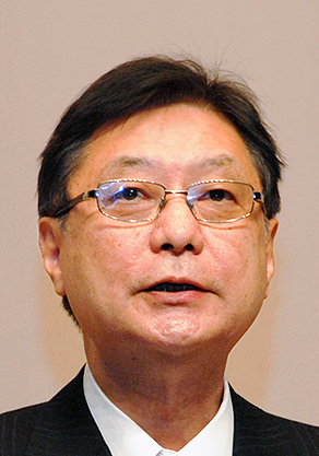
Hajime Nawata
16th President/Honorary Member
Professor Emeritus, Kyushu University
E-mail: nawata-h@mx22.tiki.ne.jp
Careers in JES
2011– Honorary Member
2007– Senior Councilor
2005–2007 16th President
2003–2007 Director (Publication)
2001–2007 President for Kyushu Regional Branch
2001–2003 Director (Publication)
1997–2001 Director (Publication)
1983– Councilor
1973– Member
Activities in JES
2001 Chair, 1st Kyushu Naibunpi Taisha Forum (Annual Meeting of JES Kyushu Regional Branch)
1998 Chair, 71st Annual Congress of JES
1996 Chair, 14th Fuji Hormone Conference (JES Summer Seminar on Endocrinology & Metabolism)
JES Awards
2002 1st JES Award
1986 6th JES Research Award
Contributions to EJ
1999–2002 Editor-in-Chief
1994–1998 Editor