2014 年 52 巻 1 号 p. 66-70
2014 年 52 巻 1 号 p. 66-70
Adrenal epithelioidangiosarcoma (AEA) is a rare neoplasm that accounts for less than 1% of sarcomas. Due to its rarity, it can easily be misdiagnosed, both by the clinician and the pathologist. Data on the patient’s occupational history was collected and analyzed. The bibliographic data was found on the PUBMED bibliographic search site after entering the word “extrahepaticangiosarcoma”. We report a case of adrenal epithelioidangiosarcoma (AEA) in a 68 yr-old Caucasian male factory worker exposed to Vinyl Chloride (VC) for 15 yr. He underwent surgery, chemotherapy and radiotherapy. Hepatic angiosarcoma is a known consequence of VC exposure, but occupational causality of extra-hepatic angiosarcoma is controversial. Extra-hepatic angiosarcomas have been reported in VC workers, but never AEA. Cancerogenic effects of VC involve all endothelial areas of the body and extra-hepatic endothelial tumors may also be caused by this substance. This is the first published report of AEA diagnosed in a worker exposed to VC.
Angiosarcoma is a rare malignant tumor (less than 1% of sarcomas), mainly localized in the skin and superficial soft tissue1,2,3). Angiosarcomas have been reported as primary neoplasms in numerous other sites, including breast, thyroid, heart, lung, pulmonary artery, liver, spleen, kidney, adrenal gland, uterus, ovary, vagina, testis, bone, and serous membranes4,5,6,7,8,9,10,11,12,13,14,15,16,17). Etiologic factors related to angiosarcomas are exposure to arsenic18), thorium dioxide19,20,21), vinyl chloride monomers22, 23), and therapeutic irradiation24,25,26,27). Since 1974, several reports have appeared regarding a distinct relationship between exposure to vinyl chloride monomers and angiosarcomas of the liver28,29,30,31,32,33,34). Adrenal epithelioidangiosarcoma (AEA) is one such rare neoplasm, as reflected by its limited documentation in the literature. It is usually a very aggressive tumor, and surgery is the treatment of choice with or without adjuvant therapy, depending on the histopathological stage and subsequent prognostic factors. The most common presenting symptoms are non-specific and include slight fever, anorexia, fatigue and chronic pain, although the disease is frequently asymptomatic, as reported by Wenig et al.2, 35,36,37). Finally bibliographic evidence is presented, found on the PUBMED bibliographic search site after entering the word “extrahepaticangiosarcoma”.

Left suprarenal mass and metastasis near the posterior abdominal wall.

Clear cells as hyperplastic adrenal tissue (bottom) and neoplastic vascular channels (top) with malignant endothelial epithelioid cells (hematoxylin-eosin ×32).

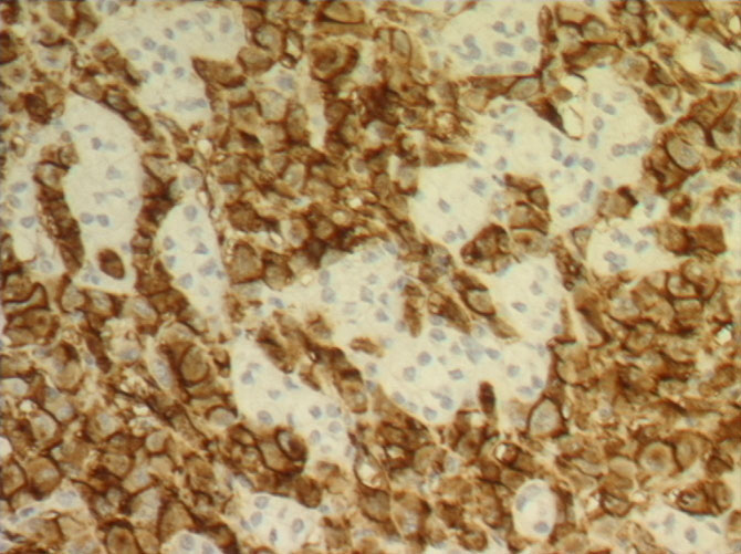
Immunohistochemical staining for CD31 antigen. Note intense brown coloration of the membrane of the endothelial neoplastic cells with residual negative adrenal tissue (×40).
In April 2011, a 68 yr old Caucasian male was admitted to the surgery clinic at the General Hospital “Perrino” in Brindisi (Italy) because of a pain in the left thorax. A CT scan showed a suprarenal mass of 7 cm with intra-parenchymal calcifications and dishomogeneous enhancement in contact with the gastric wall, left renal vein and splenic vessels (Fig. 1). On April 20, the patient underwent a laparoscopic ablation of the suprarenal mass. Subsequently, in July 2011, a CT-PET showed metastatic lesions in T4 and T5 and chemotherapy with anthracyclines was started in the Oncology Clinic. In August 2011, a bone scan was required due to diffuse skeletal pain and showed vertebral and costal metastases. Radiotherapy with a single fraction of 8 Gy to the painful portion of the chest was performed. Pain control was unsuccessful since the Visual Analogue Scale score remained constant (score 7–8). The patient died of neoplastic cachexia in November 2011.The man had worked as a compressor operator from 1962–1972 at a chemical factory in southern Italy, in a storage facility with vinyl chloride (VC). In 1991, the sector management compiled a list of workers exposed to VC after 1970. On that list the man resulted exposed to >500 ppm of VC from 1970 to 1972. From 1988 to 1993, the man worked on the production of Polyvinyl Chloride (PVC) pipes from PVC waste material. The tumor was diagnosed in 2011, 39 yr after his last direct VC exposition and 18 yr after working with PVC. The tumor was surgically excised along with surrounding adipose tissue. The adrenal gland had a nodular aspect and measured 8 × 5 × 4.5 cm, with a weight of 100 g. The cut surface showed a variegated aspect with hemorrhagic areas infiltrating the residual adrenal tissue, with a central zone of calcific fibrosis. Hematoxylin and eosin-stained sections of the adrenal gland revealed a neoplasm characterized by predominantly epithelioid malignant endothelial cells forming rudimentary vascular channels. These channels infiltrated the surrounding adrenal tissue, which was diffusely hyperplastic; they had an irregular shape and formed intercommunicating sinusoids with endothelial papillae (Fig. 2). Malignant epithelioid cells had abundant eosinophilic cytoplasm and high nuclear grade with vescicular nuclei and prominent nucleoli. In some areas the neoplastic cells had a prevalent solid pattern with slit-like vascular spaces. Wide fibrosis with microcalcifications was also present. Immunohistochemically, the neoplastic cells were strongly and diffusely positive for platelet endothelial cell adhesion molecule (PECAM-1) also known as cluster of differentiation 31 (CD31) (Fig. 3), Human von Willebrand Factor (Factor VIII-related antigen) and focally positive for CKAE1AE3. Immunostains for is a cluster of differentiation molecule (CD34), Inhibin Alpha and S100 were negative.
AEA are extremely rare neoplasms. AEA must be differentiated from other neoplasms of the adrenal gland such as adrenal cortical carcinoma, pheochromocytoma, metastatic carcinoma, and metastatic malignant melanoma, but in all cases the immunohistochemical markers are a good tool for differential diagnosis. AEA is a biologically malignant neoplasm that can infiltrate locally and metastasize37). The etiology of the epitheloidangiosarcoma remains unknown. There are only four cases described in the literature where the malignancy could be linked with prolonged exposure to arsenic-containing insecticides and presence of mesenteric fibromatosis. No other connection or correlation with a family history of adrenal neoplasms (suggesting Multiple Endocrine Neoplasia syndrome), a prior history of abdominal radiotherapy or long-term androgenic anabolic steroid treatment could be found36, 38). The disease generally affects more men than women (21 men, 8 women, 1 not specified) with a wide age range from 34 to 85 yr, predominantly patients in their 60s and 70s. The disease usually starts with pain and presence of abdominal mass, followed by significant weight loss, fever episodes and weakness. In six cases described in the literature the disease was asymptomatic but in four cases it was associated with paraneoplastic syndrome and distant metastases to bone and liver38, 39). One patient had an unusual association with Cushing’s disease while in one patient an adrenal tumor had accidentally been discovered40, 41). In the sixth asymptomatic patient, the angiosarcoma was revealed after surgery for abdominal trauma and suspected hepatic rupture was performed42). Rasore-Quartino and Kern published the first two cases in the late 1960s, but due to a lack of immunohistochemical analyses they are excluded from this study43, 44). The first case confirmed by immunohistochemical staining was published in 1988 by Kareti3). Several single case reports followed until Wenig et al. described the largest study in 1994 where nine cases of adrenal angiosarcoma were analyzed (eight new cases plus one previously published by Karety)36, 40, 45,46,47,48,49). Macroscopically the tumors varied from well-circumscribed to invasive retroperitoneal masses, solid to cystic, with size from 5 to 16 cm in diameter. All the cases described in the literature tended toward an epithelioid appearance. Among them 19 had immunoreactivity for keratins while only 3 were negative35, 36, 38, 39, 42, 46, 47, 52, 54,55,56,57,58,59). Reactivity for cytokeratin is typical of epithelioidmorphology and is believed to represent aberrant or “atavistic” expression. Human von Willebrand Factor (Factor VIII-related antigen) and platelet endothelial cell adhesion molecule (PECAM-1) also known as cluster of differentiation 31 (CD31) positivity was detected in 7 cases and 16 cases, respectively35, 36, 40,41,42, 47, 52,53,54,55,56,57,58,59). Prompt preoperative diagnosis is very complex since the tumors can appear well-circumscribed and non-contrast-enhancing, suggesting a benign, non-neoplastic formation. Their irregular histological and immunological attributes as well as their relatively low incidence can cause pathologists to mistake them for adrenal epithelial neoplasms. In clinical practice these neoplasms should always be differentiated from other vascular neoplasms, pheochromocytoma, adrenal cortical carcinoma, metastatic adenocarcinoma, metastatic malignant melanoma and other metastatic tumors as well as from benign neoplasms such as adrenal adenomas with hemorrhage and epithelioidhemangioendothelioma36, 38, 40, 50,51,52). The safest and easiest way to confirm or rule out this malignancy is by using immunohistochemistry. Endothelial-related markers (CD34, Factor VIII antigen and CD31) must be used in the antigen panel of these tumors, following their limitations in terms of sensitivity and specificity36). The adrenal angiosarcoma is a malignant neoplasm that can invade surrounding organs and tissue as well as metastasize in distant sites. Epithelioidangiosarcoma of the adrenal gland may mimic much more common primary and secondary tumors, and in view of cytokeratin positivity, especially metastatic carcinoma. Despite its rarity, knowledge of its existence is important as its pathobiologic characteristics may differ markedly from other primary and metastatic adrenal neoplasms. Because of the infrequency of this entity, optimal therapy other than surgical eradication is difficult to determine. The complete surgical resection of the adrenal gland with or without any surrounding tissue or organ infiltrated with the tumor has good outcome despite the biology of this tumor. Some cases may have been detected at an early enough stage to enable surgical cure. In view of the aggressive nature of angiosarcoma in all sites, adjuvant therapy appears justified for patients in whom complete surgical extirpation cannot be ensured. Complete eradication combined with 3- to 6-month control intervals are essential for detection of presence of local recurrence or distant metastases and their treatment with adjuvant chemo- or radiotherapy. Presence of local or distant metastases at the time of the primary detection of the tumor or in the first 6 months postoperatively is a negative prognostic parameter that shortens the overall survival of the patient. This is the first published report of adrenal angiosarcoma diagnosed in a worker exposed to VC.