尿沈渣検査は,尿中に出現する成分を尿の遠心操作にて得られた沈殿物を観察する検査である。尿沈渣の標本作成における操作が単純であるにもかかわらず,尿沈渣に出現する成分は多種多様であるため,鑑別が非常に複雑である。その要因としては,尿沈渣に出現する尿中有形成分が,ひとつの成分においても様々な形態で存在することがある。たとえばシュウ酸カルシウム結晶では正八面体型とビスケット型,コマ型などが存在し,尿細管上皮細胞に至っては基本型,特殊型と細胞形態のバリエーションが多岐にわたる。このように尿沈渣検査では成分を正しく鑑別するための知識と技術が必要である。この部では,尿沈渣に出現する基本的な尿中有形成分を鑑別する知識を習得するために,最も基本となる成分の写真について「尿沈渣検査法2010」の尿沈渣アトラスを引用(一部改編)し掲載する。また「*」でマークした写真は,尿沈渣成分の新たな情報として追加したものである.この尿沈渣アトラスを利用し,各成分の特徴を捉えることをしっかりと身につけ,今後遭遇するであろう鑑別困難な成分に対しても対処できるよう,基礎知識を学習することを目的とする。

尿細管上皮細胞 40× 無染色
Renal tubular epithelial cells 40× No staining
円柱内には小型の尿細管上皮細胞がみられる。白血球との鑑別が必要となるが,黄色調で透明感がなく細胞質辺縁構造が角状を示すものも含まれており,鑑別される。
Small tubule epithelial cells are found in the cast. Differentiation from leukocytes is necessary. They are not transparent with a yellowish tone, and cytoplasmic marginal structures are angular in shape. These characteristics help to distinguish this cell type.

尿細管上皮細胞 40× S染色
Renal tubular epithelial cells 40× S staining
Figure 3.47と同様の小型の尿細管上皮細胞である。染色性は良好で,核は濃縮状で偏在し濃青色に,細胞質は顆粒状で赤紫色に染め出されている。
Small renal tubular epithelial cells, similar to cells seen in Figure 3.47. Stainability is good; the nucleus is unevenly distributed, stained dark blue with high concentration, and the cytoplasm is granular and stained reddish purple.

尿細管上皮細胞 40× S染色
Renal tubular epithelial cells 40× S staining
最も判定しやすく,最もよく遭遇する鋸歯(きょし)型の尿細管上皮細胞である。細胞質表面構造は顆粒状で,辺縁構造は細かく凸凹を示し,核は濃縮状である。
This is the most identifiable and most frequently encountered saw-type tubular epithelial cell. The cytoplasm surface structure is granular, and the marginal structure exhibits fine unevenness. The nuclei are concentrated.

尿細管上皮細胞 40× S染色
Renal tubular epithelial cells 40× S staining
円柱内には大型鋸歯型の尿細管上皮細胞がみられる。細胞質表面構造は不規則な顆粒状で,辺縁構造は凸凹とした鋸歯状を示し,核は赤血球よりやや大きく,偏在している。
Large saw-type tubular epithelial cells are found in the cast. The cytoplasm surface structure is irregular and granular, and the marginal structure exhibits an uneven saw shape. The nuclei are slightly larger than red blood cells and are unevenly distributed.

尿細管上皮細胞 40× S染色
Renal tubular epithelial cells 40× S staining
赤紫色に良好に染め出された細胞は小~大型の鋸歯型の尿細管上皮細胞である。核は赤血球大で青色に染め出されているが,核が認められないものもみられる。
Small or large saw-type renal tubular epithelial cells is successfully stained reddish purple. The size of the nuclei is the same as red blood cells, and the nuclei are stained blue. Some cells have no nucleus.

尿細管上皮細胞 40× S染色
Renal tubular epithelial cell 40× S staining
円柱に付着してみられる細胞はアメーバ偽足型の尿細管上皮細胞である。細胞質辺縁にはアメーバ偽足状の突起がみられる。赤血球大の核を2個有している。
The cells attached to the cast is an amoeba pseudopod-type renal tubular epithelial cell. Amoeba pseudopodial protrusions are present at the cytoplasmic margin. It has two nuclei, which are the same size as red blood cells.

尿細管上皮細胞 40× 無染色
Renal tubular epithelial cell 40× No staining
アメーバ偽足型の尿細管上皮細胞である。黄色調で細胞質辺縁構造は顆粒状を示し,細胞質辺縁は深い切れ込みがあるアメーバ偽足状を呈している。
This is an amoeba pseudopod-type renal tubular epithelial cell. It has a yellowish tone. The cytoplasmic marginal structure is granular, and the cytoplasmic margin presents amoeba pseudopods with deep cuts.

尿細管上皮細胞 40× S染色
Renal tubular epithelial cells 40× S staining
染色性は良好で細胞質が赤紫色に染め出されているが,核はみられない。 中央にみられるのがFigure 3.53と同じアメーバ偽足型である。
Stainability is good, and the cytoplasm is stained reddish purple. However, no nucleus is observed. In the center, it is the same amoeba pseudopod-type as shown in Figure 3.53.

尿細管上皮細胞 40× 無染色
Renal tubular epithelial cell 40× No staining
矢印に示す細胞は尿細管上皮細胞の棘突起型である。黄色調で細胞質表面構造は細顆粒状を示し,細胞周囲には明瞭な棘状の突起がみられる。
The cell indicated by the arrow is a spinous process-type renal tubular epithelial cell. It has a yellow tone. The cytoplasmic surface structure exhibits a fine granular shape. Clear spinous protrusions are observed around the cell.

尿細管上皮細胞 40× 無染色
Renal tubular epithelial cells 40× No staining
円柱内に封入された5個の尿細管上皮細胞のうち,矢印が棘突起型である。これらの尿細管上皮細胞はビリルビン色素に着色し,濃黄色調を呈している。
Among the five renal tubular epithelial cells enclosed in a cast, the cell identified by the arrow is a spinous process type. These renal tubular epithelial cells are deep yellowish, colored by bilirubin pigment.

尿細管上皮細胞 40× 無染色
Renal tubular epithelial cells 40× No staining
矢印の3個は角柱・角錐台型の尿細管上皮細胞である。矢印①,②が尿細管内腔面側からみた正面像,③が側面像を示す。
Three arrows identify tubular epithelial cells with a prism/pyramid shape. The cells indicated by arrows ① and ② are the front view observed from the inner wall surface of the renal tubule, and ③ is the side view.

尿細管上皮細胞 40× S染色
Renal tubular epithelial cells 40× S staining
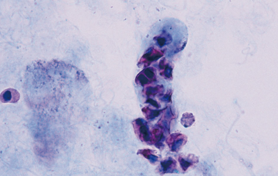
円柱内に角柱・角錐台型の尿細管上皮細胞が封入されている。細胞質は赤紫色に核は青色に良好な染色性を呈している。
Prism/pyramid-type tubular cells are enclosed in a cast. The cytoplasm is reddish purple, and the nucleus is blue. It shows good stainability.

尿細管上皮細胞 40× 無染色
Renal tubular epithelial cells 40× No staining
角柱・角錐台型の尿細管上皮細胞である。ビリルビン色素に着色され,立体感のある細胞像を示している。
These are tubular epithelial cells of the prism/pyramid type. They are colored with bilirubin pigment and exhibit a three-dimensional cell image.

尿細管上皮細胞 40× S染色
Renal tubular epithelial cells 40× S staining
Figure 3.59と同一例。角柱・角錐台型の尿細管上皮細胞はビリルビン色素に着色されている影響で,細胞質の染色態度が本来と異なっている。
The same example as Figure 3.59. Prism/pyramid-type tubular epithelial cells are colored with bilirubin pigment; therefore, the appearance of the cytoplasm with S staining is different from the general.

尿細管上皮細胞 40× S染色
Renal tubular epithelial cells 40× S staining
洋梨型の尿細管上皮細胞である。細胞質表面構造はほぼ均質状で,辺縁構造は一部不明瞭なシワ状を示している。核は白血球大で核内構造は凝集状である。
These are pear-shaped renal tubular epithelial cells. The surface structure of the cytoplasm is almost homogeneous, and the marginal structure shows partially obscure and wrinkle shape. The nucleus is the same size as a leukocyte, and the intranuclear structure is aggregated.

尿細管上皮細胞 40× S染色
Renal tubular epithelial cells 40× S staining
Figure 3.61と同様の洋梨型の尿細管上皮細胞が円柱内に封入されている。染色性は良好で,核内構造は凝集状を示し,青色に染め出されている。
Pear-shaped renal tubular epithelial cells similar to Figure 3.61 are enclosed in a cast. The stainability is good. The intranuclear structure shows agglomeration, and is stained in blue.

尿細管上皮細胞 40× 無染色
Renal tubular epithelial cells 40× No staining
矢印で囲んだ細胞は洋梨型の尿細管上皮細胞である。洋梨状を示す尿路上皮細胞と比べて細胞質は薄く,均質状から微細顆粒状である。辺縁構造は不明瞭でシワ状を呈していることが多い。
Cells surrounded by arrows are pear-shaped renal tubular epithelial cells. The cytoplasm is thin and homogeneous to fine granular compared to urothelial cells, which exhibit a pear-like shape. The marginal structure is often ambiguous and wrinkled.

尿細管上皮細胞 40× S染色
Renal tubular epithelial cell 40× S staining
Figure 3.63と同様の洋梨型の尿細管上皮細胞が円柱内に封入されている。染色性は良好で,細胞質は薄くシワ状を呈し,核は青色に染め出されている。
The pear-shaped renal tubule epithelial cells are similar to that showed in Figure 3.63 and enclosed in a cast. The stainability is good. The cytoplasm is thin and wrinkled, and the nucleus is stained blue.

尿細管上皮細胞 40× 無染色
Renal tubular epithelial cells 40× No staining
円柱に付着または封入された細胞は紡錘型の尿細管上皮細胞である。紡錘状を示す尿路上皮細胞と比べて細胞質は薄く,表面構造はほぼ均質状を示している。
The cells adhered or encapsulated in the cast are spindle-shaped renal tubular epithelial cells. The cytoplasm is thinner and the surface structure is almost homogeneous compared with urothelial cells, which have a spindle shape.

尿細管上皮細胞 40× S染色
Renal tubular epithelial cells 40× S staining
Figure 3.65と同一例。紡錘型の尿細管上皮細胞は染色性が良好で,核内構造は凝集状を示している。しばしば塩類の付着がみられ,この症例では一部に尿酸塩が付着している。
The same example as Figure 3.65. Spindle-type renal tubular epithelial cells have good staining properties, and the intranuclear structures are aggregated. Salt adhesion is often observed, and urate is partially attached in this case.

尿細管上皮細胞 40× 無染色
Renal tubular epithelial cells 40× No staining
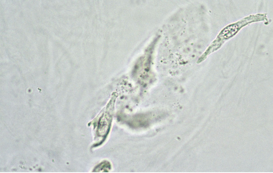
紡錘型の尿細管上皮細胞である。細胞質表面構造はほぼ均質状で,細胞質は薄くシワ状や折れ曲りがみられ,辺縁構造は不明瞭である。核は白血球大である。
These are spindle-shaped renal tubular epithelial cells. The surface structure of the cytoplasm is almost homogeneous. The cytoplasm is thin and exhibits wrinkles or folding, and the marginal structure is unclear. The nucleus is the same size as a white blood cell.

尿細管上皮細胞 40× S染色
Renal tubular epithelial cells 40× S staining
円柱に付着または封入された細胞は,洋梨型や紡錘型の尿細管上皮細胞である。細胞質の染色性は良好であるが,核は染まっていないものもみられる。
Cells adhered or encapsulated in a cast are pear- or spindle-shaped renal tubular epithelial cells. Cytoplasmic stainability is good, but some nuclei are not stained.

尿細管上皮細胞 40× 無染色
Renal tubular epithelial cells 40× No staining
集塊を構成する細胞は線維型やヘビ型が混在した尿細管上皮細胞である。細胞質は非常に薄く,表面構造はレース網目状で透明感がある。辺縁構造は不明瞭である。
The cells constituting the conglomerate are renal tubular epithelial cells of a fibrous type and snake shape. The cytoplasm is very thin, and the surface structure is lacy mesh in appearance and transparent. The marginal structure is ambiguous.

尿細管上皮細胞 40× S染色
Renal tubular epithelial cells 40× S staining
紡錘型や線維型が混在した尿細管上皮細胞が集塊でみられる。細胞質は赤紫色に,核は白血球大で青色に染め出されている。
Renal tubular epithelial cells of a spindle shape and fibrous type can be seen in a clump. The cytoplasm is stained reddish purple. The nucleus is the same size as a leukocyte and is stained blue.

尿細管上皮細胞 40× S染色
Renal tubular epithelial cells 40× S staining
集塊を構成する細胞は紡錘型や線維型が混在した尿細管上皮細胞である。レース網目状を示す細胞質は薄く赤紫色に染め出され,辺縁構造は不明瞭である。
The cells constituting the conglomerate are renal tubular epithelial cells of a spindle shape and fibrous type. The cytoplasm showing a lacy mesh in appearance is stained pale reddish purple. The marginal structure is unclear.

尿細管上皮細胞 40× S染色
Renal tubular epithelial cells 40× S staining
尿酸塩を取り囲むように集塊を構成する細胞は線維型の尿細管上皮細胞である。染色性は良好で,細胞質は赤紫色に,核は赤血球大で青色に染め出されている。
Cells constituting the conglomerate that surrounds urate are fibrous-type renal tubular epithelial cells. The stainability is good. The cytoplasm is reddish purple. The nucleus is stained blue and is the same size as a red blood cell.

尿細管上皮細胞 40× 無染色
Renal tubular epithelial cells 40× No staining
図の細胞はすべて尿細管上皮細胞である。オタマジャクシ型やヘビ型,類円形型などの種々な形態・大きさを示すが,色調や細胞質表面構造がほぼ同一であり空胞変性を示すものもある。
Cells in this figure are all renal tubular epithelial cells. Various shapes and sizes such as tadpole type, snake type, and near-circular shapes are shown. However, the color tone and surface structures of the cytoplasm are almost homogeneous. Some show vacuolar degeneration.

尿細管上皮細胞 40× S染色
Renal tubular epithelial cells 40× S staining
円柱内に円形・類円形型とともにヘビ型の尿細管上皮細胞が封入されている。細胞質は空胞状で透明感がある。核は赤血球大~白血球大で小さな核小体がみられる。
Snake-shaped renal tubule epithelial cells, along with circular and near-circular-shaped cells are encapsulated in the cast. The cytoplasm is vacuolar and transparent. The nuclei are the same size as small red blood cells or leukocytes, and some small nucleoli are seen.

尿細管上皮細胞 40× 無染色
Renal tubular epithelial cells 40× No staining
中央にみられる2個の細胞はオタマジャクシ型の尿細管上皮細胞である。ビリルビン色素に着色され,濃黄色調を呈している。核は白血球大~1.5倍大である。
The two cells in the center are renal tubular epithelial cells of the tadpole type. These are colored with bilirubin pigment exhibiting a deep yellowish tone. The size of nuclei are the same size as one or 1.5 times white blood cells.

尿細管上皮細胞 40× S染色
Renal tubular epithelial cells 40× S staining
集塊を構成する細胞は線維型やオタマジャクシ型,ヘビ型などが混在した尿細管上皮細胞である。核は赤血球大~白血球大で,クロマチンの増量はみられない。
The cells constituting the conglomerate are renal tubular epithelial cells mixed with fibrous, tadpole, and snake types. The nuclei are the size of red blood cells or leukocytes. No increase in chromatin is observed.

尿細管上皮細胞 40× 無染色
Renal tubular epithelial cells 40× No staining
集塊を構成する細胞は円形・類円形型の尿細管上皮細胞で,放射状に配列している。灰白色調で細胞質表面構造は均質状または顆粒状を示し,透明感がある。
The cells constituting the conglomerate are circular- and near-circular-type renal tubular epithelial cells and are arranged radially. Their color is grayish white, and the cytoplasmic surface structure shows a homogeneous or granular state and is transparent.

尿細管上皮細胞 40× S染色
Renal tubular epithelial cells 40× S staining
集塊を構成する細胞は円形・類円形型の尿細管上皮細胞である。核は白血球大でクロマチンの増量はみられない。細胞質には褐色のリポフスチン顆粒がみられる。
Cells constituting the conglomerate are circular- and near-circular-type renal tubular epithelial cells. The nucleus is the same size as a leukocyte, and there is no increase in chromatin. Brown lipofuscin granules are found in the cytoplasm.

尿細管上皮細胞 40× 無染色
Renal tubular epithelial cells 40× No staining
集塊を構成する細胞は円形・類円形型の尿細管上皮細胞で,放射状に配列している。灰白色調で細胞質表面構造はほぼ均質状を示し,透明感がある。
Cells constituting the conglomerate are circular- and near-circular-shaped renal tubular epithelial cells and are arranged radially. They are grayish white, and the cytoplasmic surface structure is almost homogeneous and transparent.

尿細管上皮細胞 40× S染色
Renal tubular epithelial cells 40× S staining
集塊を構成する細胞は円形・類円形型の尿細管上皮細胞で,1個の線維型がみられる。放射状配列を示す腺がん細胞との鑑別が必要となるが,核小体の増大や核異型はみられない。
The cells constituting the conglomerate are circular- and near-circular-shaped renal tubular epithelial cells, and one fibrous type cell is observed. It is necessary to distinguish it from adenocarcinoma cells, which show a radial arrangement. However, there is no nucleolus growth or atypia in this specimen.

尿細管上皮細胞 40× 無染色
Renal tubular epithelial cells 40× No staining
集塊を構成する細胞は円形・類円形型やオタマジャクシ型の尿細管上皮細胞である。細胞質は小空胞状で淡く,透明感がある。
The cells constituting the conglomerate are circular-, near-circular-, and tadpole-shaped tubular epithelial cells. The cytoplasm is small vacuolar, pale, and transparent.

尿細管上皮細胞 40× S染色
Renal tubular epithelial cells 40× S staining
顆粒成分に付着状にみられる細胞は円形・類円形型やオタマジャクシ型の尿細管上皮細胞である。細胞質は淡く透明感があり,核は偏在し膨化状だが,核形不整やクロマチンの増量はみられない。
The cells adhered to the granule component are circular-, near-circular-, and tadpole-shaped renal tubular epithelial cells. The cytoplasm is pale and transparent; the nucleus is unevenly distributed and swollen. However, no nuclear morphological irregularity or increased chromatin is observed.

尿細管上皮細胞 40× 無染色
Renal tubular epithelial cells 40× No staining
集塊を構成する細胞は円形・類円形型の尿細管上皮細胞で,リン酸塩が付着している。尿細管腔でリン酸塩が析出していたことが示唆される。
Cells constituting the conglomerate are circular- or near-circular-shaped renal tubular epithelial cells with phosphate attached. It is suggested that phosphate was precipitated in the renal tubular cavity.

尿細管上皮細胞 40× S染色
Renal tubular epithelial cells 40× S staining
集塊を構成する細胞は円形・類円形型の尿細管上皮細胞で,リン酸塩が付着している。核は膨化状で大小不同を示すが,クロマチンの増量はみられない。
The cells constituting the conglomerate are circular- and near-circular-shaped renal tubular epithelial cells with phosphate attached. The nuclei are swollen and exhibit various sizes. However, no increase in chromatin is observed.

腎組織像 10× HE染色
Kidney histology 10× HE staining
尿細管腔に線維型やヘビ型,洋梨・紡錘型の尿細管上皮細胞がみられる。
Fibrous-type, snake-shaped, and pear/spindle-shaped renal tubular epithelial cells are found in the renal tubular cavity.

腎組織像 10× Cytokeratin染色(酵素抗体法)
Kidney histology 10× Cytokeratin staining (enzyme antibody technique)
Figure 3.85と同一例。尿細管腔の線維型やヘビ型,洋梨・紡錘型などの細胞がサイトケラチン陽性を示している。
The same example as Figure 3.85. Fibrous-, snake-, and pear/spindle-type cells in the renal tubular cavity are positive for cytokeratin.

尿細管上皮細胞 40× S染色
Renal tubular epithelial cells 40× S staining
円柱内の集塊を構成する細胞は円形・類円形型の尿細管上皮細胞である。細胞質は淡く,空胞化が著しい。核は赤血球大~白血球大で偏在性を示している。
The cells that constitute the conglomerate inside the cast are circular- or near-circular-shaped renal tubular epithelial cells. The cytoplasm is pale, and there is marked vacuolation. The nuclei are the same size as red blood cells or leukocytes and are unevenly distributed.

腎組織像 20× HE染色
Kidney histology 20× HE staining
Figure 3.87と同様の空胞化が著しい尿細管上皮細胞が尿細管腔を構成している。
Renal tubular epithelial cells, which are markedly vacuolated like those in Figure 3.87, constitute the renal tubular cavity.

尿細管上皮細胞 40× 無染色
Renal tubular epithelial cell 40× No staining
顆粒円柱型の尿細管上皮細胞である。黄色調で細胞質表面構造は顆粒状を示し,核が1個みられる。
This is a granular, columnar renal tubular epithelial cell. It is yellowish, and the cytoplasmic surface structure shows a granular state. One nucleus is seen.

尿細管上皮細胞 40× S染色
Renal tubular epithelial cell 40× S staining
Figure 3.89と同様の顆粒円柱型の尿細管上皮細胞である。染色性は良好で,細胞質は赤紫色に,核は白血球大でクロマチンは核縁に凝集し青色に染め出されている。
This is a granular columnar renal tubular epithelial cell similar to Figure 3.89. The stainability is good, and the cytoplasm is reddish purple. The nucleus is the same size as a leukocyte. The chromatin is aggregated at the edge of the nucleus and is stained blue.

尿細管上皮細胞 40× 無染色
Renal tubular epithelial cell 40× No staining
空胞変性円柱型の尿細管上皮細胞である。細胞質には大小の空胞がみられ,膨化状の大きい核を有している。
This is a vacuolar degenerating cylindrical renal tubule epithelial cell. Large and small vacuoles are found in the cytoplasm. A swollen and large nucleus is seen.

尿細管上皮細胞 40× S染色
Renal tubular epithelial cell 40× S staining
Figure 3.91と同様の空胞変性円柱型の尿細管上皮細胞である。染色性は良好で,核は膨化状を示し突出しているが,クロマチンの増量はみられない。
This is a vacuolar degenerating cylindrical renal tubule epithelial cell similar to Figure 3.91. The stainability is good. The nucleus is swollen and protruded. However, no increase in chromatin is observed.

尿細管上皮細胞 40× 無染色
Renal tubular epithelial cell 40× No staining
暗褐色調の細胞はヘモジデリン顆粒を大量に含有した鋸歯型の尿細管上皮細胞である。血管内赤血球破砕症候群や発作性夜間血色素尿症などでみられる。
Dark brownish cells are saw-type renal tubular epithelial cells containing a large amount of hemosiderin granules. These are seen in vascular red blood cell fragmentation syndrome and paroxysmal nocturnal hemoglobinuria.

尿細管上皮細胞 40× Berlin blue染色
Renal tubular epithelial cells 40× Berlin blue staining
Figure 3.93と同様のヘモジデリン顆粒を大量に含有した尿細管上皮細胞は,Berlin blue染色で青藍色に染め出される。
Renal tubular epithelial cells containing a large amount of hemosiderin granules as shown in Figure 3.93 are stained blue-indigo by Berlin blue staining.

尿細管上皮細胞(丸型) 40× S染色
Renal tubular epithelial cell (round type)
40× S staining
染色性は不良もしくは淡桃色調で,核はみられないことが多い。
The stainability is poor or a pale pink tone. The nucleus is not seen in many cases.

尿路上皮細胞 40× 無染色
Urothelial cells 40× No staining
集塊を構成する円柱状の細胞は中~深層型の尿路上皮細胞である。黄色調で細胞質表面構造はザラザラしており,辺縁構造は明瞭である。このような集塊は膀胱留置カテーテルや膀胱結石によることが多い。
The cast-shaped cells constituting the conglomerate are middle-to-deep layer urothelial cells. They are yellowish, and the surface structure of the cytoplasm is rough. The marginal structure is clear. Such conglomerates are often caused by indwelling bladder catheters or bladder stones.

尿路上皮細胞 40× S染色
Urothelial cells 40× S staining
Figure 3.96と同様の中~深層型からなる尿路上皮細胞集塊である。染色性は良好で,細胞質は赤紫色に,核は青色に染め出されている。
This is a mass of urothelial cells composed of middle-to-deep layer cell types similar to that shown in Figure 3.96. Stainability is good; the cytoplasm is stained reddish purple, and the nucleus is stained blue.

尿路上皮細胞 40× 無染色
Urothelial cells 40× No staining
集塊を構成する紡錘状や円柱状の細胞は中層型の尿路上皮細胞である。黄色調で厚くみえ,細胞質表面構造はザラザラしており,辺縁構造は明瞭である(尿路結石症例)。
Spindle- or cylinder-shaped cells constituting the conglomerate are middle layer urothelial cells. It appears yellowish and thick. The surface structure of the cytoplasm is rough, and the marginal structure is clear (urolithiasis case).

尿路上皮細胞 40× S染色
Urothelial cells 40× S staining
集塊を構成する洋梨状や紡錘状の細胞は中~深層型の尿路上皮細胞である。核は赤血球大~白血球大でN/C比は小さくクロマチンの増量はみられない。
The pear- or spindle-shaped cells constituting the conglomerate are the middle-to-deep layer of urothelial cells. The nucleus is the size of red blood cells or leukocytes. The N/C ratio is small, and no chromatin increase is observed.

尿路上皮細胞 40× 無染色
Urothelial cells 40× No staining
集塊を構成する洋梨状や多辺形の細胞は深層型の尿路上皮細胞である。灰白色調で細胞質表面構造はほぼ均質状を示しており,核間距離もそろっている。
The pear-shaped and multisided cells constituting the conglomerate are deep layer urothelial cells. These are grayish white. The surface structure of the cytoplasm is almost homogeneous, and the internuclear distances are also consistent.

尿路上皮細胞 40× S染色
Urothelial cells 40× S staining
Figure 3.100と同様の深層型からなる尿路上皮細胞集塊である。染色性が不良のものもあり,新鮮な細胞であることが示唆される。核異型はみられない(膀胱留置カテーテル採尿)。
This is a conglomerate of deep layer urothelial cells similar to that shown in Figure 3.100. Some of the urothelial cells have poor stainability, suggesting that they are fresh cells. Nuclear atypia is not observed (urine collected from an indwelling bladder catheter).

尿路上皮細胞 40× 無染色
Urothelial cells 40× No staining
集塊を構成する小型の細胞はシート状配列を示しており,細胞境界が明瞭な尿路上皮細胞である。しかし,孤立散在性に排出された場合は深層型との鑑別が困難である。
Small cells constituting the conglomerate exhibit a sheet-like arrangement and are urothelial cells with distinct cell boundaries. However, it is difficult to distinguish it from a deep layer type cells when it discharged in an isolated and scattered manner.

尿路上皮細胞 40× S染色
Urothelial cells 40× S staining
Figure 3.102と類似するが,一部重なり合うように結合しており,深層型と判断される。核の大きさがほぼ揃っており,クロマチンの増量はみられない。
This is similar to Figure 3.102; however, it is combined with partial overlapping, and it is considered to be of the deep layer type. The nuclear sizes are nearly the same. No increase in chromatin is observed.

尿路上皮細胞 40× 無染色
Urothelial cells 40× No staining
集塊を構成する細胞はシート状配列を示しており,表層型の尿路上皮細胞が考えられる。黄色調で細胞質表面構造はザラザラしており,辺縁構造は角状で明瞭である。
The cells constituting the conglomerate are in a sheet-like arrangement, and they are considered surface layer-type urothelial cells. The cells are yellowish. The cytoplasm surface structure is rough, and the marginal structure is angular and clear.

尿路上皮細胞 40× S染色
Urothelial cells 40× S staining
Figure 3.104と同様,集塊を構成する細胞はシート状配列を示しており,表層型の尿路上皮細胞が考えられる。核は大きさがほぼ揃っており,クロマチンの増量もみられない。
As shown in Figure 3.104, the cells constituting the conglomerate are in a sheet-like arrangement, and they are considered to be surface layer urothelial cells. The nuclei are nearly uniform in size, and there is no increase in chromatin.

尿路上皮細胞 40× 無染色
Urothelial cells 40× No staining
集塊を構成する細胞は深層から表層に向かっての分化傾向を示す尿路上皮細胞である。小さな核小体は目立つが核の大きさはほぼ揃っている (膀胱留置カテーテル採尿) 。
Cells constituting the conglomerate are urothelial cells showing a tendency of transformation from the deep layer to the surface layer cells. Small nucleoli are conspicuous, but the size of the nucleus is nearly uniform (urine collected from an indwelling bladder catheter).

尿路上皮細胞 40× S染色
Urothelial cells 40× S staining
Figure 3.106と同様,集塊を構成する細胞は深層から表層に向かっての分化傾向を示す尿路上皮細胞である。核は赤血球大~白血球大でクロマチンの増量もみられない。
As shown in Figure 3.106, the cells constituting the conglomerate are urothelial cells exhibiting a transforming tendency from the deep layer to the surface layer cells. The nucleus size is same as a red blood cell or a leukocyte. There is no increase in chromatin.

尿路上皮細胞 40× 無染色
Urothelial cells 40× No staining
乳頭状集塊を構成する細胞は中~深層型の尿路上皮細胞である。新鮮な細胞では細胞間の結合性が強く,灰色調で細胞質表面構造はほぼ均質状を示す。
The cells constituting the papillate agglomeration are middle-to-deep layer urothelial cells. Binding between the cells is strong in fresh cells. They appear gray tone, and the cytoplasmic surface structure is almost homogeneous.

尿路上皮細胞 40× S染色
Urothelial cells 40× S staining
Figure 3.108と同様,乳頭状集塊を構成する細胞は中~深層型の尿路上皮細胞である。核は白血球大で核間距離も揃っており,クロマチンの増量もみられない。
As shown in Figure 3.108, the cells constituting the papillate agglomeration are middle-to-deep layer-type urothelial cells. The nuclei are the same size as white blood cells. The distance between the nuclei is uniform, and there is no increase in chromatin.

尿路上皮細胞 40× 無染色
Urothelial cells 40× No staining
乳頭状集塊を構成する細胞は中~深層型の尿路上皮細胞である。黄色調で細胞質表面構造はザラザラしており,細胞境界は明瞭である。核は白血球大で大きさもほぼ揃っている。
The cells constituting the papillate agglomeration are middle–deep layer urothelial cells. These are yellowish. The surface structure of the cytoplasm is rough, and the cell boundary is clear. The size of nuclei are same as white blood cells size and are nearly uniform.

尿路上皮細胞 40× S染色
Urothelial cells 40× S staining
集塊を構成する細胞は表~深層型の尿路上皮細胞である。核は軽度の大小不同を示しているがクロマチンの増量はみられない。
The cells constituting the conglomerate are surface-to-deep layer urothelial cells. The nuclei show mild anisokaryosis but do not exhibit an increase in chromatin.

尿路上皮細胞 40× 無染色
Urothelial cell 40× No staining
大型で多核化を示す表層型の尿路上皮細胞である。黄色調で厚く,細胞質には空胞様にみえる窪みが多数みられ,辺縁構造は多辺から多稜形で角張っている。
This is a large-sized surface layer urothelial cell showing multinucleation. These are yellowish and thick. The cytoplasm has many depressions appearing as vacuoles, and the marginal structure is multisided, forms multiple ridges, and is angular.

尿路上皮細胞 40× S染色
Urothelial cell 40× S staining
Figure 3.112と同様の大型で多核化を示す表層型の尿路上皮細胞である。核は濃染し大小不同を示すが,細胞質に対する核1個の割合が低い。
This is a surface layer urothelial cell showing a large, multinucleated status similar to that shown in Figure 3.112. The nuclei are heavily stained and show difference in size, but the N/C ratio is low.

尿路上皮細胞 40× 無染色
Urothelial call 40× No staining
大型で多核化を示す表層型の尿路上皮細胞である。濃い黄色調で細胞質辺縁構造は明瞭で角張っている。
This is a large-sized surface layer urothelial cell exhibiting multinucleation. It is deep yellowish, and the cytoplasmic marginal structure is clear and angular.

尿路上皮細胞 40× S染色
Urothelial cell 40× S staining
表層型の尿路上皮細胞である。3個の核は大きく,目立つ核小体を3~4個有しているが,クロマチンの増量および核小体の増大はみられない。
This is a surface layer urothelial cell. Three nuclei are large and have 3 or 4 conspicuous nucleoli. No increase in chromatin or nucleolus is observed.

尿路上皮細胞 40× 無染色
Urothelial cells 40× No staining
円柱状や紡錘状を示す細胞は主に中層型の尿路上皮細胞である。ビリルビン色素に沈着され濃黄色調である。円柱上皮細胞と比べて細胞質に厚みがあり細胞の大きさが不揃いである。
Cylindrical- and spindle-shaped cells are primarily middle layer urothelial cells. These are stained with bilirubin pigment and are dark yellow. Compared with columnar epithelial cells, the cytoplasm is thick and the cell size is irregular.

尿路上皮細胞 40× S染色
Urothelial cells 40× S staining
Figure 3.116と同一例。円柱状や紡錘状を示す中層型の尿路上皮細胞は,ビリルビン色素に沈着された影響で本来と異なった染色態度を呈している。
The same example as Figure 3.116. Middle layer urothelial cells that show a cylindrical or spindle shape exhibit different staining behavior due to the influence of the bilirubin pigment.

円柱上皮細胞 40× 無染色
Columnar epithelial cells 40× No staining
集塊を構成する細胞は柵状配列を示す円柱上皮細胞である。一端が平坦な円柱状の細胞で,細胞質表面構造はレース網目状で核は赤血球大で大きさや位置がほぼ揃っている。
The cells constituting the conglomerate are columnar epithelial cells showing a palisaded arrangement. It is a cylindrical cell with one flat end. The surface structure of the cytoplasm appears like a lacy mesh. The nucleus is the size of a red blood cell, and the size and position are almost consistent.

円柱上皮細胞 40× S染色
Columnar epithelial cells 40× S staining
Figure 3.118と同様,集塊を構成する細胞は柵状配列を示す円柱上皮細胞である。核は赤血球大で大きさが揃っており,クロマチンの増量はみられない。
As shown in Figure 3.118, the cells constituting the conglomerate are columnar epithelial cells showing a palisaded arrangement. The size of nuclei are the same as a red blood cell and uniform. The increase in chromatin is not observed.

円柱上皮細胞 40× 無染色
Columnar epithelial cell 40× No staining
短冊状を示す円柱上皮細胞である。左側の一端に線毛がみられる。細胞質は灰白色調で透明感があり,核は小さく赤血球大である(カテーテル挿入例)。
This is a columnar epithelial cell exhibiting a strip shape. A pilus is seen at one end on the left side. The cytoplasm is transparent with a grayish white tone, and the nucleus is small and is the size of red blood cells (a catheter insertion case).

円柱上皮細胞 40× S染色
Columnar epithelial cells 40× S staining
Figure 3.120と同一例。細胞集塊の一部に柵状配列がみられる。核は赤血球大で大きさが揃っており,クロマチンの増量はみられない(カテーテル挿入例)。
The same example as Figure 3.120. A palisaded arrangement is found in a part of the cell agglomerate. The size of the nucleus is the same as red blood cells and is uniform. There is no increase in chromatin (a catheter insertion case).

円柱上皮細胞 40× 無染色
Columnar epithelial cells 40× No staining
これらの細胞はすべて円柱上皮細胞である。細胞質の色調は灰白色調で透明感があり,表面構造はほぼ均質状を呈する円柱上皮細胞である(前立腺マッサージ後例)。
All of these cells are columnar epithelial cells. The color of the cytoplasm is grayish white and transparent. The surface structure of the cytoplasm is almost homogeneous (after a prostate massage case).

円柱上皮細胞 40× S染色
Columnar epithelial cells 40× S staining
Figure 3.122と同一例。集塊を構成する細胞はシート状配列をしており,単層の円柱上皮細胞が考えられる(前立腺マッサージ後例)。
The same example as Figure 3.122. The cells constituting the conglomerate are in a sheet-like arrangement; therefore, these are considered single-layer columnar epithelial cells (after a prostate massage case).

円柱上皮細胞 40× 無染色
Columnar epithelial cells 40× No staining
集塊を構成する細胞は月経時に混入した子宮内膜細胞である。子宮内膜由来の円柱上皮細胞と間質細胞とは鑑別が困難なため,円柱上皮細胞として報告する。
The cells constituting the conglomerate are endometrial cells contaminated during menstruation. Columnar epithelial cells derived from the endometrium and stromal cells are difficult to distinguish; therefore, they are reported as columnar epithelial cells.

円柱上皮細胞 40× S染色
Columnar epithelial cells 40× S staining
Figure 3.124と同一例。集塊を構成する細胞は子宮内膜細胞である。核は赤血球大~白血球大を示すが,クロマチンの増量はみられない。
The same example as Figure 3.124. The cells constituting the conglomerate are endometrial cells. The nuclei show the same size as a red blood cell or leukocyte, but there is no increase in chromatin.

円柱上皮細胞 40× 無染色
Columnar epithelial cells 40× No staining
集塊を構成する細胞は回腸上皮由来の円柱上皮細胞である。高円柱状の細胞が柵状配列を示し,灰色調で細胞質表面構造は均質状である。尿路変更術(回腸導管造設術)後例。
Cells constituting the conglomerate are columnar epithelial cells derived from the ileal epithelium. High cast cells show a palisaded arrangement and are gray in color. The surface structures of the cytoplasm are homogeneous. After urinary diversion surgery (ileal conduit diversion) case.

円柱上皮細胞 40× S染色
Columnar epithelial cells 40× S staining
Figure 3.126と同一例。柵状配列の集塊を構成する円柱上皮細胞は染色性が良好である。核は赤血球大で大きさや位置もほぼ揃っている。尿路変更術(回腸導管造設術)後例。
The same example as Figure 3.126. Columnar epithelial cells constituting conglomerates of palisaded arrangement have good stainability. The nuclei are the size of red blood cells, and the size and position are nearly homogeneous. After urinary diversion surgery (ileal conduit diversion) case.

円柱上皮細胞 40× 無染色
Columnar epithelial cells 40× No staining
尿道由来が考えられた円柱上皮細胞である。灰白色調で細胞質表面構造は均質状または淡い網目状である。網目状の細胞は核が偏在している。
These are columnar epithelial cells thought to be derived from the urethra. They are grayish white, and the cytoplasmic surface structures are homogeneous or have a pale-colored mesh. In mesh cells, the nuclei are unevenly distributed.

円柱上皮細胞 40× S染色
Columnar epithelial cells 40× No staining
Figure 3.128と同一例。無染色で網目状にみえた部分が青色に染め出されている細胞があり,粘液成分が示唆される。核はこの粘液成分により圧排されたと考えられる。
The same example as Figure 3.128. The parts that appeared in a mesh form with no staining are stained in blue with S staining. It is suggested that there are mucous components. The nucleus may have been excluded by these components.

扁平上皮細胞 40× 無染色
Squamous epithelial cells 40× No staining
集塊を構成する細胞は深層型の扁平上皮細胞である。灰色調で厚く,表面構造は均質状で光沢がある。
The cells constituting the conglomerate are deep layer squamous epithelial cells. They are thick and gray in color, and the surface structure is homogeneous and shiny.

扁平上皮細胞 40× S染色
Squamous epithelial cells 40× S staining
中央に集塊を構成する細胞は深層型の扁平上皮細胞である。左側の表層型に比べ厚く,核と細胞質の染色性は不良である。
The cells constituting the conglomerate in the center are deep layer squamous epithelial cells. They are thicker than the surface layer type observed on the left side, and the stainability of the nucleus and cytoplasm is poor.

扁平上皮細胞 40× 無染色
Squamous epithelial cells 40× No staining
集塊を構成する細胞は中層型の扁平上皮細胞である。細胞質表面構造は均質状で辺縁構造は曲線状で明瞭である。
The cells constituting the conglomerate are somatic epithelial cells of the middle layer type. The cytoplasm surface structures are homogeneous, and the marginal structures are curved and clear.

扁平上皮細胞 40× S染色
Squamous epithelial cells 40× S staining
3個の細胞からなる集塊は中層型で細胞質表面構造は均質状で一部にシワを有し,核は中心性である。周囲の表層型は細胞質が薄く均質状でシワ状を呈している。
The conglomerate composed of three cells is middle layer type. Cytoplasmic surface structures are homogeneous and partially wrinkled. The nuclei are central. The surrounding surface layer cells have a thin cytoplasm and are homogeneous and wrinkled.

扁平上皮細胞 40× 無染色
Squamous epithelial cells 40× No staining
細長い奇妙な形状を示す扁平上皮細胞である。細胞質表面構造はシワ状で一部に折れ曲がりがみられる。性周期に伴うホルモンによる変化像と考えられる。
It is a squamous epithelial cell exhibiting an elongated peculiar shape. The cytoplasm surface structure is wrinkled and partially folded. These are considered to be changes due to hormones accompanying the sexual cycle.

扁平上皮細胞 40× S染色
Squamous epithelial cells 40× S staining
Figure 3.134と同様の奇妙な形状の扁平上皮細胞である。扁平上皮がん細胞との鑑別が必要であるが,核は小さくクロマチンの増量もみられない。
This is a strangely shaped squamous cell similar to that shown in Figure 3.134. It is necessary to distinguish it from squamous cell carcinoma cells. This has a small nucleus and showed no increase in chromatin.

扁平上皮細胞 40× 無染色
Squamous epithelial cells 40× No staining
大型・多核化を示す扁平上皮細胞である。細胞質は灰白色調で厚く,表面構造は均質状で光沢を有している。
This is a squamous epithelial cell that is large and multinucleated. The cytoplasm is thick and grayish white, and the surface structure is homogeneous and glossy.

扁平上皮細胞 40× S染色
Squamous epithelial cells 40× S staining
集塊を構成する細胞は中層型の扁平上皮細胞である。細胞境界が不明瞭なため,一見大型多核様にみえる(前立腺がんのエストロゲン治療例)。
The cells constituting the conglomerate are squamous epithelial cells of the middle layer type. Because the cell boundary is unclear, the conglomerate is apparently a large multinucleated cell (estrogen treatment for prostate cancer case).

扁平上皮細胞 40× 無染色
Squamous epithelial cells 40× No staining
左上の細胞は大型化した扁平上皮細胞である。核中心性で細胞質表面には不規則なケラトヒアリン顆粒も見られる。右下の細胞も扁平上皮細胞で細胞質表面構造は均質状を示し,小空胞を多数有している。
The upper left cell is a large squamous epithelial cell. The nucleus is centered, and irregular keratohyalin granules are found on the cytoplasmic surface. The lower right cell is also a squamous epithelial cell. The cytoplasmic surface structure exhibits homogeneity, and it has multiple small vacuoles.

扁平上皮細胞 40× S染色
Squamous epithelial cells 40× S staining
Figure 3.138と同一例。これらの中層型の扁平上皮細胞は染色性が不良で細胞質表面構造はシワ状や小空胞状を示している。
The same example as in Figure 3.138. These squamous epithelial cells of the middle layer type show poor stainability. The cytoplasm surface structure appears wrinkled, and small vacuoles are observed.

扁平上皮細胞 40× 無染色
Squamous epithelial cells 40× No staining
集塊を構成する細胞は中層型の扁平上皮細胞である。細胞質表面には細かい顆粒成分が散在性にみられる。
The cells constituting the conglomerate are middle layer type squamous epithelial cells. Fine granular components are scattered on the cytoplasmic surface.

扁平上皮細胞 40× S染色
Squamous epithelial cells 40× S staining
Figure 3.140と同一例。集塊を構成する細胞は中層型の扁平上皮細胞である。尿路上皮細胞との鑑別が必要となるが,染色性は不良で細胞質表面にみられる顆粒成分は散在性に分布している。
The same example as Figure 3.140. The cells constituting the conglomerate are the middle layer squamous epithelial cells. It is necessary to distinguish these from urothelial cells. The middle layer squamous epithelial cells have poor stainability, and the granule components are scattered on the cytoplasmic surface.

扁平上皮細胞 40× 無染色
Squamous epithelial cells 40× No staining
集塊を構成する細胞は,中層型の扁平上皮細胞である。細胞質は灰白色調で厚く,表面構造は均質状を示し,小空胞を有している。
The cells constituting the conglomerate are squamous epithelial cells of the middle layer type. The cytoplasm is thick with a grayish white color, and the surface structure appears homogeneous. The cells have small vacuoles.

扁平上皮細胞 40× S染色
Squamous epithelial cells 40× S staining
Figure 3.142と同一例。染色性は著しく不良で,細胞質表面構造は均質状,小空胞状である。
The same example as Figure 3.142. The stainability is markedly poor. The cytoplasm surface structures are homogeneous, and the cells have small vacuoles.

卵円形脂肪体 40× 無染色
Oval fat bodies 40× No staining
円柱内の脂肪顆粒を有する細胞は卵円形脂肪体である。卵円形脂肪体の中には辺縁構造が不明瞭な細かい突起状を示すものがあり,大食細胞由来が考えられる。
Cells with lipid granules within the cast are oval fat bodies. Some oval fat bodies exhibit fine protrusions whose marginal structures are unclear and may be derived from macrophage cells.

卵円形脂肪体 40× S染色
Oval fat body 40× S staining
円柱内の脂肪顆粒を有する細胞は卵円形脂肪体である。核は小さく青色に,細胞質は顆粒状で赤紫色に染め出され,尿細管上皮細胞(鋸歯型)由来が考えられる。脂肪顆粒は染色されない。
The cell with lipid granules in the cast is an oval fat body. The nuclei are small and blue. The cytoplasm is granular and is stained reddish purple. It is thought to be derived from renal tubular epithelial cells (saw type). Lipid granules are not stained.

卵円形脂肪体 40× 無染色
Oval fat bodies 40× No staining
集塊を構成する細胞は卵円形脂肪体である。上皮性の強い結合を示し,尿細管上皮細胞(円形・類円形型)由来が考えられる。
Cells constituting the conglomerate are oval fat bodies. It exhibits epithelial strong binding and may be derived from renal tubular epithelial cells (circular- and near-circular shaped).

卵円形脂肪体 40× S染色
Oval fat bodies 40× S staining
Figure 3.146と同様,集塊を構成する卵円形脂肪体は尿細管上皮(円形・類円形型)由来が考えられる。核は偏在し白血球大で,クロマチンの増量はみられない。
As shown in Figure 3.146, the oval fat bodies constituting the conglomerate may be derived from renal tubular epithelium cells (circular- and near-circular shaped). The nuclei are unevenly distributed and are the size of a leukocyte; there is no increase in chromatin.

卵円形脂肪体 40× 無染色
Oval fat body 40× No staining
小さい脂肪顆粒が充満している細胞は卵円形脂肪体である。細胞質辺縁構造は不明瞭な細かい突起状を示し,大食細胞由来が考えられる。
The cells filled with small lipid granules are oval fat bodies. The cytoplasmic marginal structure exhibits unclear fine protrusions. This oval fat body appears to have been derived from macrophage cells.

卵円形脂肪体 40× S染色
Oval fat bodies 40× S staining
円柱内の脂肪顆粒を有する3個の細胞は卵円形脂肪体である。脂肪顆粒が充満し,細胞質表面構造や辺縁構造などの観察ができないため,細胞由来の判定は困難である。
Three cells with fatty granules in the cast are oval fatty bodies. Because the fatty granules are filled, it is impossible to observe the cytoplasm surface and margin structure, making it difficult to determine where the cells are derived from.

卵円形脂肪体 40× S染色
Oval fat bodies 40× S staining
集塊を構成する細胞は卵円形脂肪体である。上皮性の結合を示し,尿細管上皮細胞(円形・類円形型)由来が考えられる。核は大きいがクロマチン増量はみられない。
Cells constituting the conglomerate are oval fat bodies. These cells exhibit epithelial binding and might be derived from renal tubular epithelial cells (circular or near-circular shape). Although the nuclei are large, no chromatin increase is observed.

卵円形脂肪体 40× Sudan III染色
Oval fat bodies 40× Sudan III staining
卵円形脂肪体は,脂肪成分の証明法であるSudan III染色陽性色の赤橙色に染め出されている。脂肪成分の染色態度は脂肪の種類により異なる。
Oval fat bodies are reddish orange due to positive Sudan III staining. Sudan III staining is a method of demonstrating a fat component. The staining appearance of the fat component varies depending on the type of fat.

卵円形脂肪体 40× S染色
Oval fat bodies 40× S staining
円柱内の細胞は脂肪顆粒の含有量が少ない卵円形脂肪体である。細胞質表面構造は綿菓子状で辺縁構造は不明瞭な鋸歯状を示し,容易に大食細胞由来と判定可能である。
The cells in the cast are oval fat bodies with a low fat granule content. The cytoplasm surface structure has a cotton candy shape, and the margin structure is unclear and saw-shaped. They are easily shown to be derived from macrophages.

卵円形脂肪体 40× S染色
Oval fat bodies 40× S staining
円柱内の3個の細胞は卵円形脂肪体である。これらの細胞は表面構造が綿菓子状で,辺縁構造は不明瞭な鋸歯状を示しており,大食細胞由来と判定される。
Three cells in the cast are oval fat bodies. These cells have a surface structure of a cotton candy shape, and the margin structure is an unclear saw shape. They have been determined to be derived from macrophages.

細胞質内封入体細胞 40× 無染色
Intracytoplasmic inclusion-bearing cell
40× No staining
細胞質内には類円形を示す大小の封入体がみられる。これらの封入体は均質状で,細胞質と同系色で濃くみえ,辺縁は曲線状で明瞭である。
Near-circular-shaped large and small inclusion bodies are found in the cytoplasm. These inclusion bodies are homogeneous and appear dark, a similar color to the cytoplasm. The margins are curved and clear.

細胞質内封入体細胞 40× S染色
Intracytoplasmic inclusion-bearing cell 40× S staining
2個の核を有する細胞質内封入体細胞がみられる。大小の封入体は細胞質の染色態度と同系色で,濃赤紫色に染め出されている。
Intracytoplasmic inclusion-bearing cells with two nuclei are shown. Large and small inclusion bodies exhibit a similar staining attitude as the cytoplasm and are stained deep reddish purple.

細胞質内封入体細胞 40× S染色
Intracytoplasmic inclusion-bearing cell 40× S staining
細胞質内には円形・類円形を示す大小の封入体がみられる。封入体は細胞質の染色態度と同系色で,濃赤紫色に染め出されている。
Large and small inclusion bodies of a circular/near-circular shape are found in the cytoplasm. The inclusion bodies have a similar staining appearance as the cytoplasm and are stained deep reddish purple.

細胞質内封入体細胞 40× S染色
Intracytoplasmic inclusion-bearing cell 40× S staining
細胞質内には濃赤紫色に染め出された大小の封入体がみられる。細胞は大型で多辺形を示していることなどから,尿路上皮細胞由来が考えられる。
In the cytoplasm, large and small inclusion bodies stained deep reddish purple are found. Because the cells are large and multisided in shape, they may be derived from renal urothelial cells.

細胞質内封入体細胞 40× 無染色
Intracytoplasmic inclusion-bearing cell 40× No staining
小型の細胞質内封入体細胞は,膀胱全摘後の回腸導管による尿路変更術後の患者尿に排出されたもので回腸上皮細胞由来が考えられる。小さな封入体が1~数個みられる。 白血球との鑑別に注意が必要である。
Small intracytoplasmic inclusion-bearing cells appear to be derived from ileal epithelial cells, which were excreted in the urine following urinary tract surgery through the ileal duct after a total bladder resection. One to several small inclusion bodies are shown. Caution is required in differentiating these cells from white blood cells.

細胞質内封入体細胞 40× S染色
Intracytoplasmic inclusion-bearing cell 40× S staining
Figure 3.158と同一例。核は濃縮状,破砕状などを示し青色に,封入体は細胞質の染色態度と同系色で,濃赤紫色に染め出されている。
The same example as Figure 3.158. The nuclei exhibit a concentrated or crushed state and are blue. The inclusion bodies are stained deep reddish purple with the same staining appearance as the cytoplasm.

核内封入体細胞 40× S染色
Intranuclear inclusion-bearing cell 40× S staining
3個の核内封入体細胞がみられる。中央とその下方の細胞は核内に明瞭な封入体が形成され,左側の細胞は核内全体が封入体で満たされすりガラス状である。
Three intranuclear inclusion-bearing cells are shown. The cells at the center and below have clear inclusion bodies in the nucleus. The cells on the left side are filled with inclusion bodies throughout the entire nucleus and are ground glass-like.

核内封入体細胞 40× 無染色
Intranuclear inclusion-bearing cell 40× No staining
多核細胞の核の中央には封入体が形成されている。封入体は細胞質と同系色で濃くみえ,封入体の周囲には明庭がみられる。
An inclusion body is formed in the center of the nuclei of the multinucleated cell. Inclusion bodies appear darker in a color similar to the cytoplasm, and a halo is observed around the inclusion bodies.

核内封入体細胞 40× S染色
Intranuclear inclusion-bearing cell 40× S staining
Figure 3.161と同一例。多核細胞の核の中央には封入体が形成されている。封入体は細胞質と同系色で,濃赤紫色に染め出され,封入体の周囲には明庭がみられる。
The same example as Figure 3.161. Inclusion bodies are formed in the center of the nuclei in the multinucleated cell. The inclusion bodies are similarly colored with the cytoplasm and are deep reddish purple. A halo is found around the inclusion bodies.

核内封入体細胞 40× 無染色
Intranuclear inclusion-bearing cell 40× No staining
集塊を構成する細胞は核内封入体細胞である。それぞれの核は封入体で満たされ,すりガラス状を呈する。また中央の多核細胞は核同士が圧排像を示している。
The cells constituting the conglomerate are intranuclear inclusion-bearing cells. Each nucleus is filled with inclusion bodies and is ground glass-like. The nuclei of the multinucleated cell at the center exhibit molding.

核内封入体細胞 40× 無染色
Intranuclear inclusion-bearing cell 40× No staining
多核化した核同士は圧排像を示し,核内には無構造な封入体が形成され,封入体の周囲には明庭がみられる。クロマチンは核縁に凝集してみえる。
The multinucleated nuclei exhibit a compressed image (molding), unstructured inclusion bodies are formed in the nuclei, and halos are observed around the inclusion body. The chromatin appears to agglutinate at the edge of the nucleus.

核内封入体細胞 40× S染色
Intranuclear inclusion-bearing cell 40× S staining
Figure 3.164と同一例。多核化した核内には封入体が形成され,クロマチンは核縁に不規則に凝集している。
The same example as Figure 3.164. Inclusion bodies are formed in the multinucleated nuclei, and the chromatin irregularly aggregates on the nucleus membrane.

ヒトポリオーマウイルス感染を疑う細胞
40× 無染色
Suspected human polyomavirus-infected cell
40× No staining
N/C比大を示す細胞は,核が膨化状で丸く,核内構造はすりガラス状である。
Cells showing a large N/C ratio have a swollen and round nucleus. The nuclear structure exhibits a ground glass-like appearance.

ヒトポリオーマウイルス感染を疑う細胞
40× S染色
Suspected human polyomavirus infected cells
40× S staining
中央の2個の核を有する細胞は,核内構造がすりガラス状である。
Central cells with two nuclei have an intranuclear structure with a ground glass-like appearance.

ヒトポリオーマウイルス感染を疑う細胞
40× 無染色
Suspected human polyomavirus-infected cell
40× No staining
明らかな核の増大を示すが,核内構造はすりガラス状を示している。
The cell exhibits clear nuclear growth, but the intranuclear structure exhibits a ground glass-like appearance.

ヒトポリオーマウイルス感染を疑う細胞
40× S染色
Suspected human polyomavirus-infected cell
40× S staining
Figure 3.168と同一例。Figure 3.168と同様の細胞が円柱内にみられ,尿細管上皮細胞由来と判定することができる。
The same case as Figure 3.168. A cell similar to that shown in Figure 3.168 is found in the cast. The cell has been determined to be derived from a renal tubular epithelial cell.

ヒトポリオーマウイルス感染を疑う細胞 40× SV40免疫染色(酵素抗体法)
Suspected human polyomavirus-infected cell
40× SV40-Immunostaining (enzyme antibody technique)
Figure 3.168, 3.169と同様な細胞は,免疫染色で核がヒトポリオーマウイルスのマーカーであるSV40に陽性であることを示している。
These are cells similar to the specimens in Figure 3.168 and 3.169. The immunostained nuclei indicate that they are positive for the human polyomavirus marker SV40.

腎生検組織像 10× SV40免疫染色
Kidney biopsy histology 10× SV40-Immunostaining
Figure 3.170と同一例。尿細管腔内の尿細管上皮細胞は,ヒトポリオーマウイルスのマーカーであるSV40免疫染色で陽性を示している。
The same example as Figure 3.170. Renal tubular epithelial cells in the renal tubular cavities are positive for SV40 immunostaining, a marker of the human polyomavirus.

コイロサイト(ヒトパピローマウイルス感染を疑う細胞) 40× 無染色
Koilocytes (suspected human papillomavirus-infected
cells) 40× No staining
集塊を構成する細胞は核周囲が広く空洞化したコイロサイトであり,一部の核に核腫大がみられる。細胞の特徴から扁平上皮細胞由来と考えられる。
Cells constituting the conglomerate are koilocytes that exhibit a widely hollowed-out region around their nuclei. Nuclear enlargement is also observed in some nuclei. The cellular characteristics indicate that the koilocytes are derived from squamous epithelial cells.

コイロサイト(ヒトパピローマウイルス感染を疑う細胞) 40× S染色
Koilocytes (suspected human papillomavirus-infected
cells) 40× S staining
Figure 3.172と同一例。集塊を構成する細胞は核周囲が広く空洞化し,辺縁部が厚くみえ,一部の核に核腫大がみられるコイロサイトである。
The same example as Figure 3.172. The cells constituting the conglomerate are widely hollowed out around the nucleus. The marginal area appears thickened, and nucleus enlargement is observed in some nuclei.