第三部
尿沈渣アトラス:VII その他
2017 年 66 巻 J-STAGE-1 号 p. 154-164
詳細
2017 年 66 巻 J-STAGE-1 号 p. 154-164
尿沈渣検査は,尿中に出現する成分を尿の遠心操作にて得られた沈殿物を観察する検査である。尿沈渣の標本作成における操作が単純であるにもかかわらず,尿沈渣に出現する成分は多種多様であるため,鑑別が非常に複雑である。その要因としては,尿沈渣に出現する尿中有形成分が,ひとつの成分においても様々な形態で存在することがある。たとえばシュウ酸カルシウム結晶では正八面体型とビスケット型,コマ型などが存在し,尿細管上皮細胞に至っては基本型,特殊型と細胞形態のバリエーションが多岐にわたる。このように尿沈渣検査では成分を正しく鑑別するための知識と技術が必要である。この部では,尿沈渣に出現する基本的な尿中有形成分を鑑別する知識を習得するために,最も基本となる成分の写真について「尿沈渣検査法2010」の尿沈渣アトラスを引用(一部改編)し掲載する。また「*」でマークした写真は,尿沈渣成分の新たな情報として追加したものである.この尿沈渣アトラスを利用し,各成分の特徴を捉えることをしっかりと身につけ,今後遭遇するであろう鑑別困難な成分に対しても対処できるよう,基礎知識を学習することを目的とする。

ヘモジデリン顆粒 40× 無染色
Hemosiderin granule 40× No staining
無染色で暗褐色調の顆粒成分である。中央部はヘモジデリン顆粒を含有した尿細管上皮細胞で,背景に散在した黄色の小型顆粒がヘモジデリン顆粒である。
An unstained dark brownish granule component. In the center, there is a tubular epithelial cell containing hemosiderin granules, and the yellow small granules scattered in the background are hemosiderin granules.

ヘモジデリン顆粒 40× S染色
Hemosiderin granule 40× S staining
ヘモジデリン顆粒は,S染色では暗赤褐色に染まり,顆粒円柱の顆粒成分に類似する。このような場合は,無染色での観察やBerlin blue染色で確認する。
Hemosiderin granules stain dark reddish brown with S staining and are similar to the granular components of granular casts. In such cases, confirmation must be performed by unstained observations or Berlin blue staining.

ヘモジデリン顆粒 40× Berlin blue染色
Hemosiderin granule 40× Berlin blue staining
ヘモジデリン顆粒は,ヘモグロビンに由来する鉄を含むため,Berlin blue染色では青藍色に染まる。
Because hemosiderin granules contain iron derived from hemoglobin, they stain blue/indigo with Berlin blue staining.

桑実小体(マルベリー小体) 40× 無染色
Mulberry body 40× No staining
Fabry病の患者尿には,灰白色で透明の特徴的な渦巻状の成分が観察され,これを桑実小体(マルベリー小体)という。
In the urine of a patient with Fabry disease, a characteristic spiraling component that is grayish white and transparent is observed. It is termed a Mulberry body.

腎組織像 電顕像
Renal histology Electron microscopic image
Fabry病に特徴的な年輪状の層状蓄積物(ceramide trihexoside; CTH)がリソソーム内にみられる。
Annual ring-like layered deposits, ceramide trihexoside (CTH), characteristic of Fabry disease are found in lysosomes.

桑実細胞(マルベリー細胞) 40× S染色
Mulberry cell 40× S staining
渦巻状の特徴的な構造である桑実小体(マルベリー小体)が細胞内に多数存在するものを桑実細胞(マルベリー細胞)という。卵円形脂肪体に類似するため鑑別には注意を要する。
A Mulberry cell is a cell in which there are numerous spirally shaped structures known as Mulberry bodies. Because it is similar to an oval fat body, care must be taken during differentiation.

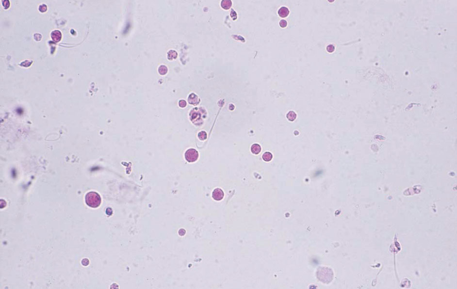
精液成分 40× 無染色
Semenal component 40× No staining
前立腺由来の性腺分泌物で,類でんぷん小体と呼ぶ。木の年輪のような層状構造を認める。
An amyloid body in prostate-derived gonadal secretions. It exhibits a layered structure similar to the annual rings of a tree trunk.

精液成分 40× S染色
Semenal component 40× S staining
類でんぷん小体は,S染色では赤色~紫色に染色される。染色像でも層状構造は確認できるが,染色後,時間が経過すると層状構造が不明瞭となる場合がある。
The amyloid body is stained red to purple with S staining. In a stained image, a layered structure may be confirmed. However, the layered structure may become unclear over time.

前立腺組織像 10× HE染色
Prostate histology 10× HE staining
腺腔内に層状構造を示す類でんぷん小体がみられる。
Amyloid bodies exhibiting a layered structure are found in the glandular cavities.

精液成分 40× S染色
Semenal component 40× S staining
精嚢由来の分泌物で,類円形で大小不同の無構造物質で,ゼラチン状を呈する。ゼラチン状の中には精子が封入されている。S染色では赤色~紫色に染色される。
Secretions from seminal vesicle, which are near-circular, nonstructural substances of different sizes, and gelatinous. Spermatozoa are enclosed in the gelatinous form. With S staining, they are stained red to purple.

精液成分 40× S染色
Semenal component 40× S staining
精嚢由来の性腺分泌物で,細長いものはろう様円柱に類似する場合がある。背景には典型的な精液成分がみられることなどから鑑別する。
A gonadal secretion derived from a seminal vesicle. When elongated, these may appear similar to waxy casts, but they can be differentiated when typical seminal components are found in the background.

精液成分 40× S染色
Seminal component 40× S staining
精嚢由来の性腺分泌物で,分泌物の辺縁は,比較的丸みを帯びていることが多いが,凹凸を認める場合もある。
A gonadal secretion derived from a seminal vesicle. In many cases, the contour of the secretions are relatively rounded; however, sometimes those are uneven.

精液成分 40× S染色
Seminal component 40× S staining
前立腺がん患者尿にみられた分泌物で,分泌物の内部には濃縮した多数の赤血球がみられる。
A seminal secretion observed in the urine of a patient with prostate cancer. A lot of concentrated red blood cells inside the secretion can be observed.

精液成分 40× S染色
Seminal component 40× S staining
細長く円柱状の分泌物であるが,角が丸みを帯びている。中央部分が桃色であるが,時間経過とともに青色に染色される。
An elongated cast-shaped secretion with rounded corners. The center portion is pink, but it becomes stained blue over time.

精液成分 40× S染色
Seminal component 40× S staining
大型の分泌物で,均一無構造を呈する。
A large secretion that exhibits homogeneity and has no structure.

精嚢組織像 40× HE染色
Seminal vesicle histology 40× HE staining
精嚢組織内に均一無構造の分泌物がみられる。
Uniform secretions with no structure are found in a seminal vesicle tissue.

精液成分 40× 無染色
Seminal component 40× No staining
大小不同の分泌物がみられる。分泌物の内部には空胞がみられる。
Secretions of different sizes containing vacuoles.

精液成分 40× S染色
Seminal component 40× S staining
細長く空胞変性円柱に類似する性腺分泌物である。背景に精子がみられることや分泌物の角が丸いことから鑑別する。
Slender secretions that are similar to vacuolar denatured casts. They can be differentiated from casts as they have rounded corners and there are spermatozoa in the background.
精液成分 40× S染色
Seminal component 40× S staining
前立腺由来の性腺分泌物で,レシチン顆粒と呼ぶ。大小不同の小型の円形成分で,赤血球や白血球と類似する。
Prostate-derived gonadal secretions, called lecithin granules. These are compact circular components of unequal size, similar to red and white blood cells.

大食細胞 40× S染色
Macrophage 40× S staining
精液成分の混入時にしばしば精子を貪食した大食細胞がみられる場合がある。
Macrophage cells that contain phagocytosed spermatozoa are often observed when a semen component is mixed into a urine sample.

大食細胞 40× S染色
Macrophages 40× S staining
急性前立腺炎患者尿にみられた大食細胞である。脂肪成分を貪食し卵円形脂肪体に類似するため鑑別に注意する。背景には精子がみられる。
Macrophages observed in the urine of a patient with acute prostatitis. They are similar to oval fat bodies because the macrophage cells phagocytize fat components. Thus, care must be taken during differentiation. Spermatozoa are seen in the background.

糞便の混入 40× S染色
Contamination by feces 40× S staining
透明なカプセル状の食物残渣で,カプセル部分は植物の細胞壁である。
Transparent, encapsulated food residues. The capsule portion is the plant cell wall.

糞便の混入 40× S染色
Contamination by feces 40× S staining
カプセル状の食物残渣である。ほとんどが肛門からの混入で乳児や女性にみられるが,憩室炎や大腸がんの膀胱浸潤などにより膀胱と腸との交通(膀胱腸瘻)がある場合には臨床的に重要な報告となりうる成分である。
Capsule-like food residue. Although most are found in infants or women due to contamination from the anus, they are clinically important if there is a passage between the bladder and the intestines (vesicointestinal fistula) caused by diverticulitis or bladder infiltration of colon cancer.

糞便の混入 40× 無染色
Contamination by feces 40× No staining
細長いカプセル状の食物残渣である。細長いものは円柱に類似する場合がある。
An elongated capsule-like food residue. Slender-shaped capsules may appear similar to casts.

糞便の混入 40× 無染色
Contamination by feces 40× No staining
豆類の棚状組織の細胞で柱状を呈する。日本人の糞便中に多くみられる。
Shelf-like cells of legumes exhibiting a columnar shape. They are commonly found in the feces of Japanese individuals.

糞便の混入 40× S染色
Contamination by feces 40× S staining
豆類の棚状組織の細胞で,豆類および味噌や豆腐の摂取により観察される。
Shelf-like cells of legumes observed in people who ate beans, miso, or tofu.

糞便の混入 40× S染色
Contamination by feces 40× S staining
細長いカプセル状の食物残渣である。カプセルの内部は顆粒円柱に類似する。
Elongated capsule-like food residue. The interior of the capsule is similar to a granular cast.

糞便の混入 40× S染色
Contamination by feces 40× S staining
植物の維管束を構成し,水分の通路となる導管である。多くはらせん状導管で,太さはさまざまである。
A duct that constitutes the vascular bundle of plants and becomes a passage for moisture. Many are spiral-shaped ducts and that thickness is various.

糞便の混入 40× S染色
Contamination by feces 40× S staining
カプセル状の食物残渣である。カプセルの内部は顆粒円柱に類似する。
A capsule-like food residue. The interior of the capsule is similar to a granular cast.

糞便の混入 40× 無染色
Contamination by feces 40× No staining
淡黄色~黄褐色の長方形~楕円形を呈する肉類由来の筋繊維である。顆粒円柱やろう様円柱と類似する。×400(対物レンズ40×)でよく観察すると均一に横筋がみられる。
A muscle fiber derived from meat appearing as a pale yellow to yellowish brown rectangle or oval shape, similar to a granular or waxy cast. When observed with ×400 (objective lens 40×), uniform, transverse stripes are observed.

糞便の混入 40× S染色
Contamination by feces 40× S staining
筋繊維で,平行する2辺を有し,円柱に類似する。糞便の混入は,背景には細菌がみられ,可能であれば採尿のやり直しを乞うとよい。
A muscle fiber with two parallel sides, similar to a cast. With fecal contamination, bacteria are found in the background. Thus, another urine sample should be collected if possible.

繊維 40× 無染色
Fiber 40× No staining
トイレットペーパーや衣類の繊維が混入する場合がある。細長いものは円柱に類似する。
Fibers of toilet paper and clothing may contaminate the sample. An elongated fiber appears similar to a cast.

繊維 40× S染色
Fiber 40× S staining
円柱に類似するが,厚みがあり辺縁と背景の境界線が明瞭である。
A fiber that appears similar to a cast; however, this fiber is thick and the boundary between the verge and the background is clear.

でんぷん粒 40× 無染色
Starch granules 40× No staining
中央に切れ込みがあり,奥歯状やバナナの輪切り状を呈する。光沢があり結晶成分と類似する。手袋や紙オムツなどのでんぷん粒が尿中に混入したものである。
With slits in the center, the granules are shaped like molars or banana slices. They are shiny and similar to crystalline components. Starch granules from gloves and paper diapers contaminated this urine.

でんぷん粒 40× 無染色
Starch granules 40× No staining
楕円形のでんぷん粒である。紙オムツからの混入である。楕円形のでんぷん粒は糞便混入時にもみられる場合がある。
Oval-shaped starch granules due to contamination from paper diapers. Oval-shaped starch granules may also be observed with fecal contamination.

でんぷん粒 40× S染色
Starch granules 40× S staining
楕円形のでんぷん粒である。紙オムツからの混入である。
Oval-shaped starch granules resulting from paper diaper contamination.

でんぷん粒 40× S染色
Starch granules 40× S staining
卵円形のでんぷん粒である。糞便の混入による。
Oval-shaped starch granules derived from fecal contamination.

混入物(紙オムツの吸水剤) 40× S染色
Contaminant (water absorptive materials of a paper
diaper) 40× S staining
紙オムツから尿を搾り出したもので,吸水剤が混入したものである。
Urine was squeezed out of a paper diaper, and the water absorptive material contaminated the urine.

混入物(紙オムツの吸水剤) 10× S染色
Contaminant (water absorptive materials of a paper
diaper) 10× S staining
紙オムツの吸水剤で,尿がゼリー状に固まる場合がある。この場合は,検査ができなくなるので可能であれば採尿のやり直しを乞うとよい。
Urine may be solidified into a jelly by a water absorbent from paper diapers. In such cases, a urine test becomes impossible. If possible, the urine sample should be recollected.

皮膚保護剤 10× S染色
Skin protective agent 10× S staining
回腸導管尿路変更術後患者尿で,しばしば皮膚開口部(Stoma)に塗布する皮膚保護剤が混入することがある。
Urine from a patient who had an ileal conduit diversion often contains skin protective agent applied to the skin around the stoma.
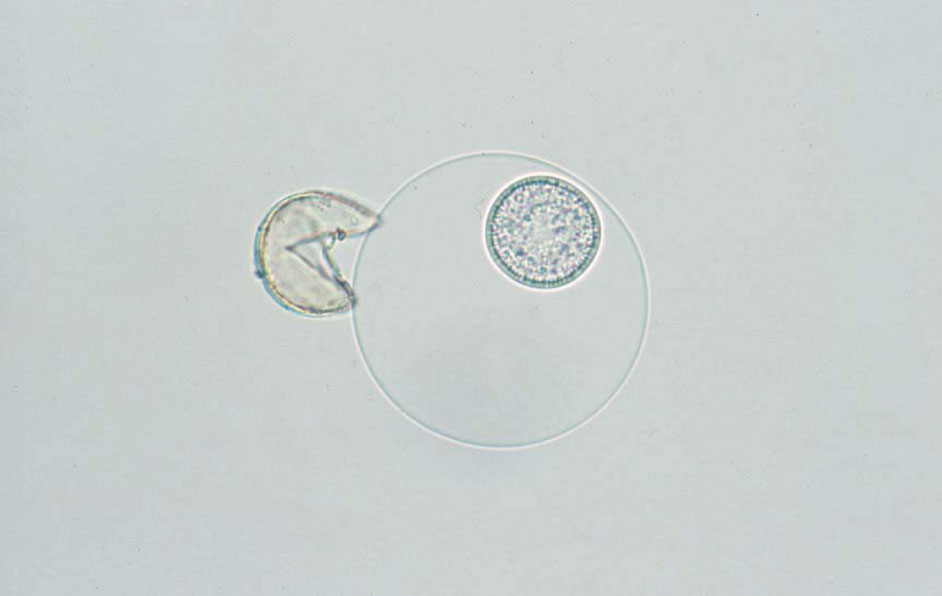
スギ花粉 40× 無染色
Cedar pollen 40× No staining
春先になると,スギ花粉の混入がみられることがある。花粉は黄褐色の球状で,1箇所突起を有する。水分を吸収すると外膜が破れ,カプセル状の成分が流出する。
In early spring, there may be contamination from cedar pollen. The pollen is observed as yellowish brown and spherical-shaped with one projection. When it absorbs moisture, the outer membrane breaks and capsule-like components emerge.

スギ花粉 40× S染色
Cedar pollen 40× S staining
S染色では赤く染まり,カプセル状の成分は,外側は青く,中央の核様成分が赤く染まる。
With the S staining, cedar pollen stains red; the capsule-like component stains blue on the outside and red in the central nucleus-like component.

ダニ 20× 無染色
Mite 20× No staining
大部分は外部からの混入であるが,持続してみられる場合,人体寄生例も報告があるので注意が必要である。大部分は一過性にみられる。
Although the majority of mite contamination is external, attention should be paid when it is found persistently, as cases of human parasitism have been reported. The majority of mite contamination cases are transient.

ダニ(卵) 40× 無染色
Mite (egg) 40× No staining
Figure 3.432と同一症例である。成虫と同時に卵がみられる場合がある。
The same case as Figure 3.432. The eggs and adult mites may be found simultaneously.

プランクトン 40× S染色
Plankton 40× S staining
淡水性プランクトンである輪虫類の混入である。
Contamination with rotifer, freshwater plankton.

プランクトン 40× 無染色
Plankton 40× No staining
淡水性プランクトンの混入である。繊毛を有し,活発に活動する。
Freshwater plankton contamination. It has cilia and moves actively.

プランクトン 40× 無染色
Plankton 40× No staining
日当たりのよい蓄尿袋内で繁殖した植物系のプランクトンである。光合成を行っており緑色を呈する。
Phytoplankton breeding in a urine collection bag at sunny place. They are photosynthetic and exhibit green.

鱗粉 40× S染色
Scale 40× S staining
蛾や蝶の羽の粉が混入したものである。
Contamination with moth or butterfly wing scales.

粘液 10× S染色
Mucous 10× S staining
女性の場合,帯下などが生殖器から,白血球,細菌,扁平上皮細胞が粘液に絡んで混入する場合がある。
In females, the flow from the genital organs may contaminate the urine. In such cases, white blood cells, bacteria, and squamous epithelial cells may be mixed in the mucus.

粘液 40× S染色
Mucous 40× S staining
粘液に多数の細菌と白血球が絡み合っている。
Numerous bacteria and white blood cells are mixed with the mucus.

脂肪球 40× S染色
Fat globules 40× S staining
扁平上皮細胞は,しばしば細胞質に脂肪変性を伴うことがある。この脂肪球は,ネフローゼ症候群などでみられる脂肪球と同様,Sudan III染色に赤く染まる。
Squamous epithelial cells are often accompanied by adipose degeneration in the cytoplasm. Similar to the globules associated with nephrotic syndrome, they stains red with Sudan III stain.

脂肪球 40× S染色
Fat globules 40× S staining
扁平上皮細胞上にみられた脂肪球である。大小不同で光沢を有する。
Fat globules found on squamous epithelial cells. They are of differing sizes and are glossy.

脂肪球 40× 無染色
Fat globules 40× No staining
外陰部に塗り薬(抗真菌薬)を流布していた患者尿で,塗り薬の混入により多数の脂肪球がみられる。光沢があり大小不同を呈する。
Urine from a patient who applied ointment (antifungal medicine) to the vulva; a large number of fat globules are observed due to contamination of the medicine. The globules are shiny and exhibit different sizes.

アーチファクト 20× S染色
Artifacts 20× S staining
カバーガラスを載せた後,カバーガラスがずれて生じるもので,平行部を有することから円柱と類似する。向きが一定なのが特徴である。
This occurs when the coverslip shifts after it is placed on a specimen. They are similar to casts because they have parallel sides, but are characterized by a consistent direction.