抄録
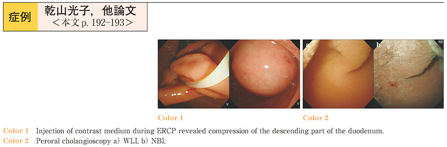
A 62-year-old woman referred to us for the management of suspected acute cholecystitis was hospitalized. Abdominal CT showed choledocholithiasis and a duodenal diverticulum. Esophagogastroduodenoscopy (EGD) revealed compression of the descending part of the duodenum in the same region as the diverticulum on the abdominal CT. ERCP was performed for the choledocholithiasis. Numerous stones were seen in the cystic lesion upon injection of contrast medium, and continuous injection revealed a cystic lesion measuring 22 mm in diameter. A diagnosis of choledochocele was made. The major duodenal papilla could not be identified on the EGD because of the ballooned choledochocele filled with contrast medium. A needle knife precut was made, and the choledochocele subsequently collapsed after the CBD stones were removed. Endoscopic hemostasis using coagulation and hemoclip was performed to stop oozing from the site of the precut. ERCP was performed again 1 week later. The major duodenal papilla was identified and EST was performed, followed by CBD stone removal. The level of pancreatic amylase in the bile was high at the first ERCP and decreased after the precut. Functional pancreaticobiliary maljunction was diagnosed, which disappeared after the precut and EST.