抄録
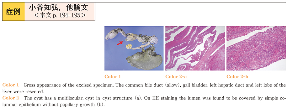
A 28-year-old woman visited our hospital with the complaints of itching and jaundice. Blood examination showed elevation of the hepatobiliary enzymes and bilirubin. Abdominal US, CT and MRI revealed cystic lesions with septae spreading contiguously to the left lobe, left hepatic duct and common bile duct. Endoscopic retrograde cholangiography (ERC) demonstrated translucent components in the bile duct, and CT immediately after the ERC suggested that one partial cystic lumen filled with contrast medium was communicating with the bile duct. Therefore, we made the diagnosis of intraductal papillary neoplasm of the bile duct (IPNB) preoperatively. However, histological examination of the resected specimens after a left hepatectomy revealed the diagnosis of mucinous cystic neoplasm (MCN) , because the ovarian-like stroma showed positive immunostaining for the estrogen receptor and progesterone receptor. At present, 10 months after the surgery, there is no evidence of tumor recurrence.
Hepatobilialy cystic neoplasms are classified into MCNs, which contain an ovarian-like stroma, and IPNB, which do not contain an ovarian-likes stroma. Although tens of cases of MCN have been reported, the reason for the existence of the stromal cells is still unclear. Our case was atypical in respect of its macroscopic appearance and its communication to the bile duct. Herein, we report this case as we believe that it will contribute to elucidation of the pathogenesis.