抄録
A 72-year-old man presented to the hospital with tonsillar swelling. Histopathological examination of a biopsy specimen from the tonsil showed diffuse proliferation of abnormal lymphocytes. Immunohistochemical analysis revealed positive staining for CD20, CD5 and Cyclin D1. These findings were compatible with the diagnosis of mantle cell lymphoma (MCL) . PET-CT revealed abnormal accumulation in the
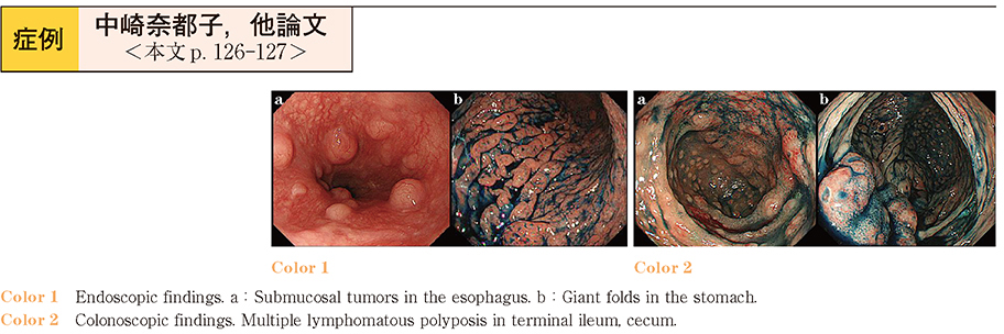
stomach and duodenum, in addition to that in the tonsils and lymph nodes. As we suspected invasion of the gastric tract by the MCL, esophagogastroduodenoscopy (EGD) was performed, which showed submucosal tumors in the esophagus and duodenum, and giant folds in the stomach. Colonoscopy showed lymphomatous polyposis in the terminal ileum, cecum and rectum.
The patient was treated with first-line chemotherapy. EGD after 2 courses of chemotherapy revealed a decrease in the size of all the lesions; the lesions reduced even further in size after 4 courses of chemotherapy, as compared to the sizes before the start of therapy. With this medical history, the patient was admitted to the hospital with an 8-month history of bloody stool. Colonoscopy revealed lymphoid follicles in the terminal ileum, cecum and rectum. Since histopathological examination of HE-stained sections of biopsy specimens showed normal lymphoid follicles, we did not perform immunohistochemical examination.
When multiple lymphoid follicles are found on endoscopy, it is important to consider the possibility of malignant lymphoma invading the gastrointestinal tract.