抄録
Patient : A 68-year-old woman.
Major complaint : Epigastric pain.
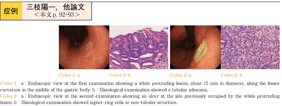
History of current disease : The patient occasionally had epigastric pain and underwent upper gastrointestinal endoscopy each year. Atrophic gastritis and Helicobacter pylori infection were diagnosed, however, the patient refused eradication therapy. Examination this year showed a white protruding lesion, about 15 mm in diameter, along the lesser curvature in the middle of the gastric body. The histopathological diagnosis was tubular adenoma.
Clinical course : Endoscopic mucosal resection was considered, however, the patient did not provide consent for the procedure. Eradication therapy was administered to prevent the development of new lesions, and first-line eraHelicobacter pyloridication was successful. Follow-up endoscopy was performed 1 month after the complication of eradication therapy, to exclude gastric cancer. An ulcer had developed at the site previously occupied by the white protruding lesion. Histopathological examination of biopsy specimens obtained from around the ulcer showed a cancer nest with signet-ring cells or non-tubular structure. Proximal gastrectomy was performed. Histopathological examination revealed a double cancer consisting of a non-solid type (por2) and a well-differentiated type (tub1) of gastric cancer. Subsequent review of the endoscopic results showed pale mucosa surrounding the white protrusion, suggesting the presence of gastric cancer around the protrusion.
Conclusion : We describe an interesting case of early-stage gastric cancer, consisting of undifferentiated carcinoma and differentiated carcinoma, that followed ulcer formation in the cancer after eradication of H. pylori.