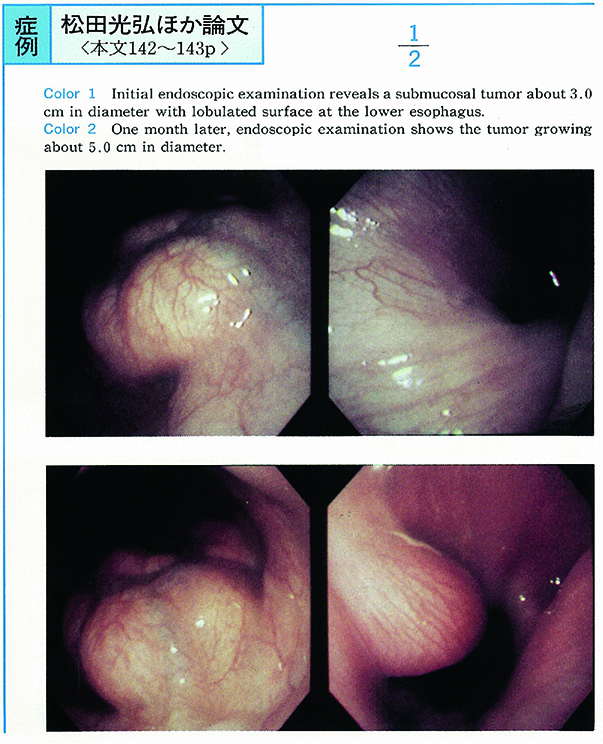
A 49-year-old male was admitted to our hospital with the chief complaint of dysphagia. Endoscopic examination revealed a submucosal tumor about 3.0cm in diameter with lobulated surface at the lower esophagus. One month later, an endoscopic examination showed the tumor growing to about 5.0cm in diameter. Endoscopic ultrasonographic examination revealed the low echoic lobulated area with calcification.
According to those findings, the tumor was suspected to be a leiomyosarcoma. Excision of the lower portion of the esophagus was performed on June 21, 1994. Pathological examination showed a leiomyoma.
A leiomyoma of the esophagus is rare. It generally has a smooth surface and grows very slow. But our case was diagnosed a leiomyosarcoma before the operation, because it had a lobulated surface and grew comparatively fast.