46 巻
選択された号の論文の65件中1~50を表示しています
掲載論文カラー写真集
-
1995 年 46 巻 p. 1-20
発行日: 1995年
公開日: 2015/05/01
PDF形式でダウンロード (25511K)
内視鏡の器械と技術
-
1995 年 46 巻 p. 56-58
発行日: 1995/06/16
公開日: 2015/05/01
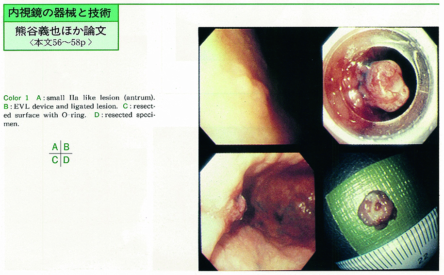 PDF形式でダウンロード (1002K)
 -
1995 年 46 巻 p. 59-61
発行日: 1995/06/16
公開日: 2015/05/01
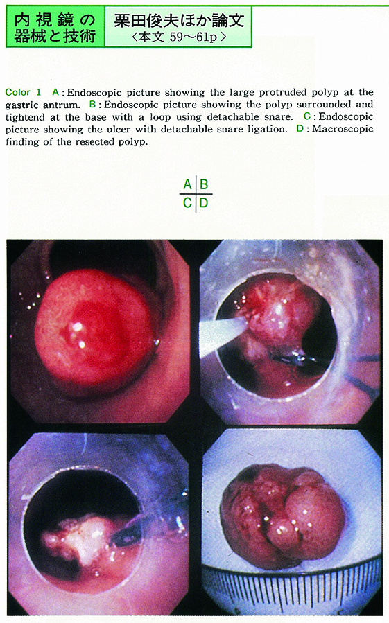 PDF形式でダウンロード (289K)
臨床研究
-
1995 年 46 巻 p. 62-64
発行日: 1995/06/16
公開日: 2015/05/01
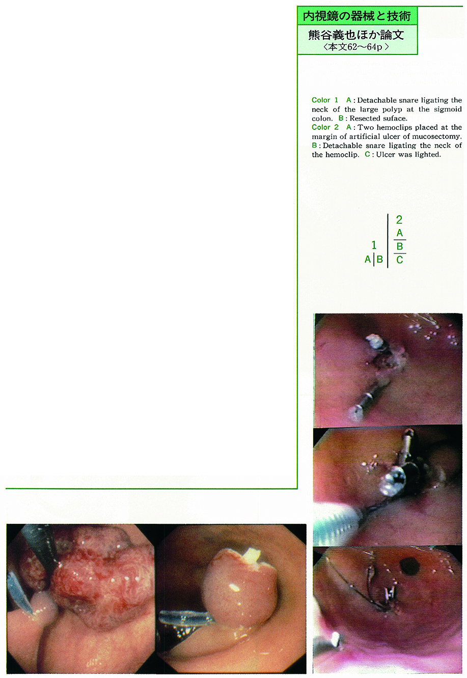 PDF形式でダウンロード (311K)
 -
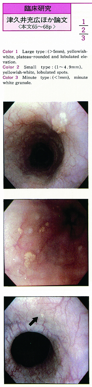
1995 年 46 巻 p. 65-68
発行日: 1995/06/16
公開日: 2015/05/01
 PDF形式でダウンロード (827K)
 -
1995 年 46 巻 p. 69-73
発行日: 1995/06/16
公開日: 2015/05/01
PDF形式でダウンロード (610K) -
1995 年 46 巻 p. 74-77
発行日: 1995/06/16
公開日: 2015/05/01
PDF形式でダウンロード (440K) -
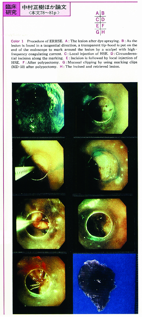
1995 年 46 巻 p. 78-81
発行日: 1995/06/16
公開日: 2015/05/01
 PDF形式でダウンロード (537K)
 -
1995 年 46 巻 p. 82-86
発行日: 1995/06/16
公開日: 2015/05/01
PDF形式でダウンロード (1043K)
症例
-
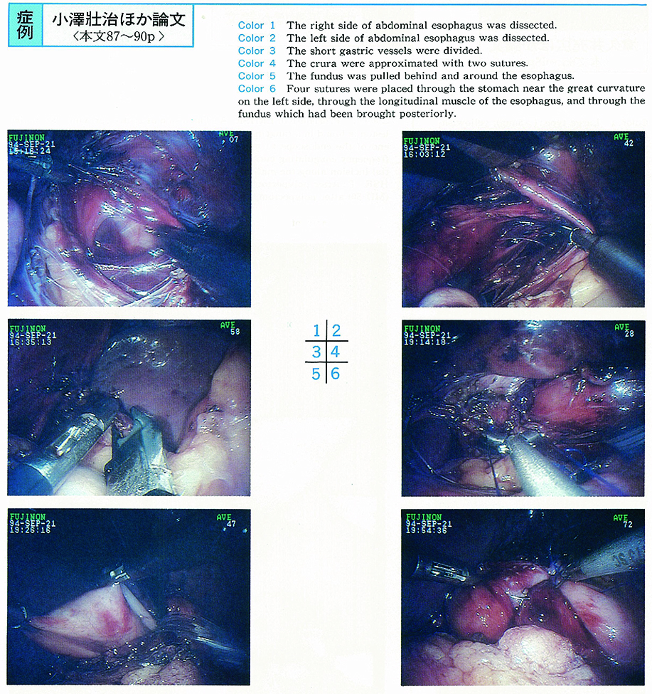
1995 年 46 巻 p. 87-90
発行日: 1995/06/16
公開日: 2015/05/01
 PDF形式でダウンロード (994K)
 -
1995 年 46 巻 p. 91-93
発行日: 1995/06/16
公開日: 2015/05/01
 PDF形式でダウンロード (412K)
 -
1995 年 46 巻 p. 94-96
発行日: 1995/06/16
公開日: 2015/05/01
 PDF形式でダウンロード (1012K)
 -
1995 年 46 巻 p. 97-100
発行日: 1995/06/16
公開日: 2015/05/01
 PDF形式でダウンロード (1775K)
 -
1995 年 46 巻 p. 101-103
発行日: 1995/06/16
公開日: 2015/05/01
 PDF形式でダウンロード (631K)
 -
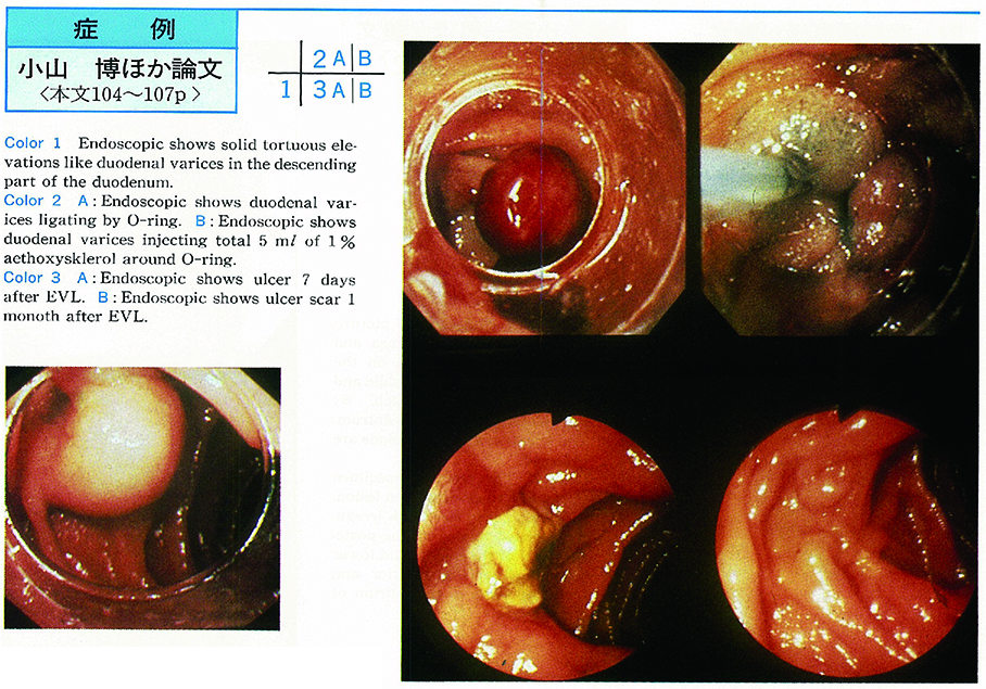
1995 年 46 巻 p. 104-107
発行日: 1995/06/16
公開日: 2015/05/01
 PDF形式でダウンロード (1770K)
 -
1995 年 46 巻 p. 108-111
発行日: 1995/06/16
公開日: 2015/05/01
 PDF形式でダウンロード (1597K)
 -
1995 年 46 巻 p. 112-114
発行日: 1995/06/16
公開日: 2015/05/01
 PDF形式でダウンロード (299K)
内視鏡の器械と技術
-
1995 年 46 巻 p. 116-117
発行日: 1995/06/16
公開日: 2015/05/01
PDF形式でダウンロード (342K) -
1995 年 46 巻 p. 118-119
発行日: 1995/06/16
公開日: 2015/05/01
PDF形式でダウンロード (214K) -
1995 年 46 巻 p. 120-121
発行日: 1995/06/16
公開日: 2015/05/01
PDF形式でダウンロード (493K)
臨床研究
-
1995 年 46 巻 p. 122-123
発行日: 1995/06/16
公開日: 2015/05/01
PDF形式でダウンロード (248K) -
1995 年 46 巻 p. 124-125
発行日: 1995/06/16
公開日: 2015/05/01
 PDF形式でダウンロード (290K)
 -
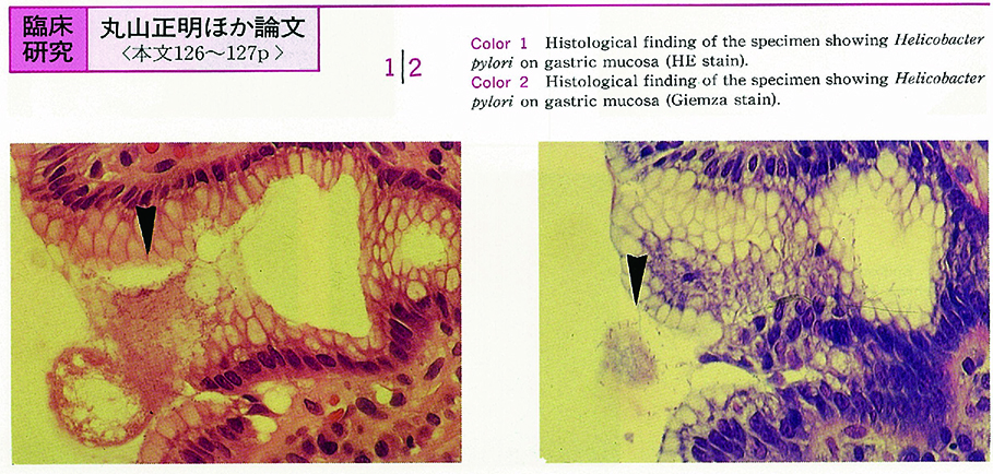
1995 年 46 巻 p. 126-127
発行日: 1995/06/16
公開日: 2015/05/01
 PDF形式でダウンロード (254K)
 -
1995 年 46 巻 p. 128-129
発行日: 1995/06/16
公開日: 2015/05/01
PDF形式でダウンロード (269K) -
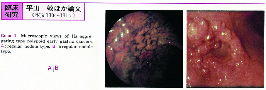
1995 年 46 巻 p. 130-131
発行日: 1995/06/16
公開日: 2015/05/01
 PDF形式でダウンロード (231K)
 -
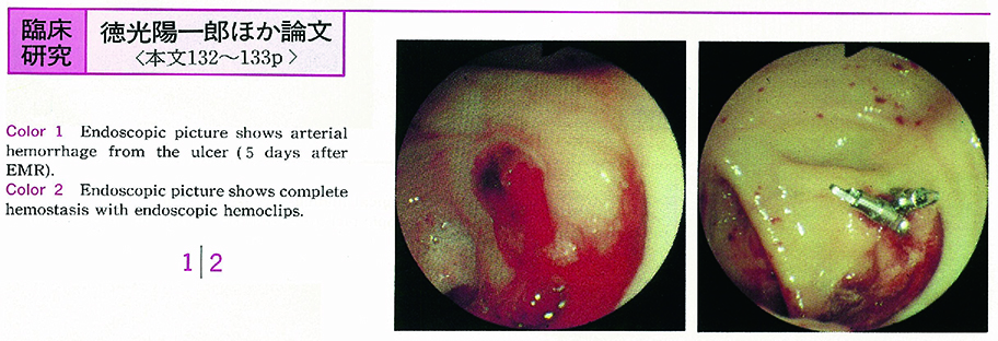
1995 年 46 巻 p. 132-133
発行日: 1995/06/16
公開日: 2015/05/01
 PDF形式でダウンロード (341K)
 -
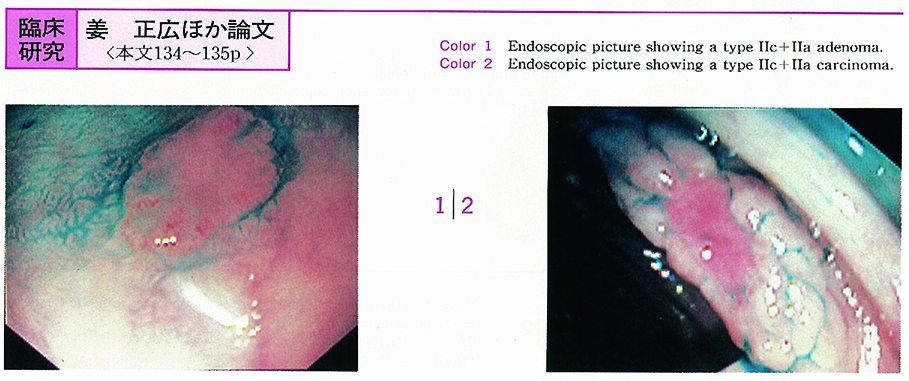
1995 年 46 巻 p. 134-135
発行日: 1995/06/16
公開日: 2015/05/01
 PDF形式でダウンロード (276K)
 -
1995 年 46 巻 p. 136-137
発行日: 1995/06/16
公開日: 2015/05/01
 PDF形式でダウンロード (581K)
症例
-
1995 年 46 巻 p. 138-139
発行日: 1995/06/16
公開日: 2015/05/01
 PDF形式でダウンロード (1579K)
 -
1995 年 46 巻 p. 140-141
発行日: 1995/06/16
公開日: 2015/05/01
 PDF形式でダウンロード (564K)
 -
1995 年 46 巻 p. 142-143
発行日: 1995/06/16
公開日: 2015/05/01
 PDF形式でダウンロード (1361K)
 -
1995 年 46 巻 p. 144-145
発行日: 1995/06/16
公開日: 2015/05/01
 PDF形式でダウンロード (958K)
 -
1995 年 46 巻 p. 146-147
発行日: 1995/06/16
公開日: 2015/05/01
 PDF形式でダウンロード (822K)
 -
1995 年 46 巻 p. 148-149
発行日: 1995/06/16
公開日: 2015/05/01
 PDF形式でダウンロード (656K)
 -
1995 年 46 巻 p. 150-151
発行日: 1995/06/16
公開日: 2015/05/01
 PDF形式でダウンロード (1362K)
 -
1995 年 46 巻 p. 152-153
発行日: 1995/06/16
公開日: 2015/05/01
 PDF形式でダウンロード (1099K)
 -
1995 年 46 巻 p. 154-155
発行日: 1995/06/16
公開日: 2015/05/01
 PDF形式でダウンロード (689K)
 -
1995 年 46 巻 p. 156-157
発行日: 1995/06/16
公開日: 2015/05/01
 PDF形式でダウンロード (1106K)
 -
1995 年 46 巻 p. 158-159
発行日: 1995/06/16
公開日: 2015/05/01
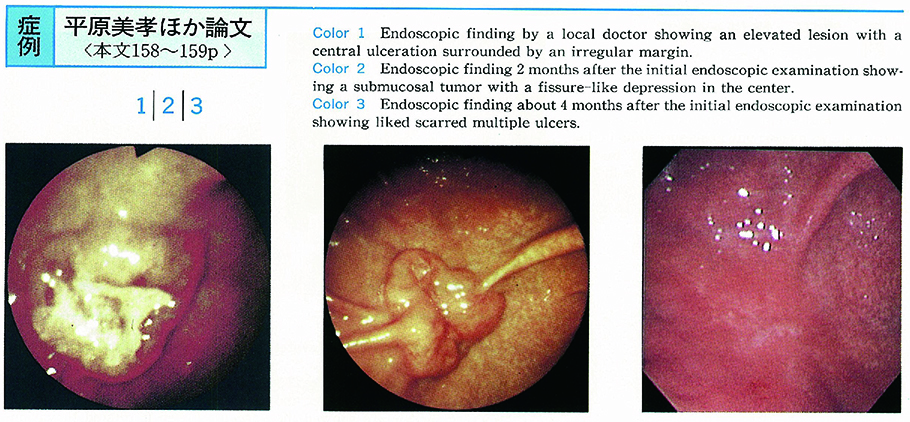 PDF形式でダウンロード (1018K)
 -
1995 年 46 巻 p. 160-161
発行日: 1995/06/16
公開日: 2015/05/01
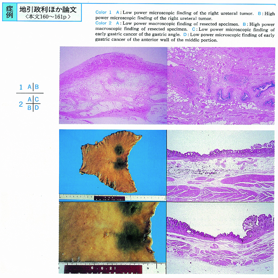 PDF形式でダウンロード (1055K)
 -
1995 年 46 巻 p. 162-163
発行日: 1995/06/16
公開日: 2015/05/01
 PDF形式でダウンロード (816K)
 -
1995 年 46 巻 p. 164-165
発行日: 1995/06/16
公開日: 2015/05/01
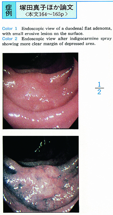 PDF形式でダウンロード (722K)
 -
1995 年 46 巻 p. 166-167
発行日: 1995/06/16
公開日: 2015/05/01
 PDF形式でダウンロード (963K)
 -
1995 年 46 巻 p. 168-169
発行日: 1995/06/16
公開日: 2015/05/01
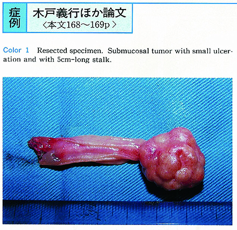 PDF形式でダウンロード (1122K)
 -
1995 年 46 巻 p. 170-171
発行日: 1995/06/16
公開日: 2015/05/01
 PDF形式でダウンロード (1176K)
 -
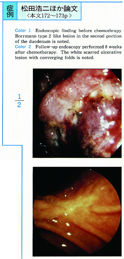
1995 年 46 巻 p. 172-173
発行日: 1995/06/16
公開日: 2015/05/01
 PDF形式でダウンロード (1088K)
 -
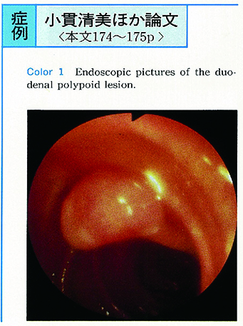
1995 年 46 巻 p. 174-175
発行日: 1995/06/16
公開日: 2015/05/01
 PDF形式でダウンロード (985K)
 -
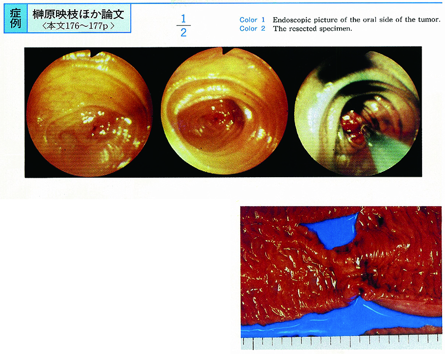
1995 年 46 巻 p. 176-177
発行日: 1995/06/16
公開日: 2015/05/01
 PDF形式でダウンロード (724K)
 -
1995 年 46 巻 p. 178-179
発行日: 1995/06/16
公開日: 2015/05/01
 PDF形式でダウンロード (1168K)
 -
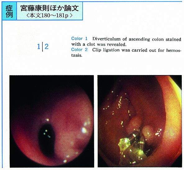
1995 年 46 巻 p. 180-181
発行日: 1995/06/16
公開日: 2015/05/01
 PDF形式でダウンロード (272K)