症例
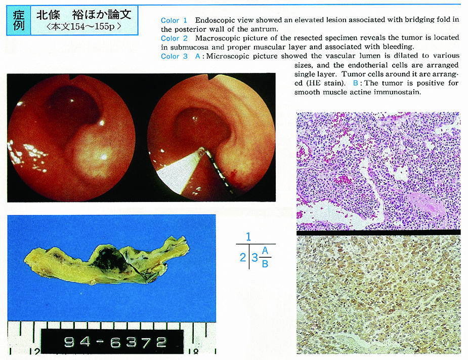
超音波内視鏡にて観察しえた胃glomus腫瘍の1例
北條 裕, 石原 学, 近藤 栄作, 有木 寿史, 貴島 佳世, 進藤 彦二, 青柳 徹二, 松崎 浩二, 尾崎 元信, 岩崎 格, 西野 執, 成木 行彦, 大塚 幸雄, 高木 純人, 辻本 志朗, 三浦 妙太
著者情報
-
北條 裕
東邦大学医学部/第1内科
-
石原 学
東邦大学医学部/第1内科
-
近藤 栄作
東邦大学医学部/第1内科
-
有木 寿史
東邦大学医学部/第1内科
-
貴島 佳世
東邦大学医学部/第1内科
-
進藤 彦二
東邦大学医学部/第1内科
-
青柳 徹二
東邦大学医学部/第1内科
-
松崎 浩二
東邦大学医学部/第1内科
-
尾崎 元信
東邦大学医学部/第1内科
-
岩崎 格
東邦大学医学部/第1内科
-
西野 執
東邦大学医学部/第1内科
-
成木 行彦
東邦大学医学部/第1内科
-
大塚 幸雄
東邦大学医学部/第1内科
-
高木 純人
東邦大学医学部/第2外科
-
辻本 志朗
東邦大学大森病院/病理
-
三浦 妙太
東邦大学大森病院/病理
ジャーナル
フリー
1995 年 46 巻 p. 154-155
詳細
- 発行日: 1995/06/16 受付日: - J-STAGE公開日: 2015/05/01 受理日: - 早期公開日: - 改訂日: -
PDFをダウンロード (689K)
メタデータをダウンロード
RIS形式
BIB TEX形式
テキスト
メタデータのダウンロード方法
発行機関連絡先
(EndNote、Reference Manager、ProCite、RefWorksとの互換性あり)
(BibDesk、LaTeXとの互換性あり)


