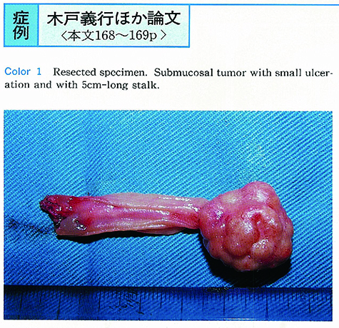
A 50-year-old woman admitted to our hospital with melena. Upper GI endoscopy and colonoscopy did not show any particular abnormality. Celiac and superior mesenteric angiography also failed to diagnosis. One hundred ml barium meal study of small intestine and hypotonic duodenography revealed duodenal tumor at its ascending part. Tumor was resected operatively and was 2cm in size with 5cm-long stalk. Pathologically this tumor was carcinoid, argyrophil-positive and argentaffin-negative. Histologic type by Soga-classification was A+C mixed type.
This case was characterized by bleeding, its shape and its location. Melena is recognized in 9% of duodenal carcinoids in Japan but carcinoid with 5cm-long stalk and originated from ascending part of duodenum is very rare.
Diagnosis of gastrointestinal bleeding, endoscopic and angiographic failed to, is difficult. In such a case barium meal study and compression method is important to detect the lesion.