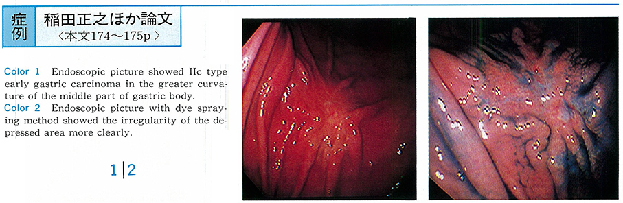
A 65-year-old woman who had an abnormality pointed out by radiological examination at gastric mass survey in November 1994, came to our center. Endoscopic and Radiological examination revealed irregular shaped depressed lesion with fold conversion in greater curvature of the middle portion of gastric body. Clinical diagnosis was type of IIc early undifferentiated gastric cancer. But biopsy specimen was differentiated tubler adenocarcinoma.
From the result of histological diagnosis, it reversed as follows. (1) The outline of lesion was not so well defined, (2) with gradually narrowed the fold, (3) a few small nodules in the depressed lesion, (4) reddish mucosa suggested the tubuler pattern of mucosed pit. These figures were matched the typical figure of defferentiated adenocarcinoma. Gastrectomy was carried out. By the histopathological study, this carcinoma was shown to be differentiated type adenocarcinoma arising from fundic gland mucosa. It is invasion was limited to the submucosal layer.