消化器内視鏡の進歩:Progress of Digestive Endoscopy
Online ISSN : 2189-0021
Print ISSN : 0389-9403
47 巻
選択された号の論文の77件中1~50を表示しています
掲載論文カラー写真集
臨床研究
  • 岩切 勝彦, 中川 義也, 琴寄 誠, 山田 久木, 杉浦 敏昭, 二神 生爾, 末岡 伸夫, 川上 明彦, 瀬底 正彦, 香川 隆男, 平 ...
    1995 年 47 巻 p. 48-51
    発行日: 1995/12/08
    公開日: 2015/05/01
    ジャーナル フリー
     胃液内総胆汁酸濃度・pHと逆流性食道炎の重症度,また胃液内総胆汁酸濃度と胃液pHの関係について検討を行った。30人の逆流性食道炎患者は重症度により,軽症(色調変化型食道炎 ; 9例),中等症(融合のないびらん潰瘍型食道炎 ; 12例),重症(融合するびらん潰瘍型食道炎 ; 9例)の3群に分類した。胃液は早朝空腹時に極力反射を防止し,胃管を胃内に挿入した。右側臥位にして1時間安静後,胃液を採取し遠心分離により粘液除去後,総胆汁酸濃度は酵素法により,pHはpHメーターにより測定した。胃液内総胆汁酸濃度は重症群の融合するびらん潰瘍型食道炎(1966±778µM,M±SE)が,軽症群(45±17µM),中等症群(43±15µM)の食道炎に比し,有意に高値であった。胃液内pHは各群に差は認めなかった。逆流性食道炎30例中7例は,酸との共存により食道粘膜障害を惹起しうる200µM以上の胆汁酸が存在した。200µM以上の胆汁酸を認めた7例中5例の胃液pHは4.0未満の酸性域を示した。以上より,高濃度胆汁酸の存在は融合を伴うびらん潰瘍型食道炎を惹起する可能性が考えられた。また胃液内pHが酸性域であっても,胆汁酸の存在は否定できなかった。
  • 木場 崇剛, 山口 肇, 白尾 国昭, 近藤 仁, 小野 裕之, 佃 博, 小松 嘉人, 清水 靖仁, 平山 敦, 小黒 八七郎, 加藤 抱 ...
    1995 年 47 巻 p. 52-55
    発行日: 1995/12/08
    公開日: 2015/05/01
    ジャーナル フリー
     国立がんセンター中央病院で1991年1月から1995年2月までの食道内視鏡的粘膜切除(EMR)を施行した40症例46病変について検討した。症例の年齢は46歳から81歳まで平均63.8歳であり,男女比は35 : 5であった。部位はImが32病変と多く,次はEiの9病変であった。組織型は全例扁平上皮癌であり,主肉眼型は0-Ⅱcが最も多く34病変(74%),0-Ⅱaが6病変(13%),0-Ⅱbが6病変(13%)で,切除方式は分割切除が31病変(67%)であり残りは一括切除であった。深達度はep,mm1が20例,㎜2が13例,mm3が5例,smが2例であった。EMRは食道粘膜癌に対する根治療法として有用であると考えられ,合併症としての狭窄はブジーを使用することにより解消可能であるため,全周性切除となる病巣に対しても積極的に行うべきである。また,完全切除と病理組織学的に判定された症例でも再発することがあるため,治療後すべての症例で十分な経過観察が必要である。今後は十分な症例を重ねた後,適応条件の拡大も検討必要があると思われた。
  • 渋谷 進, 飯田 浩行, 河島 孝彦, 青柳 啓之, 近森 文夫, 高瀬 靖広
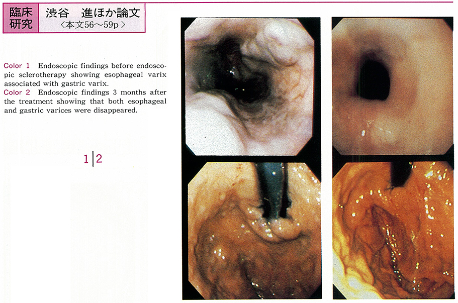
    1995 年 47 巻 p. 56-59
    発行日: 1995/12/08
    公開日: 2015/05/01
    ジャーナル フリー
     現在,左胃静脈と短胃静脈を連絡した噴門静脈叢は,食道静脈瘤の供血路の1つとして重要となっている。そこで今回われわれは,初回硬化療法時に2-5時方向および7-11時方向の食道静脈瘤から静脈瘤内注入された血管造影剤添加硬化剤にて得られた背臥位および側臥位の食道静脈瘤造影像(以下EVIS)から,左胃静脈および短胃静脈と噴門静脈叢の関連について検討した。対象は食道静脈瘤症例7例である。その結果,2-5時方向の食道静脈瘤から硬化剤を注入した場合には,左胃静脈の関与した噴門静脈叢が全例に,7時-11時方向の場合には短胃静脈が関与した噴門静脈叢が全例に認められた。また,左胃静脈が関与した噴門静脈叢は胃壁の背側,短胃静脈のものは腹側を中心に存在した。噴門静脈叢には胃壁近傍および胃壁外のものが認められた。また,左胃静脈および短胃静脈が関与した噴門静脈叢は全例で互いに連絡していた。治療前に内視鏡所見でみられた食道静脈瘤に連なった胃静脈瘤は治療後全例で消失した。以上から,噴門静脈叢には胃壁内のものと胃壁外のものとがあり,左胃静脈および短胃静脈が関与した噴門静脈叢同士が連絡する部位は胃壁内であると思われた。今後,この両方向からのEVISは噴門部の門脈血行路の解明に有用な手段となると思われた。
  • 大谷 吉秀, 大上 正裕, 吉田 昌, 久保田 哲朗, 熊井 浩一郎, 北島 政樹
    1995 年 47 巻 p. 60-63
    発行日: 1995/12/08
    公開日: 2015/05/01
    ジャーナル フリー
     胃粘膜下腫瘍のなかで,頻度の高い悪性腫瘍である平滑筋肉腫に対する腹腔鏡下手術の適応と方法について述べる。教室における胃粘膜下腫瘍に対する腹腔鏡下胃局所切除術の適応は,①腫瘍径2から5cmで,噴門もしくは幽門輪にかからない,②腫瘍の増大傾向を認め,悪性腫瘍が疑われる。なお,組織学的診断が確定している場合はそれぞれの治療方針に従うとしている。手術は腹腔鏡下に,2本の鉗子で腫瘍近傍の胃壁を把持挙上して,自動縫合器を用いてsurgical marginを十分確保して楔状切除を行った。これまで7例の平滑筋肉腫に本法を施行した。全例手術翌日から経口摂取を開始し,術後5から7日で退院した。胃筋原性腫瘍の特徴として,病理組織学的診断が得にくいこと,内視鏡による切除が困難なこと,良性腫瘍の可能性もあり侵襲の大きい開腹手術の決断が下されにくいこと,などがあげられる。そのような観点から,われわれの試みている腹腔鏡下胃局所切除術は,開腹手術に比べ低侵襲性で,安全,確実であり,症例を選択することによって平滑筋肉腫に対して有用な治療法となりうる。
  • 谷 雅夫, 井上 晴洋, 神戸 文雄, 斎藤 直也, 竹下 公矢, 遠藤 光夫
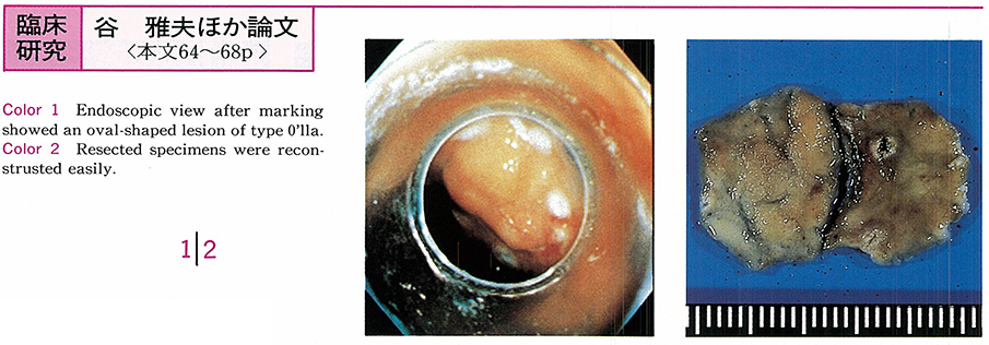
    1995 年 47 巻 p. 64-68
    発行日: 1995/12/08
    公開日: 2015/05/01
    ジャーナル フリー
     われわれは早期胃癌の内視鏡治療として,高分化型,2cm以下の0Ⅱa病変,1cm以下ul(-)の0Ⅱb病変,1cm未満ul(-)の0Ⅱc病変を絶対適応として,粘膜切除術(EMRC法)を第一選択として施行している。1.5cm未満の隆起型や明瞭な陥凹型病変では一括切除を行うが,1.5cm以上の隆起型,わずかな陥凹型や平坦型病変では,十分な断端を得るために計画的分割切除を行っている。分割切除に際しては,1個1個の標本を大きくマーキングを含めて切除することが再構築にとって重要で,また1個の標本中に病変の多くを含めることが病理組織学的検索にとって重要である。現在までにEMRC法で切除した胃腫瘍性病変50例62病変中,41病変は一括切除例,17病変は計画的分割切除例,4病変は一括切除を試みて遺残し,追加切除を加えたものであった。再構築が可能と考えられた病変は,計画的分割切除で17病変中14病変(82%),追加切除で0%であった。3-34ヵ月の経過観察中に遺残再発を認めたものは,一括切除例で41病変中1病変(2.4%),追加切除例で4病変中1病変(25%)の2病変であり,計画的分割切除例には相対適応例も含めて遺残再発を認めていない。粘膜切除の理想は一括切除であるが,病変によってはEMRC法による積極的な計画的分割切除が遺残再発の防止につながると考えられた。
  • 栗原 直人, 熊井 浩一郎, 大谷 吉秀, 大上 正裕, 久保田 哲朗, 北島 政樹
    1995 年 47 巻 p. 69-72
    発行日: 1995/12/08
    公開日: 2015/05/01
    ジャーナル フリー
     早期胃癌に対する治療法を選択するにあたり,内視鏡による病変の範囲や深達度診断が重要である。教室ではsm癌症例に対して標準術式(D2郭清)を行い,m癌に対しては開腹縮小手術(D1郭清),腹腔鏡下手術(lesion lifting法による腹腔鏡下胃局所切除術,腹腔鏡下胃内粘膜切除術),内視鏡的粘膜切除術などを適応基準により選択している。このような治療法の選択に重要なm癌の深達度診断精度向上を目的として20MHzの細径プローブ(リニア型,ラジアル型)を用いた超音波内視鏡検査(EUS)を行った。48症例に対して病理組織学的診断との対比からEUS深達度診断に検討を加えた。m癌の深達度正診率は全体として88.9%であるが,従来のリニア型プローブと比較して,最近開発されたラジアル型プローブを用いた正診率に向上がみられた。外科病理学的検討では肉眼型で隆起型,組織型で高分化型腺癌,INFαの症例で高い正診率が得られた。EUSにてm癌と診断したが,術後病理組織学的にsmと診断された症例は3例あり,病型としては陥凹型,組織型はtub2-porであった。m癌に対する治療法の選択にあたり術前診断として病変の大きさ,肉眼型,組織型を明らかにするとともに,より高い正診率が得られる20MHz細径プローブを用いたEUSは,壁深達度診断に有用であると考えられた。
  • 石田 博, 末岡 伸夫, 平川 恒久, 小林 正文
    1995 年 47 巻 p. 73-77
    発行日: 1995/12/08
    公開日: 2015/05/01
    ジャーナル フリー
     Helicobacter pylori(以下H. pylori)陽性十二指腸潰瘍90例を対象にプロトンポンプ阻害剤と抗生剤の併用による除菌療法を行った。lansoprazole(以下LPZ)は30mg1日1回6週間投与した。LPZ投与開始2週間後にdual therapyでは,amoxicillin(AMPC)500-750mgを1日2回またはclarithromycin(CAM)200mgを1日2回,10日間併用した。Triple therapyではさらに同時にmetronidazole(MNZ)250mg1日2回,10日間を追加した。除菌判定は服薬終了4週以降に行い,CLOテスト,鏡検法,培養法いずれも陰性のものを除菌(eradication)とした。除菌成績は①LPZ+AMPC(1,500mg)群27例中13例(48%),②LPZ+CAM群14例中8例(57%),③LPZ+AMPC(1,500mg)+MNZ群8例中7例(87%),LPZ+CAM+MNZ群14例中13例(92%)であった。副作用は90例中7例(7.8%)にみられ,薬疹4例,下痢2例,金属味1例であった。Triple therapyの除菌成績は良好で,特にLPZ+CAM+MNZ群の3剤はいずれも臨床常用量であり,重篤な副作用もなく,除菌効果と安全性を兼ね備えた優れた除菌療法と思われた。除菌成功12カ月後に内視鏡による経過観察可能例は31例あり,2例(6.4%)がH. pylori再陽性となった。「簡易法」によるphenol red内視鏡検査を除菌療法の前後に行い,除菌結果と変色パターンを比較検討した。除菌療法前は,幽門前庭部は94%(66/70)がphenol red液にて変色され,diffuse例が多かった。胃体部は症例の45%(32/70)が小彎側を中心にregionalに変色され,木村,竹本の萎縮腺境界より幽門側が明瞭に変色する例があった。十二指腸潰瘍例において,胃内pHの調整なしにphenol red内視鏡検査により,中間帯および幽門腺領域のH. pyloriは検出可能と思われた。
  • 斉藤 健一, 大木 一郎, 小川 晃, 長廻 紘
    1995 年 47 巻 p. 78-81
    発行日: 1995/12/08
    公開日: 2015/05/01
    ジャーナル フリー
     内視鏡的ポリペクトミーによって得られたtubular adenoma 16例,tubulovillous adenoma7例,mucosal cancer6例の腫瘍マーカー(CEA,CA19-9,PCNA,p53)の局在を免疫組織学的に検索し,大腸ポリープのmalignant potentialityを検討した。腫瘍マーカーごとに染色率をみると,CEA染色の陽性率はtubular adenoma 81.3%,tubulovillous adenoma 85.7%,mucosal cancer 100%であり,CA19-9染色では同じ順序で62.5%,71.4%,83.3%であった。PCNA染色の陽性率は同じ順序で56.2%,88.9%,100%であった。p53の陽性率はtubular adenoma 0%,tubulovillous adenoma 11.1%,mucosal cancer 66.7%であり,p53染色に関してはmucosal cancerとtubular adenomaは推計学的に有意差(p<0.05)を認めた。CEA,CA19-9,PCNAは各群間に推計学的有意差を認めなかった。p53は大腸腺腫の癌化の重要な役割を果たす可能性があることが示唆され,諸家の報告とも一致した。腫瘍マーカーの染色は大腸ポリープのmalignant potentialityを知る1つの研究手段であり,腫瘍マーカー染色を組み合わせることによってmalignant potentialityを考察することが可能となると思われる。
  • 山城 正明, 飯沼 元, 横田 敏弘, 横山 正, 小野 裕之, 佃 博, 清水 靖仁, 平山 敦, 斎藤 大三, 近藤 仁, 白尾 国昭, ...
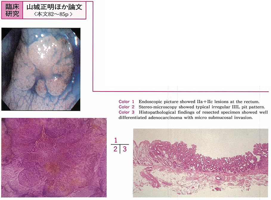
    1995 年 47 巻 p. 82-85
    発行日: 1995/12/08
    公開日: 2015/05/01
    ジャーナル フリー
     1991年1月より1995年4月までの4年4カ月間に,国立がんセンター中央病院にて実体顕微鏡観察が施行された大腸腫瘍性病変は263病変あり,そのうち工藤らの腺口形態でいうⅢL型pit patternと判定したものは213病変あった。213病変のⅢL型pitを,①腺管の分布,②形,③大小不同の3点からregular ⅢL型(R-ⅢL型)とirregular ⅢL型(IR-ⅢL型)に亜分類し,病理組織所見との比較検討を行った。R-ⅢL型では172病変中腺腫が153病変(90.9%),m癌が19病変(9.1%)あった。一方,IR-ⅢL型では41病変中腺腫が25病変(61.0%),m癌が12病変(29.2%),sm癌が4病変(9.8%)あり,R-ⅢL型に比しIR-ⅢL型は,癌の合併率とsmの比率において悪性度が高く,また腫瘍径が6mm以上で,sm癌の頻度が16.7%(4/24)と高率であった。ⅢL型pit patternをR-ⅢL型とIR-ⅢL型に亜分類することは,pit patternの診断において大腸腫瘍性病変の質的診断をより正確に行うことであり,また拡大電子スコープを用いた内視鏡診断および内視鏡的治療の適応決定に対して有用と考えられた。
  • 川本 智章, 井戸 健一, 中澤 義明, 礒田 憲夫, 穂積 正則, 鈴木 孝典, 長嶺 伸彦, 井岡 達也, 花塚 和伸, 関根 豊, 木 ...
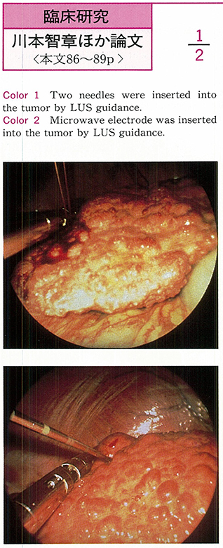
    1995 年 47 巻 p. 86-89
    発行日: 1995/12/08
    公開日: 2015/05/01
    ジャーナル フリー
     われわれは,超音波腹腔鏡(LUS)を用いて腹腔鏡下エタノール注入療法(LEIT)および腹腔鏡下マイクロ波凝固療法(LMCT)を行い,良好な成績を得ている。どちらの方法も局所麻酔下で行うことに特徴がある。LEITでは,腫瘍径が2cm以下の単発例が最もよい適応である。多発例の場合には2cm以下で,腫瘍数が3個以下を適応としている。一方,LMCTでは4cm以下の大きさの腫瘍を適応としている。LEITでは,穿刺針を何本か刺入することにより,比較的大量のエタノールを安全に注入できるため,1回の操作で腫瘍の広い範囲を治療することができる。また,LMCTも侵襲が少なく,凝固による十分な壊死効果が得られる。いずれの方法もLUSを用いるため,他の画像診断では捉えることのできない小肝細胞癌に対して,特に有用な治療法であると考えられる。肝細胞癌の治療には,従来は主として外科的切除,TAE,PEITなどの方法しかなかった。しかし現在では,これに腹腔鏡下の治療が加わり,選択の幅が広がった。肝細胞癌の腹腔鏡的治療は,安全性はもちろん,入院期間の短縮,早期の経口摂取あるいは歩行開始が可能であり,癌結節の完全壊死も得られるため,cost benefitあるいはQOLは十分高いものが得られ,今後ますます発展が期待される。
  • 中島 俊之, 西谷 篤史, 柿沼 徹, 小坂 健一, 岩堀 泰基, 大久根 浩, 藤盛 健二, 吉岡 久, 大西 久仁彦, 藤原 研司, 中 ...
    1995 年 47 巻 p. 90-93
    発行日: 1995/12/08
    公開日: 2015/05/01
    ジャーナル フリー
     進行膵癌84例および肝硬変376例を対象に,門脈圧亢進症にしばしば認められる食道・胃静脈瘤および胃粘膜病変の観察を,上部消化管内視鏡検査にて行った。膵癌例における静脈瘤の出現は20%に認められ,膵体部に多くみられた。また形態は,F1が76%を占め,F3はみられなかった。膵癌例における胃粘膜病変の出現は7%に認められた。肝硬変例における静脈瘤と胃粘膜病変の出現率では,静脈瘤は70%に,胃粘膜病変は35%に認められた。静脈瘤の形態はF1が38%であった。静脈瘤例における胃粘膜病変の出現率を膵癌例と肝硬変例で比較すると,膵癌例では24%に,肝硬変例では49%に認められ,膵癌例では肝硬変例に比して有意に低率(P<0.05)であった。この違いは,膵癌例では静脈瘤の形態が軽度のものが多かったこと,肝外性の門脈系変化ではcavernous transformationなどのcollateral circulationが生じやすいため,胃粘膜血行動態に及ぼす影響が少ないことによると考えられた。
  • 山田 英夫, 西須 孝, 蜂巣 忠, 高橋 和久, 山縣 正庸, 守屋 秀繁, 神津 照雄, 磯野 可一
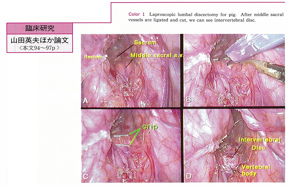
    1995 年 47 巻 p. 94-97
    発行日: 1995/12/08
    公開日: 2015/05/01
    ジャーナル フリー
     われわれは,大腸切除術low anterior resectionおよび,ヘルニア修復術extraperitoneal approachの手技を応用した,新しい腹腔鏡下手術(整形外科領域の腰椎椎間板切除術lumbar discectomyおよび前方椎体間固定術anterior interbody fusion)の手技の開発と確立を行った。椎間板L5-Sに対してはabdominal approachで,椎間板L2-5に対してはextraperitoneal approachにて行った。腹腔鏡手術に習熟した外科医にとって椎間板の露出は容易であり,scopeにて椎間板を真正面にみえる術野は,整形外科医にとっては顕微鏡下手術を行っているかのごとく視野良好で,安全確実に腰椎椎間板切除術lumbar discectomyおよび前方椎体間固定術anterior interbody fusionが行えた。腹腔鏡下手術に習熟した外科医と腰椎手術に習熟した整形外科医の連携により,本術式は安全かつ良好な治療成績が得られるものと推測された。
症例
  • 荻原 英夫, 小笹 芳子, 細井 董三, 岡田 利邦, 中井 呈子, 矢沢 知海, 中村 尚志, 阿美 克彦, 木村 泰司, 大倉 康男, ...
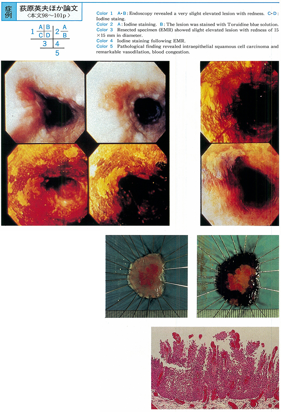
    1995 年 47 巻 p. 98-101
    発行日: 1995/12/08
    公開日: 2015/05/01
    ジャーナル フリー
     食道の深達度mの0-Ⅱa型病変の多くは白色ないし褪色調を呈しており,赤色調の病変はsm癌を疑うのが常識となっているが,今回われわれは赤色調のepの0-Ⅱa型病変を経験したので報告する。症例は60歳男性。特に自覚症状はなく,定期検診目的に当クリニックで上部消化管内視鏡検査を受けたが,上切歯列より約30cmの左壁に発赤調の丈の低い隆起性病変が発見され,生検にて扁平上皮癌の診断が得られた。発赤調ではあるが,表面は顆粒集簇様で弧の変形が認められないことから,深くてもm2までと診断し,東京医科歯科大学第1外科にてEMRを施行した。病理検査の結果は15×15mmの0-Ⅱa型で,深達度はep,組織型はsquamous cell carcinoma(SCC),ly(-),v(-)であった。
  • 田村 光広, 浮田 雄生, 松本 宙明, 神津 隆弘, 角田 二郎, 石井 俊也, 中島 俊一, 石塚 俊一郎, 吉岡 秀樹, 五十嵐 良典 ...
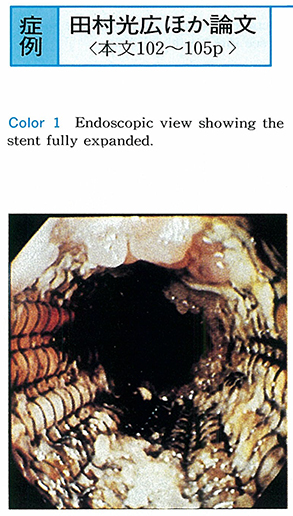
    1995 年 47 巻 p. 102-105
    発行日: 1995/12/08
    公開日: 2015/05/01
    ジャーナル フリー
     99歳男性が嚥下困難を主訴に来院。食道造影および上部消化管内視鏡で,中部食道に不整な辺縁を持つ全周性狭窄が認められた。生検の結果は高分化型扁平上皮癌であり,食道進行癌と診断した。年齢などより手術適応なしと判断し,金属ステント(Ultraflex®,Microvasive社製)を使用して狭窄解除を施行した。嚥下困難は改善し,術直後を除き疼痛などの自覚症状もなかった。術後3カ月の時点でステントの開存状態は良好で,挿入位置の移動もなかった。同ステントは従来のプラスチック・ステントに比べ,①挿入時の操作がより容易,②柔軟なため屈曲部への使用が可能,③固定性が良好,④術後再狭窄に対する再治療が可能,⑤自覚症状が少ないなどの点で優れていた。
  • 谷 穣, 三治 哲哉, 緑川 昌子, 半田 豊, 森田 重文, 大野 博之, 吉田 肇, 鶴井 光治, 三坂 亮一, 川口 実, 斉藤 利彦 ...
    1995 年 47 巻 p. 106-108
    発行日: 1995/12/08
    公開日: 2015/05/01
    ジャーナル フリー
     残胃縫合部に発生した切断神経腫について報告する。症例は49歳男性。既往歴として20歳時,胃潰瘍にて広範囲胃切除術(Billroth Ⅱ法)をうけている。6年前に近医での内視鏡検査で胃腺腫と診断され,内視鏡的粘膜切除術(以下EMR)目的に入院した。内視鏡所見では体上部小彎前壁側に隣接する2つの隆起性病変(口側に胃腺腫,肛門側に粘膜下腫瘍様の小隆起病変)を認めた。これらに対してEMRを施行し,両病変を一括切除した。病理組織的には口側の腺腫は中等度異型で,focal carcinomaは認められなかった。肛門側の隆起は,切断された神経線維と結合組織がからみあって増生し,それが一塊となって腫瘤状に結節を形成しており,切断神経腫と診断した。隆起を呈する切断神経腫については胆道系の報告はみられるが,胃ではわれわれの検索範囲内には認められず,極めてまれと思われ,今回若干の考察を加えて報告した。
  • 月岡 佳久, 円岡 寿, 伊藤 喜三男, 日高 康夫, 片桐 敏郎, 荻野 達夫, 堀向 文憲
    1995 年 47 巻 p. 109-112
    発行日: 1995/12/08
    公開日: 2015/05/01
    ジャーナル フリー
     虚血性大腸炎は,緊急内視鏡検査時に発見される最も頻度の高い疾患の1つである。患者は68歳,男性。S状結腸癌のため3年前にS状結腸切除術を受けており,その経過観察目的に大腸X線検査予定となった。大腸X線検査の前処置として,粉末クエン酸マグネシウム1包とピコスルファートナトリウム10mlを服用し,大量下痢のあと腹痛,血便が出現し,緊急大腸内視鏡検査を施行した。吻合部口側より横行結腸中部まで出血やびらんを認め,粘膜全体も浮腫状を呈していた。臨床症状,緊急大腸内視鏡検査,大腸X線検査および病理学的所見などにより虚血性大腸炎と診断した。3日ほどで症状は軽快し,一過性型の虚血性大腸炎であった。大腸検査の際,本疾患の可能性も念頭に置き前処置を行う必要があると思われる。
  • 篠澤 功, 佐藤 順, 楢原 義之, 杉浦 敏昭, 渡 淳, 水野 杏一, 横井 公良, 森山 雄吉, 辰口 篤志, 藤森 俊二, 山門 進 ...
    1995 年 47 巻 p. 113-116
    発行日: 1995/12/08
    公開日: 2015/05/01
    ジャーナル フリー
     症例は73歳男性。嘔気,粘液便,意識障害を主訴に入院した。入院時血液検査では,電解質異常と脱水を認め,補液による補正を行う必要があった。大量の粘液便排泄を認めたため,大腸内視鏡検査を施行し,歯状線直上より口側に約12cmに及ぶ全周性の絨毛構造を呈する扁平隆起性病変を認め,病理組織学的に絨毛腺腫(villous adenoma)であり,electrolyte depletion syndrome(EDS)を伴った直腸絨毛腺腫と診断した。絨毛腺腫からの水分,電解質分泌にはprostaglandin E2(以下PGE2)の関与が示唆されており,本例に対しPGE2合成阻害薬であるindomethacinを投与したが,無効であった。その後も大量の粘液便排泄は続き,Miles手術を施行し,EDSは改善した。EDSを伴った絨毛腺腫の本邦報告例は1971年から1995年までに26例であり,若干の文献的考察を加え報告する。
  • 横山 孝典, 中島 洋, 松本 茂藤子, 松尾 英男, 佐藤 悦久, 川村 直弘, 斎藤 光浩, 今村 真紀子, 徳植 秀樹, 柴山 淳, ...
    1995 年 47 巻 p. 117-120
    発行日: 1995/12/08
    公開日: 2015/05/01
    ジャーナル フリー
     症例は52歳,大酒家の男性。輸血歴なし。約30年前アルコール性肝障害を指摘されたが,飲酒を続けていた。平成5年4月健康診断にてHCV抗体陽性を指摘され,肝機能障害精査目的にて当科に紹介入院した。腹腔鏡検査にて肝ドーム付近に5-10mmの暗紫色円形の平皿状陥凹を散在性に認めた。ポルフィリン体の検査では,尿中ウロポルフィリン(以下URO),コプロポルフィリン(以下COPRO)の増加を認めた。約1年後再度腹腔鏡検査を施行し,生検組織に紫外線を照射したところ蛍光発色を認めた。その後,尿中UROは342μg/g・cr,尿中COPROは23μg/g・crとなった。晩発性皮膚ポルフィリン症(以下PCT)の場合は尿中URO50%以上,尿中COPRO10%以下になるとされ,本例もこれに一致する。皮膚症状を欠いたPCTの診断に腹腔鏡検査が有用であった1例を経験したので報告した。
内視鏡の器械と技術
  • 中村 真一, 光永 篤, 村田 洋子, 鈴木 茂, 林 直諒
    1995 年 47 巻 p. 122-123
    発行日: 1995/12/08
    公開日: 2015/05/01
    ジャーナル フリー
    We perform sclerotherapy using 1% Aethoxysklerol for F0-F1 level esophageal varices. We consider bleeding and pain after sclerotherapy to be harmful.
    We therefore mixed powdered sodium alginate and thrombin, and tried to inject the mixture using Alto Shooter, to esophageal varices after sclerotherapy. The injection of the mixture was effective for stopping bleeding and reducing pain after sclerotherapy. The mixture could be fixed firmly to the mucosa. The hemostatic effect of thrombin and the mucosal protection effect of sodium alginate, could be improved using a mixture of these drugs. There have been no side effects or trouble.
    In conclusion, the injection of a mixture of sodium alginate and thrombin was easy and minimally invasive. It was therefore a useful method for the prevention of bleeding and pain after sclerotherapy. We can perform sclerotherapy safely in the outpatient clinic with these methods.
臨床研究
  • 静 雅美, 山田 俊彦, 柿崎 暁, 小林 出, 石原 弘, 阿部 毅彦, 飯塚 春太郎, 小島 明, 桜井 誠司, 小野里 康博, 高木 ...
    1995 年 47 巻 p. 124-125
    発行日: 1995/12/08
    公開日: 2015/05/01
    ジャーナル フリー
    Therapeutic effect of endoscopic variceal ligation (EVL) for esophageal varices was analyzed. Subjects were 32 cases treated by EVL and 42 cases by endoscopic injection sclerotherapy (EIS) . These cases were followed up from January 1993 to January 1995 and red color signs were positive in all cases.
    Twenty-six cases (84%) were effective with no red color sign at one month after EVL. The relapse rate of these effective cases was 24.9% at six months, and 60.4% at 12 months. These results were similar to those by EIS. In cases in which the findings of the esophageal varices were of form F2 or above, relapse rate at 12 months was 95.2% on EVL, and 60% on EIS. As for prognosis, EVL had higher relapse than EIS (p<0.05) . Risk of complication was less in EVL than in EIS therapy.
    These results suggest that EVL is as effective as EIS. However, in cases of form F2 or above, efficacy of EVL treatment was shorter than that of EIS. Therefore, EVL and EIS therapy should be combined.
  • 矢部 諭, 岩堀 泰基, 吉田 典子, 西蔵 ツワン, 下地 克典, 中島 俊之, 佐々木 一圭, 藤原 研司
    1995 年 47 巻 p. 126-127
    発行日: 1995/12/08
    公開日: 2015/05/01
    ジャーナル フリー
    The aim of the present study was to find the usefulness of gastric aspirated urea concentration in detection of the efficacy of eradication of H pylori (HP) by comparing with gastric ammonia concentration and serum pepsinogen I/II ratio.
    A total of 32 subjects (15 with gastric ulcer, 15 with duodenal ulcer, and 2 with gastro-duodenal ulcer) were infected with HP. HP infection was detected by microscopic examination of slides stained by Gram's method and treated with Pfeiffer fluid. All patients received lansoprazole (30mg, every morning) and ecabet sodium (2g, twice a day) for 6 or 8 weeks. Patients also received amoxicillin (750mg, three times a day) for last 2 weeks. The clearance and eradication rates were 100% (20/20) and 60% (12/20) , respectivery.
    The eradicated group had significantly higher gastric urea and serum pepsinogen I/II levels, and significantly lower gastric ammonia level than before eradication and the non-eradicated group. There were few subjects overlapping in gastric ammonia and serum pepsinogen I/II levels, but no subjects overlapping in gastric urea level in the eradicated and the non-eradicated groups. Measurement of gastric urea level may provide a useful tool for detecting the efficacy of HP eradication.
  • 成木 行彦, 瓜田 純久
    1995 年 47 巻 p. 128-129
    発行日: 1995/12/08
    公開日: 2015/05/01
    ジャーナル フリー
    We evaluated serum pepsinogen I (PG I) and II (PG II) assay, and 13C-Urea breath test (13C-UBT) in a diagnosis of Helicobacter pylori (HP) after teatment with lansoprazole and clarithromycin. Forty two patients included 10 gastric ulcer, 7 duodenal ulcer, 8 gastroduodenal ulcer and 17 chronic gastritis, treated with 30mg of lansoprazole and 600mg of clarithromycin in a day for 14 days.
    Serum PG I and PG II levels reduced and PG I/II ratio increased in effective cases after therapy significantly. There were not refractory cases in range of more than 31% of reduction rate of PG I, 41% of PG II and 161% of increasing rate of PG I/II ratio. However, 1/3 of effective cases did not demonstrate so much change in PG I and PG II levels. 13C-UBT of effective cases reduced within normal range of Δ13C after therapy. Δ13C of refractory cases did not decrease into normal range.
    Although measurement of serum PG I and PG II was not adequate in a diagnosis of HP after therapy, 13C-UBT examination is not risky and can be useful in observing the eradication of HP.
  • 秋本 真寿美, 橋本 洋, 北村 容子, 榊田 聖子, 安達 由美子, 安部 康二, 土屋 まり子, 渡辺 七六, 石黒 久貴, 栗原 毅, ...
    1995 年 47 巻 p. 130-131
    発行日: 1995/12/08
    公開日: 2015/05/01
    ジャーナル フリー
    We studied on : (1) the Helicobacter pylori (Hp) eradication rate and side effects (2) the association between eradication and inflammatory reaction of gastric mucosa and (3) the correlation between serum anti-CagA antibody titer and gastric diseases. Patients formed two groups : twenty-one patients in group A recieved lansoprazole (L) 30mg, plus clarithromycin (CAM) 400mg, for two weeks ; twenty-six patients in group B were treated with L 60mg, plus CAM 800mg for two weeks.
    Results : (1) Using a 13C-urea breath test instead of a conventional method including CLO, culture and histology, measured eradication rates fell from 43% to 19% in group A, from 58% to 31% in group B. Side effects rose in frequency as dosage increased. (2) After eradication, levels of IL-8 and myeloperoxidase (MPO) in gastric mucosa improved. There was no correlation between inflammation severity before eradication and eradication rate. (3) Serum anti-CagA antibody titer rose in patients with open ulcers and was significantly high in Hp-infected patients with. Changes in antibody titer after eradication may reflect changes in microorganism volume and host reaction.
  • 松倉 則夫, 恩田 昌彦, 秋谷 行宏, 大川 敬一, 白川 毅, 瀧田 雅仁, 吉行 俊郎, 加藤 俊二, 長谷川 博一, 樋口 勝美, ...
    1995 年 47 巻 p. 132-133
    発行日: 1995/12/08
    公開日: 2015/05/01
    ジャーナル フリー
    A new triple therapy for eradication of HP in 24 patients of peptic ulcer (13 duodenal and 11 gastric ulcers ; 18 male and 6 female ; 18-74 years, mean 49) were undergone with PPI+AMPC+ecabet sodium. We used 30mg id or bid of lansoprazole, 0.5g tid of amoxicillin and 1.0g ecabet sodium bid for 2 weeks. Eradication of HP infection was diagnosed by 13C-UBT and detection HP DNA in the gastric juice using PCR method. Eradication rate was 22/24 (92%) by 13C-UBT and 19/24 (79%) by PCR method. Side effect was diarrhea which was found in 1/24 (4.2%) patients.
  • 大草 敏史, 藤木 和彦, 高清水 一善, 鈴木 伸治, 桜沢 俊秋, 堀内 亮郎, 下井 謙吾, 本田 公男, 有明 浩一郎, 榎本 靖子 ...
    1995 年 47 巻 p. 134-135
    発行日: 1995/12/08
    公開日: 2015/05/01
    ジャーナル フリー
    To evaluate the potential benefit of a 2-week regimen including lansoprazole, various antibiotics and ecabet sodium for eradication of H. pylori from the gastric mucosa.
    Ninety eight patients with H. pylori infection with gastric ulcer (33 patients, mean age 56) , duodenal ulcer (23 patients, mean age 48) , and chronic gastritis (42 patients, mean age 56) were included in a randomised open pilot study ; they were treated with lansoprazole 30mg od + amoxicillin 500mg qid (LPZ+AMPC) , lansoprazole 30mg od + amoxicillin 500mg qid + ecabet sodium 1g bid (LPZ+AMPC+ES) , lansoprazole 30mg od + roxithromycin 150mg bid (LPZ+RXM) , or lansoprazole 30mg od + clarithromycin 400mg bid (LPZ+CAM) for 2 weeks. Endoscopy was performed before and 3 months post-treatment. The presence of H. pylori was assessed in ulcer-border, antral and corporeal biopsies by rapid urease test (CLO test) and histology (Acridine-orange stain+HP monoclonal-antibody stain) .
    H. pylori eradication was observed in 27/43 (63%) in the LPZ+AMPC group, in 22/29 (76%) in the LPZ+AMPC+ES group, in 4/19 (21%) in the LPZ+RXM group, and in 3/7 (43%) in the LPZ+CAM group. Six patients reported side effects including ; diarrhea (4) , urticaria (1) in the LPZ+AMPC group, and diarrhea (1) in the LPZ+RXM group. Because of the highest eradication rate and no side effect of the LPZ+AMPC+ES therapy, we recommend it as first-line anti H. pylori treatment for a patient with H. pylori infection.
  • 小松 嘉人, 斉藤 大三, 平山 敦, 佃 博, 木場 崇剛, 小野 裕之, 横田 敏広, 白尾 国昭, 近藤 仁, 吉田 茂昭, 加藤 勝 ...
    1995 年 47 巻 p. 136-137
    発行日: 1995/12/08
    公開日: 2015/05/01
    ジャーナル フリー
    Helicobacter pylori (HP) has been reported to associate with gastric cancer and the eradication of HP would be of great value to reduce the incidence of gastric cancer. Although several treatment, including antibiotics and proton pump inhibitors, are utilized for the HP infected patients and effective, these drugs also produce considerable side effects.
    We have reported the growth inhibitory effect of epigallocatechin gallate (EGCG) , a main constituent of Japanese green tea, on HP in vitro. Then we examined the effect of Sunphenon containing EGCG on the eradication of HP, clinically. Seven HP positive volunteers or patients without gastric dieseases were treated with Sunphenon 600-700mg daily for 4 weeks. Endoscopy was performed before and at the end of the therapy and HP was detected by culture with two pair biopsy specimens taken from the antrum and upper body of stomach. Whereas the reduction of volume of HP was revealed in 4 out of 7 (57%) , this results was not satisfactory. This reason was considered to be based on the rapid disappearance of Sunphenon from the gastric lumen. It is necessay to develop the method for remaining Sunphenon in stomach for the sufficint eradication of HP, in future.
  • 齋藤 洋子, 福富 久之
    1995 年 47 巻 p. 138-139
    発行日: 1995/12/08
    公開日: 2015/05/01
    ジャーナル フリー
    In the cancer screening center of Ibaraki prefectural mass survey society, the upper gastrointestinal endoscopy is performed for the checked up people with stomach cancer screening test. To evaluate our way to do the upper gastrointestinal endoscopy without examinees' uncomfortableness, we studied 919 cases using the questionnaire. A nurse or endoscopic technician asks examinees about the questionnaire (Table 1) . Most examinees who were composed of 682 males and 237 females, came to this society for the first time (Fig. 1) . In our center, examinees have a look at premedications and endoscopic examination in a video-program before examination. During examination, the endoscopist explains the endoscopic findings to them.
    In conculsion, : (1)Almost examinees agreed with examinations (Table 2) , (2)The analysis of questionnaire showed that examinees, took endoscopic examination with little uncomfortableness (Table 3) . We conclude that for painless examination, it is important for examinees to understand how the examination is done.
  • 家富 克之, 三好 和夫, 崎村 恭也, 半田 祐一, 乾 純和
    1995 年 47 巻 p. 140-141
    発行日: 1995/12/08
    公開日: 2015/05/01
    ジャーナル フリー
    We studied the factors that ease pain during an endoscopic examination. The results are as follows : 1) no restriction of eating on the previous day or drinking water on the day of examination, 2) flavored local anesthetics (anestheflavor) and music anesthesia, 3) kind treatment of patients by endoscopists, 4) kind words and care of trained nurses, and 5) intravenous administration of 5mg of diazepam. It is important to do these things properly to ensure painless endoscopic examination.
  • 鈴木 荘太郎
    1995 年 47 巻 p. 142-143
    発行日: 1995/12/08
    公開日: 2015/05/01
    ジャーナル フリー
    Gastrointestinal (GI) endoscopy has been recognized one of hard painful examinations. Recently, mechanical and technical advances made GI endoscopy easy and safe clinical procedures for detection and therapeutics to GI tumors.
    Although these developments of clinical diagnosis and treatment, psychological and physical factors in the examination and premedication in GI endoscopy for the patients have not been yet resolved. On the other hands, many endoscopists tend to use more and more sedative and opiate drugs in routine endoscopy. This report evaluated that subjective symptoms complained by patients and objective symptoms given by endoscopists, endoscopic technicians and nurses whether the premedication is necessary or not in routine GI endoscopy.
    Any other drugs using for endoscopic premedication would not be desired more than 80% of all patients recieved endoscopy were suggested in this paper. Then acceptance of GI endoscopy without any kinds of premedications was more than 80% respectively. Results of high agreement rate on endoscopy, and no desire of premedications could be achieved by endoscopists through instructions including informed consent and professional attendance by endoscopic technicians and nurses with the qualification. And additional effects can be accomplished by using with TV endoscopy which could be seen the endoscopic picture during the own examination and use of well fitted mouthpiece.
  • 六倉 俊哉, 国本 英治, 小川 善秀, 三輪 博久
    1995 年 47 巻 p. 144-145
    発行日: 1995/12/08
    公開日: 2015/05/01
    ジャーナル フリー
    We analysed the therapeutic result of endoscopic hemostasis for 75 cases of bleeding gastric ulcer from January 1994 to May 1995. Ethanol infusion was tried 118 times, and HSE infusion or clipping were tried 21 times each. In all 75 cases, endoscopic hemostasis was successful for the first time, but in 5 cases rebleeding was noted and 3 cases underwent operative therapy. In most cases, ethanol infusion was effective, but in some cases with a large exposed vessel or spurting bleeding, it was difficult to stop bleeding only with ethanol infusion, and clipping or HSE infusion or other method was needed.
    Recently we tried continuous ethanol infusion therapy for 8 of those difficult cases. In this method, we inject 5 to 10 ml of ethanol directly into the exposed vessel continuously and very slowly over 2 to 10 minutes, and the ethanol injected into the vessel is never washed out by bleeding. So the effect to make thrombus is remarkable, and the hemostasis was successful and rebleeding was not noted in all 8 cases. Huge ulcer formation were noted in those cases because of extensive intraarterial thrombosis, but the healing of such artificial ulcers were very rapid and no trouble was aroused by those ulcers.
    In conclusion, continuous ethanol infusion therapy was thought to be effective therapeutic modality for bleeding gastric ulcer especially with large exposed vessel and spurting bleeding.
  • 安部 孝, 伊藤 慎芳
    1995 年 47 巻 p. 146-147
    発行日: 1995/12/08
    公開日: 2015/05/01
    ジャーナル フリー
    For 6 years, since 1988 to 1994, we have been performed medical treatment of the 143 gastric lesions, which were diagnosed of early gastric cancer or adenoma, by the endoscopic mucosal resection (EMR) .
    Method : We have performed the EMR by the Tada's method. We have peformed the EMR by two panendoscopes before 1992, but after 1992, by one 2-channels therapeuetic scopes.
    Results : (1)In this study, 60 gastric cancer lesions were examined, in which 42 lesions were already diagnosed as gastric cancer before EMR. Diagnosis of 11 lesions, which were 12% of adenoma, were corrected to the early gastric cancer after EMR. On the 7 lesions, which were suspected of gastric cancer, final diagnosed of the early gastric cancer by EMR. 48/60 lesions (80%) were intramucosal cancer.
    (2)By the 2 panendoscopes method, pathological complete resection rate and no recurrence rate were 63%, 85%. Simularly, by the 2-channels therapeutic scope, there were 60%, 96%. In the two EMR methods, there was no significant difference. There were 6 recurrenced cases, all these cases were occured in the pathological incomplete resected cases. Only one complication was post EMR bleeding case.
    In conclusion, EMR is effective and safety method for early gastric cancer.
  • 三森 教雄, 村井 隆三, 安藤 博, 衛藤 謙, 笹屋 一人, 楠山 明, 岩本 公和, 長山 瑛, 伊坪 喜八郎
    1995 年 47 巻 p. 148-149
    発行日: 1995/12/08
    公開日: 2015/05/01
    ジャーナル フリー
    We evaluated the laparoscopic wedge resection (LWR) and percutaneus transgastricwall mucosal resection (PTEMR) for early gastric cancer. LWR indicated anterior, greater curveture and partial posterior gastricwall lesions. Under general anesthesia, 3-5 tracars are inserted abdominal wall. After crumping jejunum at Treiz ligament, two needles (18G) are injected percutaneus transgastricwall around the lesion. A suture is inserted through the needle and catched that by looped stainless wire from another site. Then two points lifting of the gastric wall is made. Same way, another two needles are inserted around the lesion for 4 points cross lifting of the gastricwall and wedge resection was perfomed with autosuture device.
    PTEMR is a procedure for gastric mucosal resection under gastroendoscopy using laparoscopic forceps and scissors through gastrostomy. The lesion is grasped by the forceps and resected by a snare with electrocautery under gastroscopic view.
    We have perfomed 7 lesions of 6 cases by PTEMR, and 2 cases by LWR for early gastric cancers.
症例
  • 池上 恒雄, 大原 昇, 佐藤 秀一, 土橋 展子, 杉山 茂樹, 羽田 哲也, 片本 哲郎
    1995 年 47 巻 p. 150-151
    発行日: 1995/12/08
    公開日: 2015/05/01
    ジャーナル フリー
    Liver cirrhosis (type B) with an esophageal varix was diagnosed in a 53-year-old male in November 1992. On February 2, 1995, epigastralgia suddenly developed. After seven days, endoscopic examination revealed peeled mucosa on the surface of a varix alongside a longitudinal ulcer in the lower half of the esophagus. After thirty days, the varix became flat and a white scar developed.
    Esophageal submucosal dissection is considered to be a disorder classified between Mallory-Weiss syndrome and Boerhoave syndrome. Common causes are endoscopic injection sclerotherapy, foods, drugs, and pemphigus. It is sometimes idiopathic. Eighty-five cases of esophageal submucosal dissection have been reported in Japan, but none of them was followed by the disappearance of an esophageal varix similar to this case.
  • 高田 丈, 卜部 元道, 溝渕 昇, 林田 康男, 榊原 宣
    1995 年 47 巻 p. 152-153
    発行日: 1995/12/08
    公開日: 2015/05/01
    ジャーナル フリー
    Case 1 : A 49-year-old man was treated with antibiotics for the removement of a nail. After two weeks of treatment, he visited our hospital with a complaint of epigastralgia. On the endoscopic examination, the diagnosis was esophageal candidiasis, grade II of Kodsi's classification.
    Case 2 : A 63-year-old man with a gastric polyp was resected by endoscopic polypectomy. The yearly endoscopic examination of this time, the diagnosis was a lower esophageal candidiasis, grade I of Kodsi's classification.
    The diagnosis of esophageal candidiasis is comparativly easy because the endoscopic finding is characteristic. The increase of candida of the digestive system causes candidemia and dissemination of candidiasis. There is a report that the mortality rate of acute dissemination of candidiasis is about 40%. So early treatment is important for candidiasis.
    In case 1, intravenous administration of fluconazole 200mg/day and case 2, oral administration of fluconazole 150mg/day were started. One week later, a diffuse white coat and a white elevated lesion disappeared from both cases on the endoscopic examination.
  • 山田 久木, 岩切 勝彦, 杉浦 敏昭, 大国 真一, 渡 淳, 水野 杏一, 琴寄 誠, 二神 生爾, 中川 義也, 小林 正文
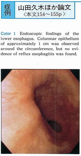
    1995 年 47 巻 p. 154-155
    発行日: 1995/12/08
    公開日: 2015/05/01
    ジャーナル フリー
    A 64-year-old woman was hospitalized with heartburn and precordial pain. Cardiovascular disease was not detected. Therefore, we investigated the gastrointestinal tract. Conventional esophageal manometry and upper gastrointestinal series were normal. On endoscopy, columnar epithelium of approximately 1 cm was found around the circumference of the lower esophagus, but there was no acute reflux esophagitis. On Bernstein test, heartburn appeared, but precordial pain did not occur. On esophageal balloon dilatation, there was a sense of pressure, but precordial pain did not occur. Because heartburn occurred on Bernstein test, we considered the possibility of gastroesophageal reflux (GER) disease, and investigated 4-hour intraesophageal pH monitoring. GER occurred on several occasions during the first hour of the postprandial period, and 2 episodes of GER were accompanied by heartburn. During the first hour of the postprandial period, the percent time pH<4.0 was 9.6%.
    The cause of precordial pain was unclear, but the existence of GER was established. So we administered proton pump inhibitor (PPI) to confirm the diagnosis and for treatment. After oral administration of PPI, heartburn and precordial pain completely disappeared. We concluded that the cause of precordial pain in this case originated with GER.
  • 大谷 健一, 河村 修, 関口 利和, 青木 祐子, 山田 拓郎, 宮崎 誠, 堀越 勤, 草野 元康, 森 昌朋, 細村 泰夫
    1995 年 47 巻 p. 156-157
    発行日: 1995/12/08
    公開日: 2015/05/01
    ジャーナル フリー
    We reported a case of esophageal squamous cell papilloma treated with endoscopic mucosal resection. A patient was a 41-year-old man who had a flat elevated lesion (40x20mm) with granular surface on the posterior wall of the upper esophagus. Endoscopic resection was performed on February 25, 1993. Histological findings revealed squamous cell papilloma. Immunohistochemistry for human papilloma virus using immunoperoxidase technique was negative. DNA of human papilloma virus in resected specimen (dott blott technique) was negative. HPV infection was excluded.
    However, three cases of squamous cell papilloma with malignant changes have been previously reproted. Therefore, a papilloma which size is more than 10mm and one showing the change in size should be actively resected under the endoscopy.
  • 山下 浩二, 佐藤 薫隆, 為我井 芳郎, 小松原 登, 二階堂 孝
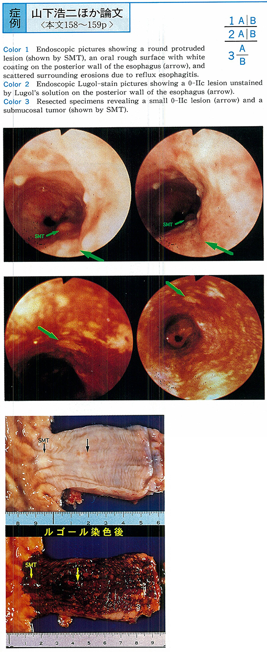
    1995 年 47 巻 p. 158-159
    発行日: 1995/12/08
    公開日: 2015/05/01
    ジャーナル フリー
    A 45-year-old male complaining of anorexia and heartburn was diagnosed by endoscopic findings that 0-IIc lesion with ulcer and the surrounding scattered erosion due to the reflux esophagitis on the lower esophagus (Ei) . The submucosal invasion was suspected and the total thoracic esophagectomy with reconstruction was performed. The operational findings were Ei-Im-Post, 0-IIc, Pl (-) , N (-) , M (-) , Stage I, R3. The resected specimen revealed superficial carcinoma coexisting with a submucosal tumor in the lower part of the esophagus (Ei) . Microscopic findings showed only 2mm sized 0-IIc lesion was moderately differentiated squamous cell carcinoma with submucosal microinvasion (sm) and n0, ly0, vo, ow (-) , aw (-) , ew (-) . It also revealed the submucosal tumor was leiomyoma (5x3mm) which seemed to arise from the muscularis propria. The patient was discharged with no postoperative complications.
    Only 2mm in size of the 0-IIc esophageal carcinoma had sm1 microinvasion. It was very rare case, but it provided the question about the indication of the endoscopic mucosal resection (EMR) . The post-operative pathological examination of the resected specimen is important, but also it is more important to preoperatively diagnose the acurate depth of invasion.
  • 海野 潤, 白須 達也, 相磯 光彦, 野村 智久, 奥山 尚, 荒井 咲子, 林田 憲正, 国府田 静生, 田所 昌夫, 石河 利隆
    1995 年 47 巻 p. 160-161
    発行日: 1995/12/08
    公開日: 2015/05/01
    ジャーナル フリー
    A 69-year-old man was referred to our hospital for the purpose of endoscopic treatment of the early gastric carcinoma located at EGJ. As it was type IIa well differentiated adenocarcinoma, which was thought to be intramucosal, EMR was performed. The resected specimen was 8mm in diameter.
    Early gastric carcinoma in a fundic gland area, espacially near EGJ, is tend to be histopathologically well defferentiated. The more close to EGJ the location is, elevated type is the more frequently found than depressed type. So it is possible that early gastric carcinoma near EGJ is misdiagnosed as adenomatous polyp. In fact, this case was misdiagnosed as benign polyp 2 years before EMR. However significant degrees of atypism was already existed in that biopsy specimens retrospectively. We should pay attention to a gastric polyp at EGJ, because it may be an early carcinoma and if it is, in case of an indication of EMR, EMR should be performed.
  • 楠原 健朗, 青野 茂昭, 大野 建, 中村 直子, 下屋 正則, 松田 浩二, 笹木 淳司, 東納 重隆, 岩井 淳浩, 岩下 悦郎, 川 ...
    1995 年 47 巻 p. 162-163
    発行日: 1995/12/08
    公開日: 2015/05/01
    ジャーナル フリー
    A 65-year-old female visited our hospital due to the epigastralgia. An endoscopic examination for the upper gastrointestinal tract revealed the foreign body stuck in the gastric wall. It was safely removed by an endoscope and ascertained a fish bone. The fish bone was 25mm long and 11mm of it was buried in the gastric wall.
    According to the literature, most of patients don't realize they have swallowed foreign bodies by mistake. Adults rarely swallowed them and foreign bodies rarely stay in the stomach. Besides, a fish bone rarely becomes a cause of foreign bodies in the stomach. On this occasion, we encountered an adult case of a foreign body, a fish bone, stuck in the gastric wall and succeeded to remove it endoscopically for the treatment.
  • 光野 雄三, 加藤 善久, 伊佐山 浩通, 山田 尚士, 皆川 信幸, 日吉 徹, 関谷 裕之, 中田 良, 吉次 通泰, 庵 政志, 田中 ...
    1995 年 47 巻 p. 164-165
    発行日: 1995/12/08
    公開日: 2015/05/01
    ジャーナル フリー
    We report here our experience of patient treated initially with famotidine and subsequently with proton pump inhibitor omeprazole. Protein levels rose to normal range after 4 months and remained within normal limits, but upper endoscopy still showed enlarged gastric folds after 5 months. Conservative therapy with PPI and H2-RA must be tried prior to surgery.
  • 加藤 圭介, 恩田 昌彦, 加藤 俊二, 廣瀬 洋一郎, 白川 毅, 秋谷 行宏, 土屋 喜一, 松倉 則夫, 徳永 昭, 山下 精彦
    1995 年 47 巻 p. 166-167
    発行日: 1995/12/08
    公開日: 2015/05/01
    ジャーナル フリー
    A 61-year-old man was admitted to other hospital on January 13, 1995 with the complaints of vomiting and diarrhea. After admission, the hematemesis was happened to be shock state by blood loss on the 28th hospital day. Emergency gastric endoscope was revealed the bleeding from esophageal ulcer all around in upper part of EC junction, multiple gastric ulcers in lesser curvature of the angle, posterior wall of the upper body, duodenal ulcer in anterior wall of bulbs were found.
    During conservative therapy, the hematemesis was found again with shock state, and the patient moved to our hospital. Serum gastrin level (450 pg/ml) , pepsinogen I level (114 ng/ml) were higher in average and Helicobacter pylori IgG antibody was positive (81U/ml) . We reported the case of multiple esophageal, gastric and duodenal ulcer that was resisted against proton pump inhibitor with repeats of shock by rebleeding due to the highly suspected gastrinoma.
  • 和泉 元喜, 鳥居 明, 木村 薫, 生方 博子, 加藤 慎一, 有泉 雅博, 戸田 剛太郎
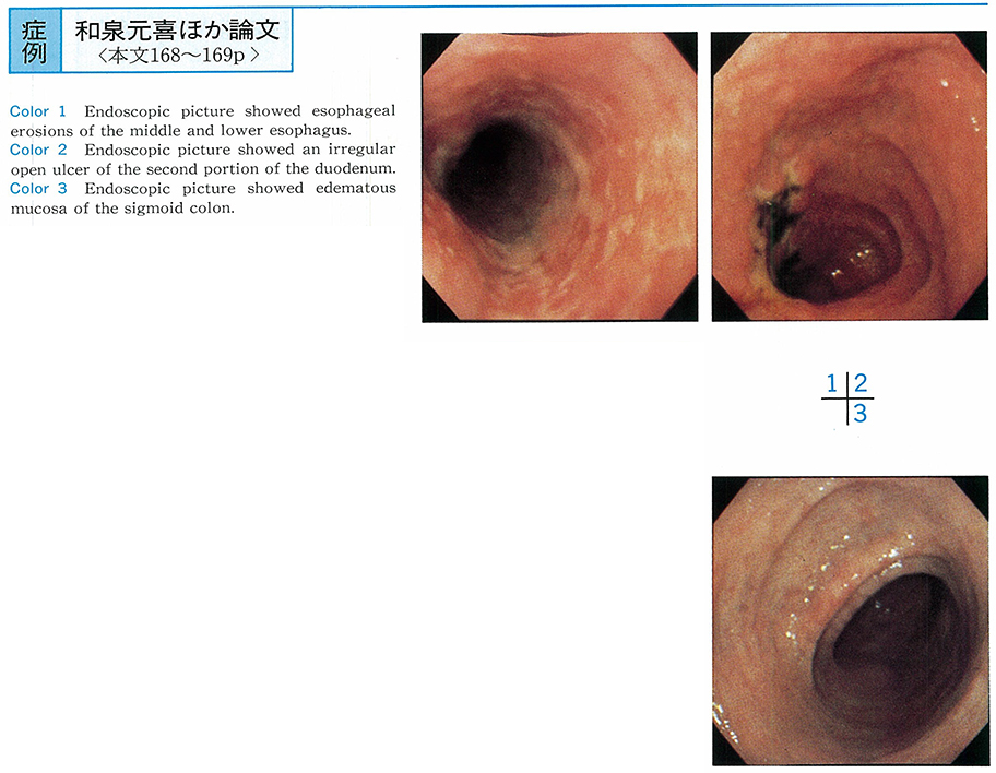
    1995 年 47 巻 p. 168-169
    発行日: 1995/12/08
    公開日: 2015/05/01
    ジャーナル フリー
    A 71-year-old man was admitted to our hospital complaining of nausea and waterly diarrhea. Physical examination on admission showed low grade fever, lower abdominal pain and increasing bowel sound. Laboratory data on admission showed leukocytosis (21,700/mm3) and hypoproteinemia (4.3g/dl) . Occult blood of stool was positive, but stool culture showed no significant bacteria.
    Esophagogastroduodenoscopy showed erosions from middle to lower esophagus, edematous mucosa of the 1st and 2nd portion of duodenum and irregular open ulcers coated with white fur on the 2nd portion of the duodenum. But gastric mucosa was normal. Colonoscopy showed endematous and reddened mucosa of Bauhin's valve, sigmoid colon and rectum, but no abnormal findings were recognized from ascending to descending colon. Histological examination of biopsy specimens of these lesions showed only infiltration of inflammatory cells in the mucosal layer and fibrosis in the submucosal layer, but no granuloma was found.
    We suspected Crohn's disease because of endoscopic findings and clinical course, and so prednisolone 30 mg/day and salazosulfapyridine 3 g/day were given, clinical course and endoscopic findings were improved. But pathologically diagnostic creteria for Crohn's disease were not satisfied and considering of his age, ischemic change was most suspected. So careful follow-up of his clinical course and repeated endoscopic examination are important for establishment of the diagnosis.
  • 三輪 博久, 小川 善秀, 六倉 俊哉
    1995 年 47 巻 p. 170-171
    発行日: 1995/12/08
    公開日: 2015/05/01
    ジャーナル フリー
    A 53-year-old woman was admitted for further examinations for gastric polypoid lesion which looked like submucosal tumor by X-ray examination. X-ray films revealed elevated lesion of 2cm in diameter with bridging folds located on the anterior wall of the middle gastric body close to the lesser curvature. Endoscopically, elevation had bridging folds, covered with normal gastric mucosa and a red small depression was observed on the top. The laparoscopy assisted partial resection of the stomach was performed. Laparoscopically, a white, elastic hard nodule was observed on the serosal surface. The excised specimen revealed a submucosal tumor, 23x20x12mm in size. The heterotopic gastric mucosa with cystic changes was microscopically observed in the submucosal layer extending to submucosal layer. No malignancy was detected. No acinar nor duct structures ware seen, which is pathognomonic for ectopic pancreas. No recurrence was seen 1 year after resection.
  • 鴨志田 佐智子, 山口 俊一, 小峰 文彦, 荒川 泰行, 竹内 勝啓, 宮川 正秀, 阿部 政直, 高本 雄幸, 上田 仁, 仁木 基裕, ...
    1995 年 47 巻 p. 172-173
    発行日: 1995/12/08
    公開日: 2015/05/01
    ジャーナル フリー
    A 60-year-old male with past medical histories of hypertension and diabetes mellitus for 13yrs, had epigastric discomfort. He was pointed out abnormality on upper gastrointestinal fiberscope. Upper GIF revealed multiple elevated lesions from cardia to lower body of the stomach, biopsy of which is carcinoid tumor. As total gastrectomy was performed, resected stomach had 9 tumors, maximum size of which was 42×35mm in diameter.
    Multiple gastric carcinoid tumor which occupies about 10% of all gastric carcinoid tumors, is commonly reported to be about 10mm in diameter. It has usually metastasis if above 10mm in size. In summary, we report a rare case of carcinoid tumor characterized by large tumor size and no metastasis.
  • 稲田 正之, 徳重 浩一, 大倉 康男, 八巻 悟郎, 野本 一夫, 志賀 俊明, 仲谷 弘明, 加茂 章二郎, 鬼倉 秀世, 西沢 護
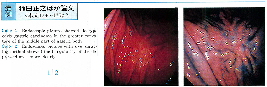
    1995 年 47 巻 p. 174-175
    発行日: 1995/12/08
    公開日: 2015/05/01
    ジャーナル フリー
    A 65-year-old woman who had an abnormality pointed out by radiological examination at gastric mass survey in November 1994, came to our center. Endoscopic and Radiological examination revealed irregular shaped depressed lesion with fold conversion in greater curvature of the middle portion of gastric body. Clinical diagnosis was type of IIc early undifferentiated gastric cancer. But biopsy specimen was differentiated tubler adenocarcinoma.
    From the result of histological diagnosis, it reversed as follows. (1) The outline of lesion was not so well defined, (2) with gradually narrowed the fold, (3) a few small nodules in the depressed lesion, (4) reddish mucosa suggested the tubuler pattern of mucosed pit. These figures were matched the typical figure of defferentiated adenocarcinoma. Gastrectomy was carried out. By the histopathological study, this carcinoma was shown to be differentiated type adenocarcinoma arising from fundic gland mucosa. It is invasion was limited to the submucosal layer.
  • 中村 靖幸, 高橋 宣胖, 柵山 年和, 下野 聡, 水谷 央, 山本 真司, 井上 一成, 大西 健夫, 玄 智傭, 横山 正人
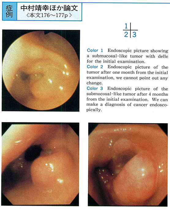
    1995 年 47 巻 p. 176-177
    発行日: 1995/12/08
    公開日: 2015/05/01
    ジャーナル フリー
    A 85-year-old woman visited to our hospital with epigastralgia as her complaint. Submucosal-like tumor was found on the antrum of stomach endoscopically. The biopsy specimen was taken from the tumor. However pathologic diagnosis of malignancy was not obtained, so endoscopic examination was performed every month carefully. After 4 months later from the first examination, the surface of tumor altered to be irregular endoscopically, and the biopsy was successful to diagnose the gastric cancer. Frequent examination may be useful for diagnosis of gastric cancer resembling submucosal tumor.
  • 石塚 裕人, 大谷 吉秀, 久保田 哲朗, 熊井 浩一郎, 北島 政樹
    1995 年 47 巻 p. 178-179
    発行日: 1995/12/08
    公開日: 2015/05/01
    ジャーナル フリー
    Case 1 : This 46-year-old man underwent subtotal gastrectomy in 1991 for two IIc lesions. Endoscopy in 1994 revealed a polypoid lesion in the posterior wall of the gastric remnant, and biopsy results were positive for adenocarcinoma. The lesion was resected by endoscopic mucosal resection (EMR) . Pathological findings revealed well differentiated adenocarcinoma limited to the mucosal layer and the surgical margin was negative.
    Case 2 : This 75-year-old man underwent subtotal gastrectomy for a IIa lesion in 1992. Endoscopy in 1994 revealed a polypoid lesion at the anastomotic site, and biopsy results were adenocarcinoma. Endoscopic ultrasonography (EUS) indicated that cancer was limited to the mucosal layer and EMR was performed. Pathological findings confirmed well differentiated adenocarcinoma limited to the mucosal layer and the surgical margin was negative.
    In the Keio University Hospital, we have strict guidelines for local resection of primary gastric cancer : (1) the lesion must be limited to the mucosal layer, (2) protruding lesions must be smaller than 2.5 cm in diameter, (3) depressed lesions must be smaller than 1.5 cm in diameter and Ul (-) . The indications for EMR are even more limited ; smaller than 1 cm in diameter.
    We have operated on 11 cases of early gastric remnant cancer thus far, and experienced only 1 case of lymph node metastasis in a submucosal cancer of a patient operated for gastric cancer 29 years before the second operation. There have been 124 reported cases of early cancer in the remnant stomach in Japan, and only 3 cases had lymph node metastasis, all submucosal cancers. For these reasons, we can regard EMR for early cancer in the remnant stomach as a good alternative to operation when performed along the same guidelines as primary early gastric cancer.
  • 森 一博, 古谷 亮, 西野 執, 尾崎 元信, 吉利 晃治, 長山 徹, 伊原 文恵, 飯田 和成, 石原 学, 吉谷 和男, 中島 利子
    1995 年 47 巻 p. 180-181
    発行日: 1995/12/08
    公開日: 2015/05/01
    ジャーナル フリー
    A 77-year-old female was admitted diagnosed with pyloric stenosis due to Borrmann type IV gastric cancer, 8 cm in size.
    Abdominal ultrasound and CT scan revealed marked irregular hypertrophy of the gastric antrum with ascites. Bloodwork reveald serum IAP level of 727µg/ml. Our diagnosis did not indicate surgical treatment, however, endoscopic treatment using Nd-YAG laser irradiation of 6,988 joules (50W, 0.5sec) with local injection of 4mg Lentinan was performed each month for three months.
    The patient was able to eat soft rice gruel within 5 days of the initial treatment without experiencing nausea or vomiting. Following the third treatment, upper GI series revealed partial tumor reduction, and bloodwork results indicated serum IAP level of 373µg/ml. The patient exhibited positive recovery (QOL) for 8 months, then declined due to carcinoma pleuritis and peritonitis. The patient died 12 months after the initial endoscopic treatment. Autopsy revealed diffuse hypertrophy of the gastric antrum with signet ring cell carcinoma.
  • 瀬底 正彦
    1995 年 47 巻 p. 182-183
    発行日: 1995/12/08
    公開日: 2015/05/01
    ジャーナル フリー
    A 39-year-old man visited my clinic because of persisitent epigastric pain. He ate raw fish“shime saba”4 days ago, and had epigastralgia since next morning. Physical examination was non-remarkable except for tenderness on epigastric region. Ultrasonography showed gall bladder debris. Endoscopic examination of upper GI tract revealed dome-shaped mucosal nodule with erosions and Anisakis penetrating the mucosa of the duodenal bulb. A warm was successfully removed with using biopsy forceps. Epigastric pain disappeared soon thereafter.
    Anisakiasis is common parasitic infection of GI tract, but rarely found in duodenum (0.64%) . To my knowledge, this is the fifth case of duodenal anisakiasis reported in Japan.
feedback
Top